Colour vision II
1/37
There's no tags or description
Looks like no tags are added yet.
Name | Mastery | Learn | Test | Matching | Spaced | Call with Kai |
|---|
No analytics yet
Send a link to your students to track their progress
38 Terms
what is the trichromatic (young-helmholtz theory)
•The theory proposes that some combination of 3 channels can explain discrimination of colour.
•Each channel would give rise to a specific colour sensation.
•Each channel would have its own spectral sensitivity function
explain the disadvantages of the trichromatic (young-helmholtz) theory (4)
cannot explain presence of 4 unique hues: red, yellow, green and blue
Humans never name colours reddish-green, or bluish-yellow
Cannot explain colour after-images
Cannot explain how dichromats can perceive yellow and white
explain what the Opponent (Hering) colour theory is (4)
proposed 3 retinal substances which was consistent with the trivariance of vision.
3 channels that respond in opposition / operate opponently:
one works in blue yellow opponency / red green opponency/ brightness processing (light and dark)
Not tenable at the level of the retina, but possible post receptorally through neural processing
what does the opponent colour theory allow / advantages of this theory OVER the trichromatic theory (2)
allowed for presence of 4 unique colours in perception (red, blue, green and yellow)
explains after colour images (see pictures)
explain this diagram/how each channel is created
red - green channel = subtract red and green outputs from each other creates a red-green response
blue - yellow channel = Add red and green outputs and subtract from the blue / compare with the blue
achromatic / luminance channel = add L and M
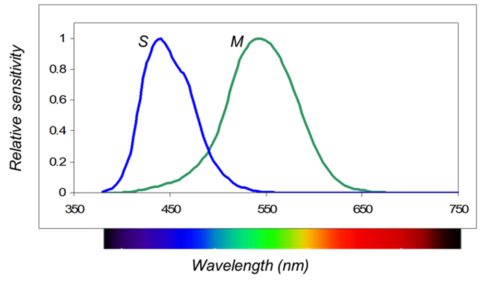
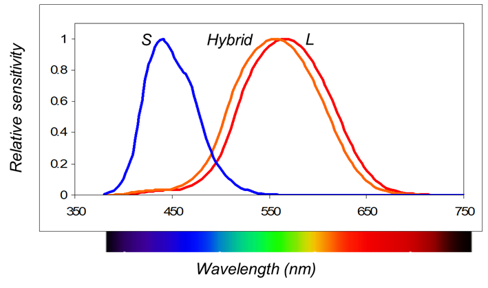
what are the 2 sub-systems of color vision
S-(M+L) channel
(L-M) channel
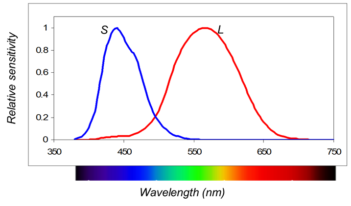
explain the S - (M+L) channel (4)
–Blue-yellow channel
–Compares the signal in S cones with that in L and M cones
–Uses S bipolar cells and bistratified ganglion cells (K cells)
–Synapse at LGN in cells of interlayers (konio cells) and V1 into layers 3 and 4A of V1
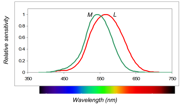
explain the (L-M) channel (4)
–Red-green channel
–Compares the signals of the M and L-wave cones
–Uses midget bipolars and ganglion cells (P cells)
–Synapse at LGN in parvocellular layers (3, 4, 5, 6); and V1 into 4cb
(color processing becomes more complex at V1 than at the LGN)
stage colour theory - what are opponent cells and where are they found
found in retina and visual cortex
nerve cells in the retina and visual cortex that react to colours (on and off switches)
such cells respond differently depending on the target colour
For retinal ganglion cells the receptive fields showed differences in the centre-surround organisation depending on colour.
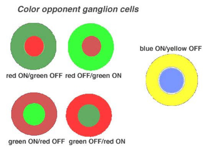
🔴🟢 Red ON / Green OFF – This cell activates when it sees red and turns off when it sees green.
🟢🔴 Green ON / Red OFF – This one is the opposite.
🔵🟡 Blue ON / Yellow OFF – Activates with blue, turns off with yellow.
These cells help your brain figure out exactly what colour you're seeing by comparing what turns "on" and what turns "off".
explain how color vision deficiencies [CVD] are classified (2)
congenital and acquired
congenital = Congenital are present from birth and result from an inherited anomaly with the photopigments or cone/rod structure.
acquired = result from an ocular disease process
percentage of males and females that exhibit a colour vision anomaly
8% of males and 0.5% of females
state and explain the names of the different colour vision deficiencies (1) (1:3) (1:3)
monochromacy = cannot see colours as they only have one photopigment
dichromacy = have 2/3 cone photopigments / without one of them:
–Protanopia
–Deuteranopia
–Tritanopia
Anomalous trichromacy = have 3 photopigments but one is anomalous:
–Protanomaly
–Deuteranomaly
–Tritanomaly
explain what protanopia, deuteranopia and tritanopia is (dichromacy)
protanopia = red deficient - absence of red photopigment/ L cones (red sensing)
deuteranopia = green deficient - absence of green photopigment/ M cones (green sensing)
tritanopia = blue deficient - absence of blue pigment/S cones (blue sensing)
explain what protanomaly, deuteranomaly and tritanomaly is - anomalous trichromacy
protanomaly = red photopigment is anomalous / faulty = red looks duller and harder to tell red from green
deuteranomaly = green photopigment is anomalous/faulty = greens look more like reds so harder to tell apart
tritanomaly = blue photopigment is anomalous/faulty = hard to tell difference between blue and yellow
what are monochromats (5)
–The only true ‘colour blind’ individuals.
–Have never experienced colour perception.
–Can match any colour with a single primary colour.
–They do so through the luminosity channel (brightness match).
–Very rare
explain the typical (rod) monochromats (5)
–Have only rods: there are thought to be no cone photoreceptors.
–Low visual acuity - fovea has no cones
–Nystagmus present - oscillation of the eyes
–Prefer dim light levels
–Vλ function is the same as the dark-adapted eye – V’λ - only have dark spectral sensitivity curves
explain the atypical (cone) monochromats
–Have only blue cones and rods.
–Crude colour perception under low/mesopic light where both are rods and cones are operating.
what are / explain dichromats (3)
Can match any colour/wavelength of light using two primary colours
Absent cone-type or photopigment (L, M or S) but the same number of cones as in ‘normal colour vision’ - same acuity as they have the same number of cones
Dichromacy conveys a selective advantage: dichromats can defeat camouflage designed to confuse colour-normal
explain protanopia (5) - what it is, when it occurs, its effects etc.
Protanopia is the absence of L cone photopigment / red - missing L sensitivity
Protanopia occurs when there is an absence of an L-cone photopigment gene or when this gene is replaced by a hybrid gene which codes for a photopigment whose spectral sensitivity is identical to that of a normal M-cone photopigment.
Protanopes have reduced sensitivity to long wavelength light.
red yellows and greens look the same to them - cannot tell the differences between them
red regions will appear darker
explain deuteranopia (5)
•Deuteranopia is the absence of M-cones / green - missing M sensitivity
•Deuteranopes have either no M-cone photopigment gene or a hybrid gene which has the spectral sensitivity of the normal L-cone photopigment gene.
•Deuteranopes have normal spectral sensitivity - lot of overlap between M and L
reds will be a similar brightness to normal as they still have red curve and blue curve
confuse colours
explain tritanopia (6)
•The tritanopia is the absence of S-cones/ blue - missing S sensitivity
•The result of genetic defects in the S-cone photopigment gene.
•Tritanopes have normal spectral sensitivity.
•Inherited as an autosomal dominant trait.
•Tritanopia is rare = most likely assume it is due to disease not genetics as very rare
Male and female equally affected
what is and explain small field tritanopia
•Seen in everyone with normal colour perception/vision
•The S cones are absent from the central 20 mins of arc of the fovea - in middle of fovea there are no blue cones so we behave like a tritanope
•Hence producing a colour vision defect for targets of 20 mins of arc or less - if target is 20 mins of arc or less then observer will behave like a tritanope
what is a key characteristic of a dichromat and explain this (4)
they possess a neutral point
neutral point = a region in the spectrum that is perceived as colourless (i.e. wavelength that is indistinguishable from white) / one spectral locus that matches perfectly white - one wavelength
this is because of confusion lines - define groups of colours confused by the dichromats and can be shown on the CIE chromaticity diagram.
in normal vision there is no wavelength that matches white
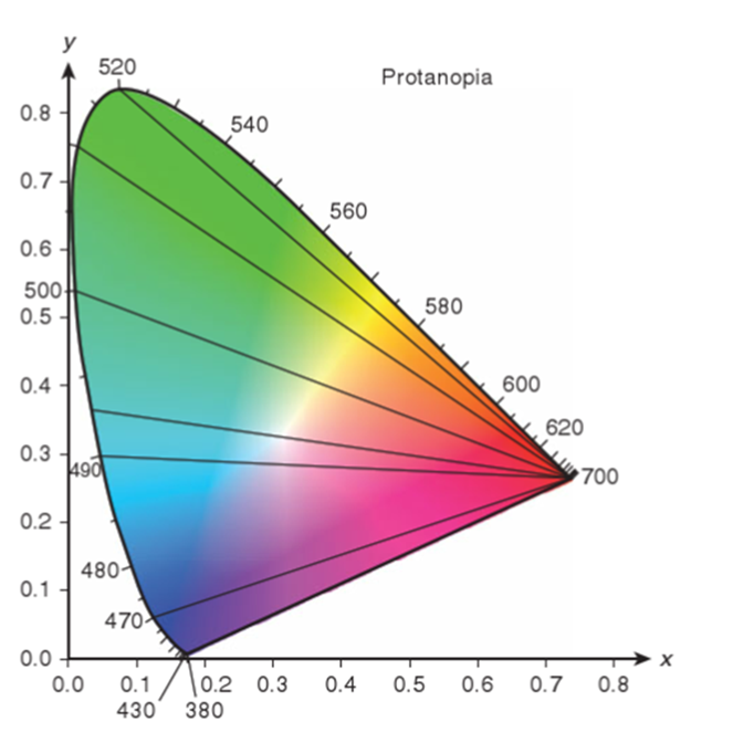
explain confusion lines on a CIE diagram for someone with protanopia and what is the neutral point (5)
•The confusion lines shown join points on the CIE chromaticity diagram that appear to have the same colour for the particular dichromat - these colours look the same for someone with protanopia
•Protanopes can distinguish up to 18 colours (Pitt, 1935) including yellow, white and blue (there should be the same number of lines).
•Normal trichromats can distinguish about 150 spectral hues/different colours
Neutral Point – of a protanope – this point looks white to them (not to others) (492nm)
as they are missing red they cannot see all the colours and confuse some colours - confuse blues,oranges and pinks
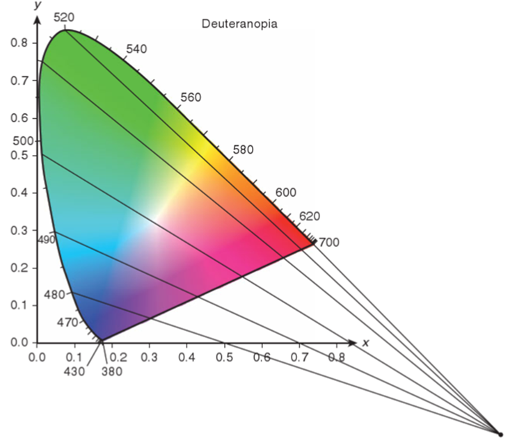
explain confusion lines on a CIE diagram for someone with deuteranopia and what is the neutral point (4)
Deuteranopes can distinguish 28 colours (there should be the same number of lines).
Normal trichromats can distinguish about 150 spectral hues
confuse green, yellow and reds - any colour that lies along those lines
neutral point = 498nm - this wavelength looks white to them - confused with white
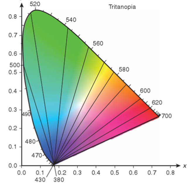
explain confusion lines on a CIE diagram for someone with tritanopia and what is the neutral point (4)
Tritanopes are thought to perceive about 10-11 colours.
Although their perception of colour is reduced, dichromats cannot be classed as true colour-blind as they can distinguish at least some colours.
confuse blues and greens, reds, pinks and violets
neutral point - 570nm - yellow is confused as white to them
explain how anomalous trichromats have trichromatic vision that differs from normal people trichromatic vision (2)
•however, requires a different mixture of primaries to a normal in order to obtain a colour match (so for a normal to make a colour theres a certain amount of red and x colour they would mix together whereas for a protan they would want more red same with deutan and tritan would want more green and blue) anomalous trichromats won’t acceot a normal match
•This is the result of two out of the three cone spectral absorption curves being very similar.
explain why anomalous trichromats have a great degree of variability in their colour vision / varies greatly among them
–Some may have colour discrimination that is nearly as good as that of normals, whereas others may resemble dichromats
how are anomalous trichromats accurately identified and classified (3)
with the use of an anomaloscope that allows the evaluation of an individual’s Rayleigh matches
i.e., the proportions of red (~679nm) and green (~ 544nm) light need to be mixed to match a yellow (~ 589nm).
can tell if they are a dichromat or an anomalous trichromat
explain protanomaly (3)
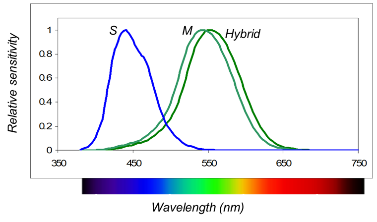
They have 3 cone photopigments but the L/red pigment is replaced by an M-like hybrid pigment - dodgy green pigment
red is anomalous and replaced by the hybrid so they want more red as they have a reduced L/red sensitivity
Protanomals are therefore less sensitive to long wavelength light than normals
explain deuteranomaly (4)
•M pigment/green is replaced by an L-like (red) hybrid pigment.
have reduced M sensitivity
•Deuteranomals have normal spectral sensitivity functions.
•Deuteranomaly is the most common form of congenital colour deficiency
explain the patterns of inheritance of red/green defects - defects in which the M or L cones are affected (5)
–X-linked Recessive.
–Gene carried by mother.
–Usually exhibited by boys.
–8% of males (1 in 12) more common as only need pne
–0.5% of females (1 in 200) less as both XX has to have it
explain occurrance of blue/yellow defects in males and females (2)
much rarer occurring equally in male and females.
As tritanopia is rare it is important to always consider ocular pathology as a possible cause – assume disease before genetics
what are acquired defects the result of
Can be the result of a general or ocular disease, side-effect of medications, toxic poisoning or head trauma
explain predictability of acquired defects
•Acquired defects are unpredictable, they may progress or regress and may not affect both eyes/entire visual field equally
always test monocularly as they do not affect both eyes equally
explain the general rule of acquired colour defects - but many exceptions to the rule (2)
•Defects at the level of the photoreceptors or in pre-retinal media are more commonly associated with tritan deficiencies - blue/yellow
•Defects of the post-receptoral layers (inner retina and ganglion cells i.e. optic nerve) cause a predominantly red-green deficiency.
explain the differences between congenital and acquired CVD’s