Biochem Exam 2
 Call Kai
 Learn
Learn Practice Test
 Spaced Repetition
 Match
Match Flashcards
 Knowt Play
1/260

There's no tags or description
Looks like no tags are added yet.
Name | Mastery | Learn | Test | Matching | Spaced | Call with Kai | Chat |
|---|
No analytics yet
Send a link to your students to track their progress
261 Terms

Myoglobin (Mb)
small protein lacking β structure completely
amino acids are part of eight α helices
hyperbolic binding, high O2 affinity (p50=2.8torr)
His F8 (Proximal Histidine)
The residue that forms the 5th coordination bond directly to the central Fe(II) iron atom
His E7 (Distal Histidine)
Forms a hydrogen bond to the bound O2 molecule

polypeptide chain and heme
what is myoglobin made out of

heme
iron-containing porphyrin derivative
prosthetic group that helps bind oxygen
prosthetic group
an organic compound that allows a protein to carry out some function that the polypeptide alone cannot perform
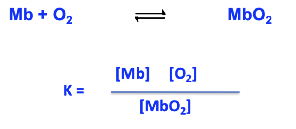
K: dissociation constant
How can binding of O2 to myoglobin be described?
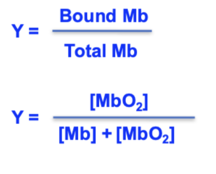
fractional saturation (Y)
the proportion of myoglobin molecules that have bound O2

Y = (pO2) / (K+pO2)
fractional saturation equation

hyperbola
whats the shape of the saturation (Y) vs pO2 plot for myoglobin?
saturation
point at which all virtually all myoglobin molecules have bound to O2
Y is half maximal, pressure is at 50%
K is the concentration of concentration of O2 at which ____ aka _____
760
how many torr in 1 atm
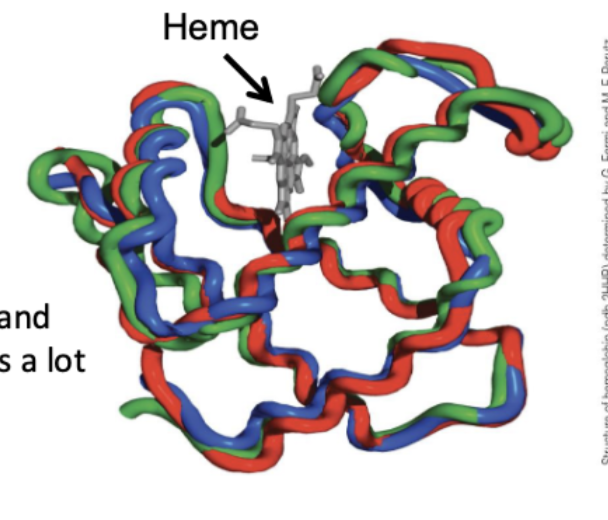
hemoglobin (Hb)
heterotetramer (2α 2β chains; overlapped 3D structure)
all have a heme group in a hydrophobic pocket, a His F8 that ligands the Fe(II) ion, and a His E7 that forms a hydrogen bon to O2.
sigmoidal curve; cooperative binding; lower O2 affinity (p50 = 26 torr)
invariant residues
those that are identical in all the globins
are essential for the structure and/or function of the proteins and cannot be replaced by other residues
found in all vertebrate hemoglobin and myoglobin
hemocratit (40% F; 45% M)
measures oxygen carrying capacity of the blood
(blood volume occupied by RBC)
anemia
too few RBCs

sigmoidal
whats the shape of the saturation (Y) vs pO2 plot for hemoglobin?

26 torr
pO2 at which hemoglobin is half-saturated (p50)
pO2 corresponds to the part of the hemoglobin binding curve where the O2 affinity falls off most sharply
why is hemoglobins oxygen delivery system efficient?
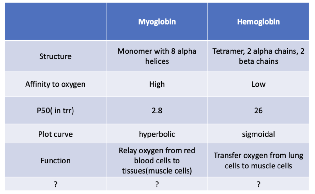
memorize
myoglobin hemoglobin table

rotation of one αβ unit relative to the other
how do oxygenated and deoxygenated states of hemoglobin change conformation?
T (tense)
deoxyhemoglobin state
low O2 affinity
R (relaxed)
oxyhemoglobin state
high O2 affinity
T → R
1st oxygen molecule binding switches the conformation from ___
R → T
1st oxygen releasing switches conformation
allosteric proteins
proteins with multiple binding sites ex. hemoglobin
he binding of a small molecule (called a ligand) to one site alters the ligand-binding affinity of the other sites
O2 pressure, H+ concentration
factors that affect oxygen binding

they become more acidic and release H+
what happens to the N and C terminus groups when oxygen binds to hemoglobin
Bohr effect
the reduction of hemoglobin's oxygen-binding affinity when the pH decreases (H+ increases) is known as the
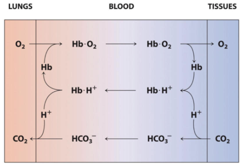
Hemoglobin picks up O2 in the lungs.
In the tissues, H+ derived from the metabolic production of CO2 decreases hemoglobin's affinity for O2, thereby promoting O2 release to the tissues.
Back in the lungs, hemoglobin binds more O2, releasing the protons, which recombine with bicarbonate to re-form CO2.
describe oxygen transport with Bohr effect
lungs
where does hemoglobin pick up O2?
tissues, decreases
in the ____ H+ derived from the metabolic production of CO2 _____ hemoglobin's affinity for O2
lungs, bicarbonate
in the ___ hemoglobin binds more O2, releasing the protons, which recombine with ____ to re-form CO2.
2,3-bisphosphoglycerate (BPG)
Allosteric regulator that binds strictly in the central cavity of the T state, stabilizing deoxy-Hb and lowering O2 affinity
stabilizes the deoxy conformation of hemoglobin; O2 would bind too tightly
why is BPG important?
T (deoxy) state
in what state does BPG bind in the central cavity of hemoglobin?

reduces hemoglobin's O2 affinity by stabilizing the deoxy conformation
effect of BPG
dont like BPG
Why fetus hemoglobin has higher affinity than adult hemoglobin?
sickle cell hemoglobin (Hb S)
Individuals with two copies of the defective gene
debilitating condition that predominantly affects populations of African descent
he hydrophobic valine residues(blue) on hemoglobin S are optimally positioned to bind to this patch
this intermolecular association leads to the rapid aggregation of hemoglobin S molecules to form long, rigid fibers
describe sickle cell hemoglobin
Hemoglobin S (Hb S)
Caused by a single point mutation (β6 Glu→Val)
Exposed valine causes hydrophobic aggregation into rigid fibers in deoxy state
mutant hemoglobin C
makes the red blood cells a bit more rigid than normal, leading to mild anemia
Thalassemias
Genetic disorders resulting from a reduced rate of synthesis of α or β globin chains
Globular proteins
hemoglobin and myoglobin are
actin filaments, intermediate filaments, microtubules
3 structural proteins
extracellularly
fibers of the protein collagen provide structural support (intra/extra)

actin monomer
adenosine triphosphate (ATP) slips into a pocket on the protein
ribose hydroxyl groups and the phosphate groups form hydrogen bonds with the protein
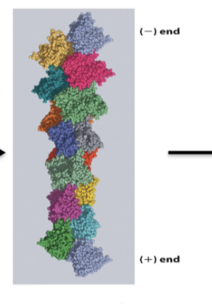
F-actin (filamentous actin)
a double chain of subunits in which each subunit contacts four neighboring subunits
the end with the ATP site is known as the (−) end,
and the opposite end is the (+) end

Actin Filament Assembly
Subunits usually add more rapidly to the (+) end, which therefore grows faster than the (−) end
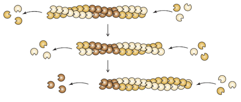
Polymerization of actin monomers
Reversible process, so the polymer undergoes constant shrinking and growing as subunits add to and dissociate from one or both ends of the microfilament
treadmilling
when the net rate of addition of subunits to one end of an actin filament matches the net rate of removal of subunits at the other end, the polymer is said to be ___
Capping, branching, and severing proteins
how is assembly and disassemby of actin filaments regulated
structural support, cell movement, tensile force
functions of actin filaments (3)
tubulin
forms microtubules
microtubule
bout three times thicker and much more rigid than actin because it is constructed as a hollow tube

protein tubulin (α,β)
microtubule structure
guanine nucleotide (GTP or GDP)
what does tubulin bind

protofilament
end-to-end association of tubulin dimers form a short linear

positive (terminating in β tubulin)
which end of tubulin grows faster
at both ends
what end of tubulin does disassembly happen?
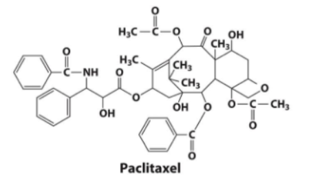
Paclitaxel (taxol)
drug stabilizes microtubules, preventing depolarization

colchicine
drug depolarizes microtubules
intermediate filaments
strictly structural, no cell motility
interact with actin filaments and microtubules via cross-linking proteins
dead remnants of epidermal cells
where are intermediate filaments most prominent?
keratin
best known intermediate filament
soft keratins
help define internal body structures,
hard keratins
in, hair, and claws
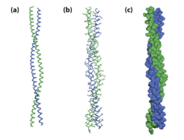
coiled coil (dimer of α helices)
structure of an intermediate filament
100
how many residues in the α helical chain of a coiled coil?

7 residue repeating; first and fourth residues are predominantly nonpolar
amino acid sequence of intermediate filaments consists of
disulfide
what bond cross links keratin fibers
collagen
extracellular structural protein most abundant in animals

every 3rd amino acid is glycine; rest 30% are proline and hydroxyproline
structure of collagen
its a chiral
why does collagens dominant amino acid have to be glycine?
scurvy
lack of vitamin C (ascorbate)
collagen contains too few hydroxyproline residues and hydroxylated lysine residues, so the resulting collagen fibers are relatively weak.
hydroxyproline and hydroxylated lysine
what amino acids are low in scurvy?

catalyzes proline reaction (collagen can’t form without it)
what does ascorbate (vitamin C) do?
covalently (chem modified)
how are collagen molecules cross-linked?
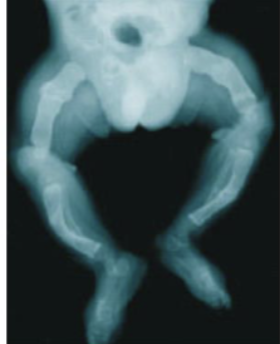
osteogenesis imperfecta
Defects in collagen type I (the major form in bones and tendons) cause the congenital
disease
motor proteins
act on structural elements such as actin filaments and microtubules to generate the movements that allow the cells to reorganize their contents, change their shape, and even crawl or swim
use ATP to carry out mechanical work
ATP hydrolysis → mechanical energy
myosin works with actin to produce movement by converting

2 large polypeptides form 2 globular heads, 1 tail
muscle myosin structure
actin and adenine nucleotide
each head of myosin contains a binding sit for

neck
what acts as a lever and is stiffened by small light chains (2 polypeptides) in myosin?
In a series of steps that include protein conformational changes and the hydrolysis of ATP, the myosin head releases its bound actin subunit and rebinds another subunit closer to the (+) end of the actin filament.
describe the process of myosin rebinding
thick filament
myosin tails associate to form
thin filaments
heads each consist of an actin filament and actin-binding protein
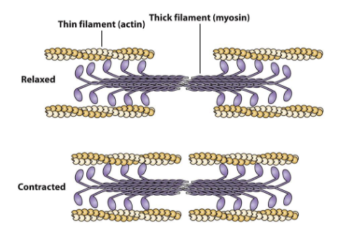
contraction
when myosin acts on actin to shorten muscle

The conversion of ATP to ADP + Pi triggers conformational changes in the myosin head that are communicated to the actin-binding site as well as to the lever (the neck region).
The chemical reaction of ATP hydrolysis thereby drives the physical movement of myosin along an actin filament.
In other words, the free energy of the ATP hydrolysis reaction is transformed into mechanical work
Describe the Myosin-Actin Reaction Cycle
cytokinesis, transport, tension rods
myosin works with actin for what three things?
Usher syndrome
the most common form of deaf-blindness in the United States
is characterized by profound hearing loss, retinitis pigmentosa (which leads to blindness), and sometimes vestibular (balance) problems.
myosin type VIIa
genetic mutations identified in Usher syndrome
premature stop codons, amino acid substitutions, and deletions—all of which compromise the protein's function
microtubule tracks
what does kinesin move along?
Kinesin
a large protein and has two large globular heads and a coiled-coil tail domain.
light chains
situated at the opposite end of the kinesin protein, bind to proteins in the membrane shell of a vesicle(cargo)

Kinesin moves its cargo toward the (+) end of a microtubule along a single protofilament. Other microtubule-associated motor proteins appear to use a similar mechanism but move toward the (−) end of the microtubule.
The motor activity of kinesin requires the free energy of ATP hydrolysis. However, kinesin cannot follow the myosin lever mechanism because its head domains are not rigidly fixed to its neck regions.
In kinesin, a relatively flexible polypeptide segment joins each head to an α helix that eventually becomes part of the coiled-coil tail. In myosin, the lever is a long α helix that extends from the head to the coiled-coil region and is stiffened by the two light chains.
describe the kinesis reaction cycle
processivity
Kinesin is therefore said to have high ____
its cargo can be moved long distances without being lost.
High processivity is advantageous for a transport engine because
immune system
potentially recognize, respond to, and remember millions or billions of antigens