Week 1 (8/25) - Endocrine System
1/56
There's no tags or description
Looks like no tags are added yet.
Name | Mastery | Learn | Test | Matching | Spaced |
|---|
No study sessions yet.
57 Terms
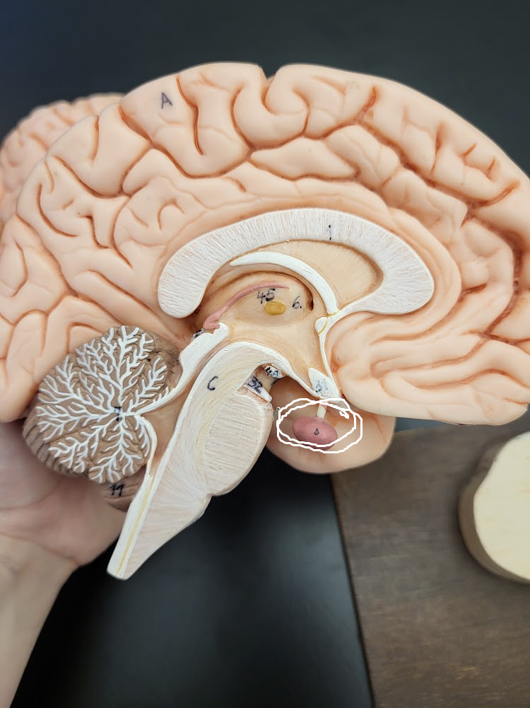
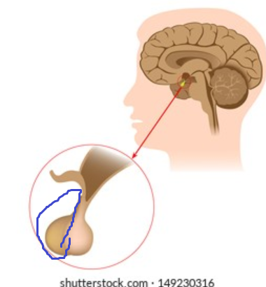
pituitary gland/hypophysis
“dot” coming from a funnel under the hypothalamus that serves partially as a neural communicator and partially as an endocrine communicator
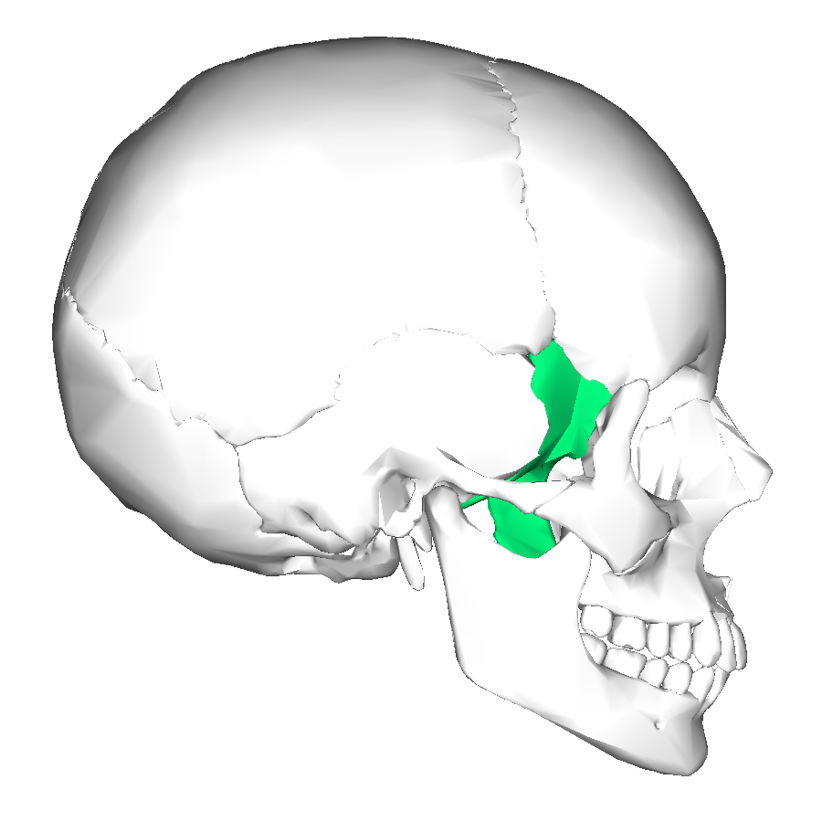
sella turcica
landmark on the sphenoid bone where the pituitary gland sits

sphenoid bone
skull bone visible in the eye cavity and under the zygomatic process, contains the sella turcica landmark
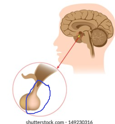
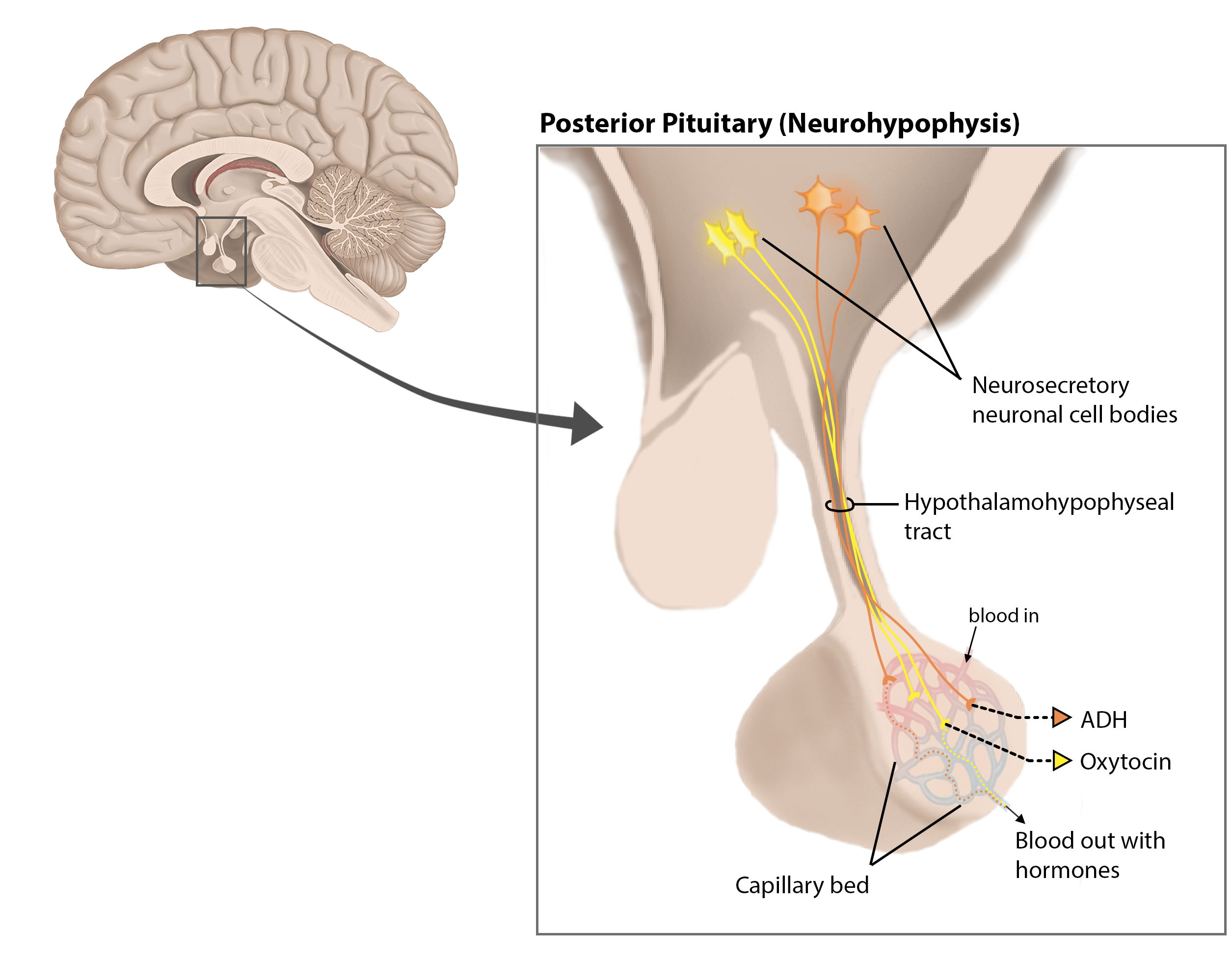
posterior pituitary/neurohypophysis
side of the pituitary gland which contains more neurons and is whiter in color from myelin coating
internal carotid artery
branch of the common carotid artery that runs under/into the brain
inferior hypophyseal artery
inferior artery on the top of the posterior pituitary gland

posterior hypophyseal veins
veins “below” the inferior hypophyseal artery on the posterior pituitary gland

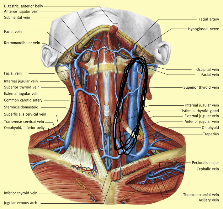
internal jugular vein
vein of the neck that is on the inside, near the internal carotid artery, and thicker than the external one
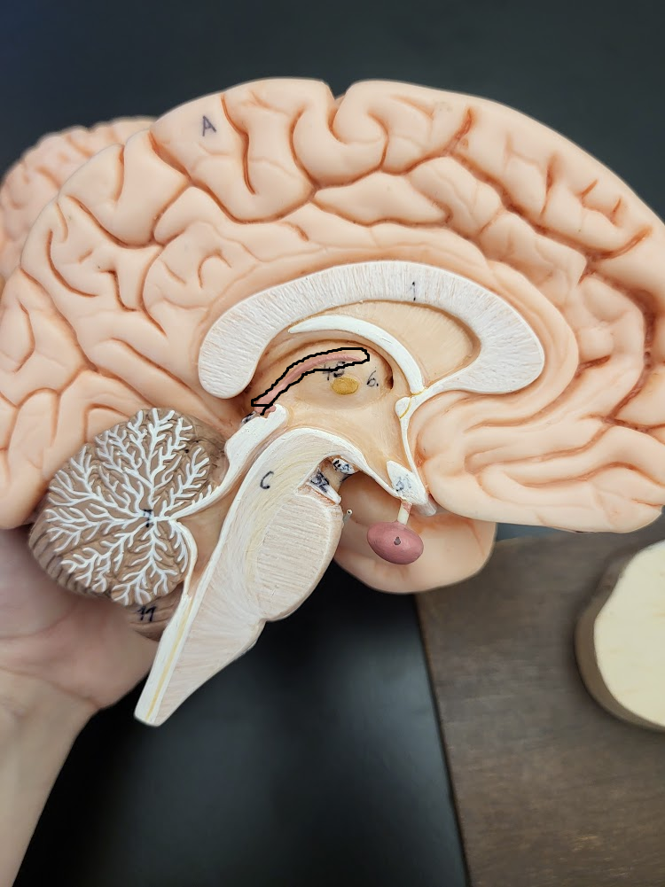
hypothalamic-hypophyseal tract
bundle of axons in the infundibulum that run down from the hypothalamus tissue
anterior pituitary/adenohypophysis
outer side of the pituitary gland that is pink and more glandular
portal system
2 capillary plexuses (beds) connected by veins
hypophyseal portal system
superior hypophyseal artery > primary capillary plexus > hypophyseal portal vein > secondary capillary plexus > anterior hypophyseal veins
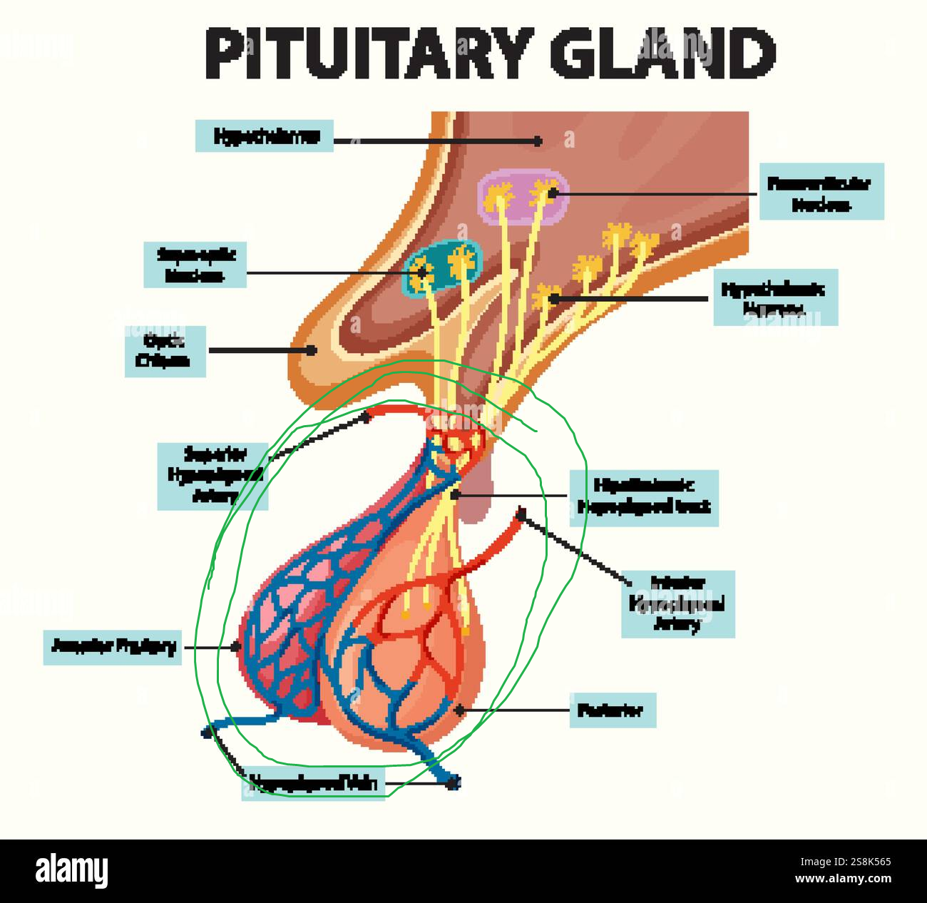
superior hypophyseal artery
1st in hypophyseal portal sys, supplies anterior pituitary, located at the very top where the lobe connects (higher than inferior one)

primary capillary plexus
2nd in hypophyseal portal sys, part of anterior pituitary, located at the top of where superior hypophyseal artery connects to lower veins

hypophyseal portal vein
3rd in hypophyseal portal sys, part of anterior pituitary, located between primary and secondary capillary plexuses
secondary capillary plexus
4th in hypophyseal portal sys, part of anterior pituitary, located in bulk of the anterior lobe

anterior hypophyseal veins
last in hypophyseal portal sys, part of anterior pituitary, pop out the end of the lobe

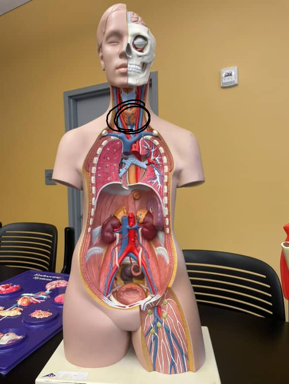
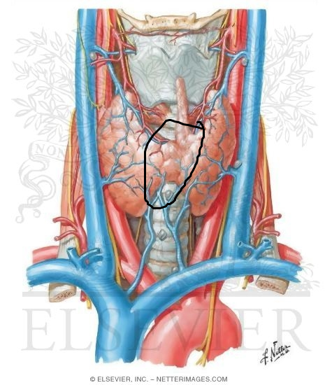
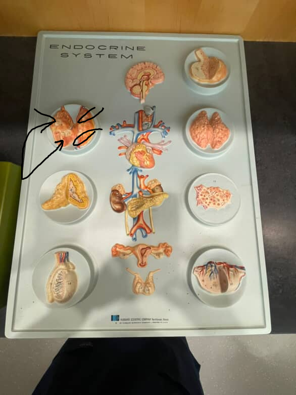
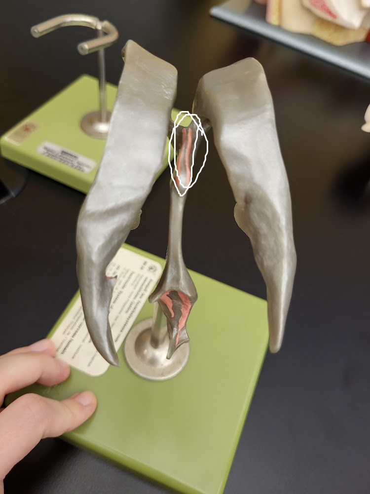
thyroid gland
butterfly shaped gland in the neck on the trachea, right below the larynx
right and left lateral lobes
either side of the thyroid gland
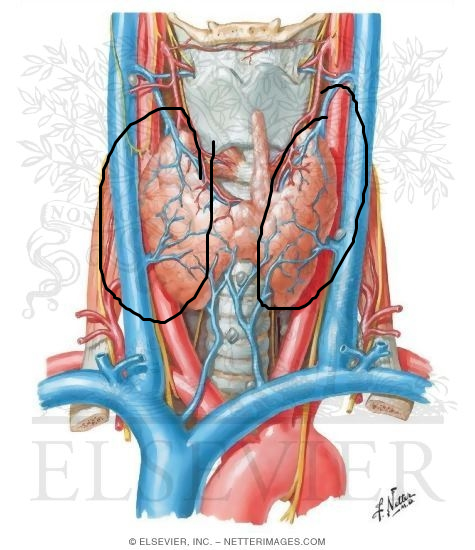
isthmus
region connecting lobes of the thyroid gland
trachea
windpipe under and continuing on below the thyroid gland

larynx
voicebox just superior to the thyroid gland

external carotid artery
branch of the common carotid artery on the side of the face that branches to the left, does not supply into the brain, enters top of thyroid gland

parathyroid glands
4 amber colored dots on the back view of the thyroid gland
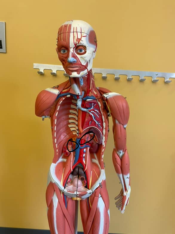
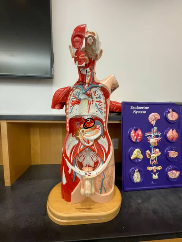
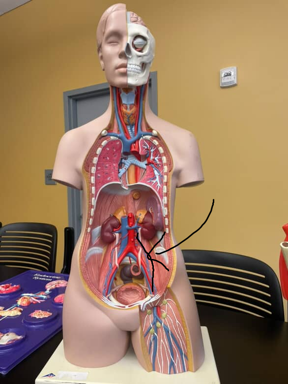
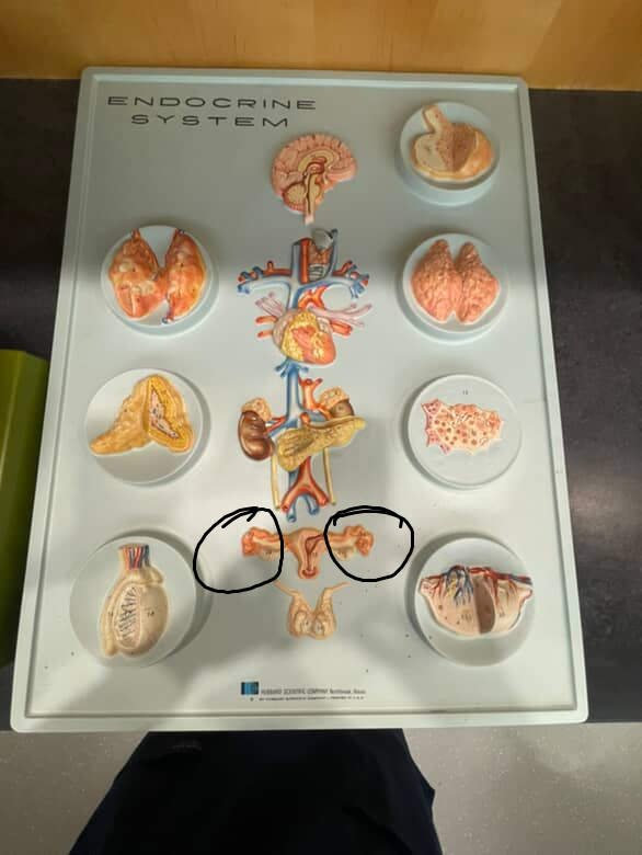
adrenal glands
located over top of the kidneys

kidneys
bean shaped organs in the back of the abdomen, under adrenal glands
suprarenal arteries
branches off abdominal aorta that branch into the adrenal glands

suprarenal vein
veins that stick into the adrenal glands
adrenal medulla
the inner part of the adrenal gland that is mostly a knot of nervous tissue
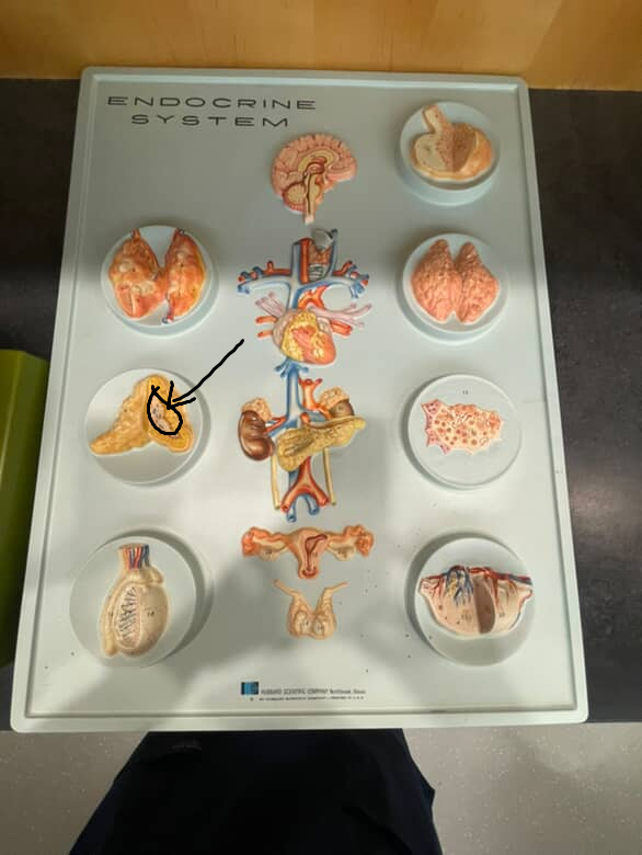
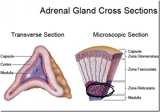
adrenal cortex
the yellow, fatty outer part of the adrenal glands with 3 zones

zona glomerulosa
outer part of the adrenal cortex, producing mineralcorticoids
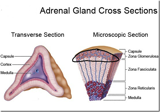
zona fasciculata
middle zone of adrenal cortex, making glucocorticoids
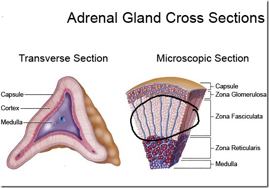
zona reticularis
outer zone of the adrenal cortex, making gonadocorticoids
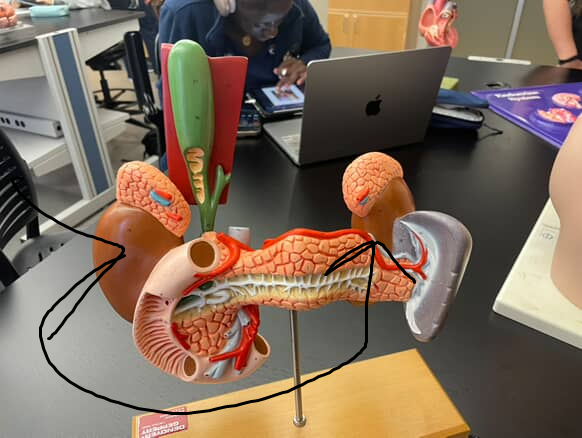
pancreas
tadpole shaped organ near the liver, yellow and fatty

duodenum
top of the small intestine, encircles the head of the pancreas
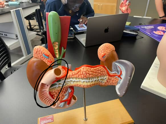
head of pancreas
part of pancreas that is thicker and near the duodenum

tail of pancreas
thinner part of the pancreas, bordering the spleen

celiac trunk/axis
trunk or diverging point on the abdominal aorta, near the pancreas/duodenum interssection
hepatic portal vein
vein that diverges out like a v from the liver

gonads
refers to sex endocrine organs
ovarian artery
gonadal artery in females that descends form the abdominal aorta and supplies the ovary from the top, under suspensory ligaments
testicular artery
gonadal artery in males that descends from the abdominal aorta and supplies the testicle from the side/top

ovarian vein
vein that descends from near the kidneys and enters the top of the ovaries

testicular vein
vein that descends from near the kidneys and enters the top of the testes

ovaries
female gonads, producing ova and hormones
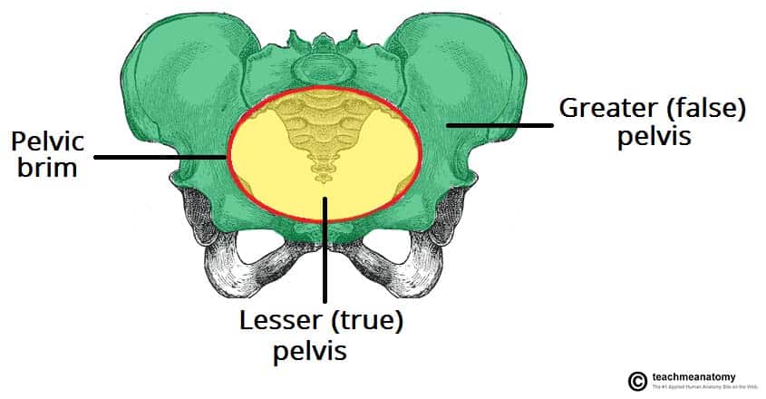
true pelvis
portion of pelvis inferior to the pelvic brim
uterine tubes
tubes over top of the ovaries

uterus
triangle shaped cavity where the uterine tubes come together

testes
male gonads, located in scrotum sack under the penis

scrotum
skin pouch surrounding the testes

vas deferens
sperm cord connecting to testes
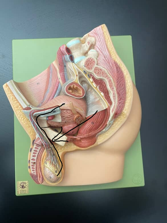
penis
male sex organ over top of the testicles

spermatic cord
cord connecting to the testes, surrounds the vas deferens

pineal gland
pinecone shaped gland hanging from the roof of the third ventricle in the diencephalon
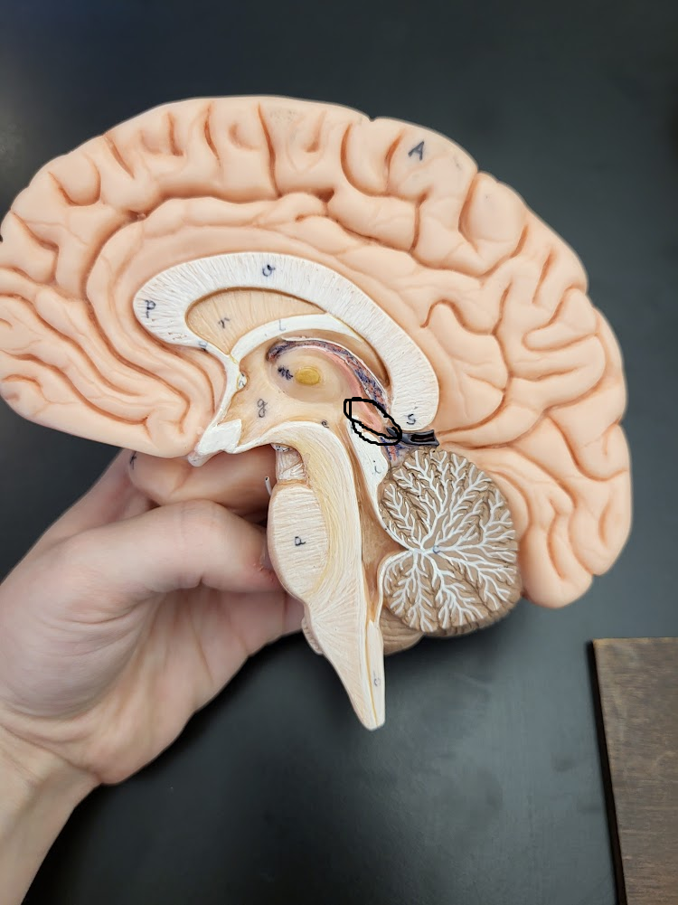
epithalamus
brain region that contains choroid plexus & the pineal gland
third ventricle
“middle part” of the brain’s ventricular system, containing a “hole” in the middle
superior colliculus
smaller, more superior bump in the diencephalon, under the two big ovals (or just under the pineal gland in a side view)