Salivary Glands

0.0(0)
studied byStudied by 0 people
learnLearn
examPractice Test
spaced repetitionSpaced Repetition
heart puzzleMatch
flashcardsFlashcards
Card Sorting

1/6

encourage image

There's no tags or description

Looks like no tags are added yet.

Study Analytics
Name
Mastery
Learn
Test
Matching
Spaced

No study sessions yet.

7 Terms

1
New cards

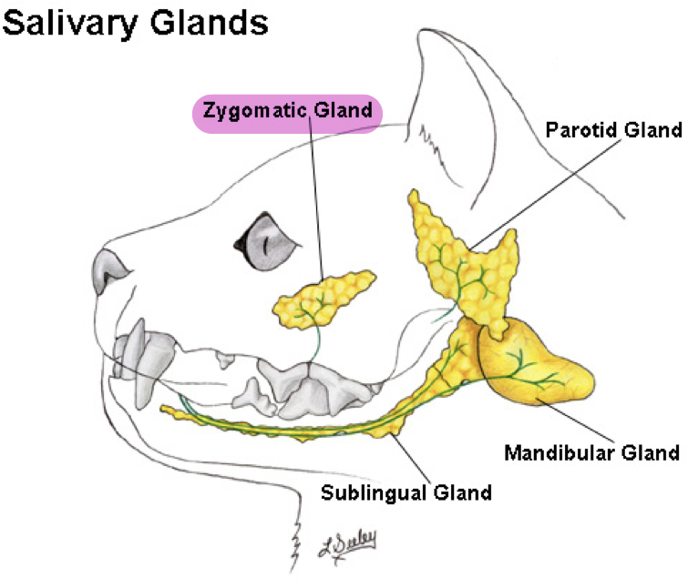
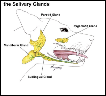
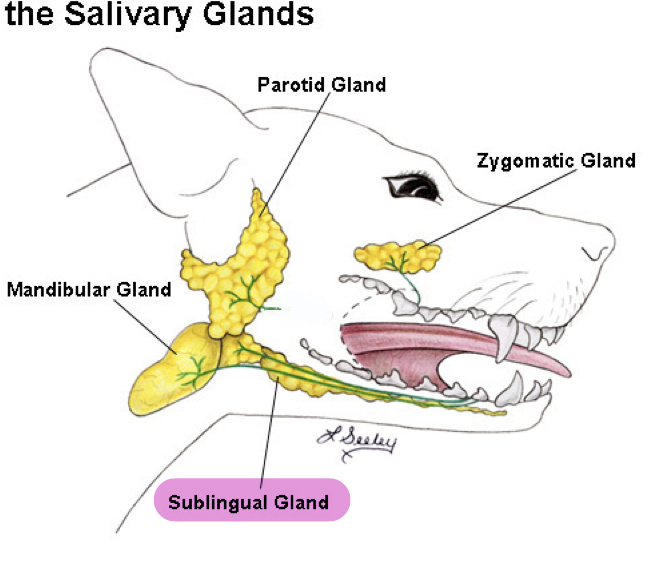
What are the 4 salivary glands?

  • Parotid

  • Mandibular

  • Sublingual

  • Zygotmatic

2
New cards

Salivary Glands

  • Aids in

  • Acts as

  • Contains which enzyme

    • Function

    • Present in

    • Absent in

Aids in: Formation of bolus

Acts as: Lubricant during swallowing

Contains which enzyme: Amylase

  • Function: Hydrolysis and digest of starch

  • Present in: Pigs

  • Absent in: Ruminants and dog/cats

3
New cards

Parotid Gland

  • Color

  • Location

    • Dorsally

    • Ventrally

    • Medially

  • In horse makes contact with

  • Duct opens where

Color: Lighter red

Location: Fills retromandibular fossa

  • Dorsally: Base of ear

  • Ventrally: Extend into neck and intermandibular space

  • Medially:

    • Common carotid artery

    • External jugular vein

    • Hyoid bones and its muscles

    • Branches of facial and trigeminal nerves

    • Lymph nodes

In horse makes contact with: Guttural pouch

Duct opens on: Buccal vestibule

<p><strong><u>Color:</u> </strong><span style="color: red"><strong>Lighter red</strong></span></p><p><strong><u>Location:</u> </strong>Fills <span style="color: red"><strong>retromandibular fossa </strong></span></p><ul><li><p><strong><u>Dorsally:</u> </strong><span style="color: red"><strong>Base</strong></span><strong> </strong>of <span style="color: red"><strong>ear</strong></span></p></li><li><p><strong><u>Ventrally</u>: </strong><span style="color: red"><strong>Extend</strong></span><strong> </strong>into <span style="color: red"><strong>neck</strong></span><strong> </strong>and <span style="color: red"><strong>intermandibular space </strong></span></p></li><li><p><strong><u>Medially:</u> </strong></p><ul><li><p>Common carotid artery </p></li><li><p>External jugular vein </p></li><li><p>Hyoid bones and its muscles </p></li><li><p>Branches of facial and trigeminal nerves </p></li><li><p>Lymph nodes</p></li></ul></li></ul><p><strong><u>In horse makes contact with</u>: </strong><span style="color: red"><strong>Guttural pouch</strong></span></p><p><strong><u>Duct opens on:</u> </strong><span style="color: red"><strong>Buccal vestibule </strong></span></p>
4
New cards

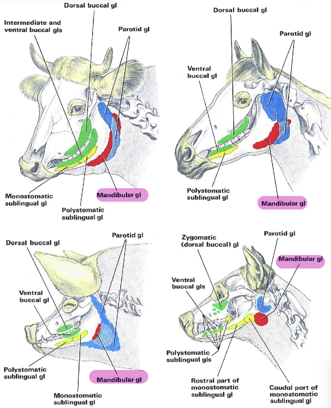
Shape of Parotid Gland in Different Species

  • Carnivores

  • Pig

  • Ruminent

  • Horse

Carnivores: Small and triangular

Pig: Large and triangular

Ruminant: Club shaped

Horse: Large and fills retromandibular fossa
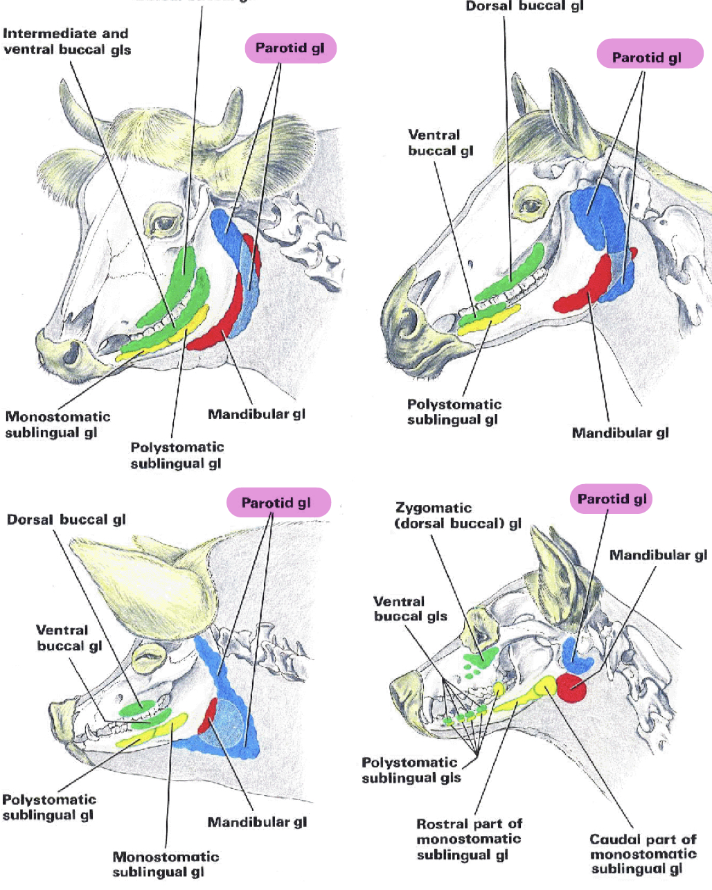
<p><strong><u>Carnivores:</u> </strong><span style="color: red"><strong>Small</strong></span><strong> </strong>and <span style="color: red"><strong>triangular</strong></span></p><p><strong><u>Pig:</u> </strong><span style="color: red"><strong>Large</strong></span><strong> </strong>and <span style="color: red"><strong>triangular</strong></span></p><p><strong><u>Ruminant:</u> </strong><span style="color: red"><strong>Club shaped</strong></span></p><p><strong><u>Horse:</u> </strong><span style="color: red"><strong>Large</strong></span><strong> </strong>and fills<span style="color: red"> <strong>retromandibular fossa </strong></span></p><p></p>
5
New cards

Mandibular Gland

  • Location

  • Partly covered by

  • Shape and number compared to parotid in

    • Dog

    • Pig

  • In ruminants

  • In horse

  • Duct open on

Location: Between basihyoid and wing of atlas

Partly covered by: Parotid gland

Shape and number compared to parotid in

  • Dog: Oval, more than parotid

  • Pig: Oval, less than parotid

In ruminants: Larger and extend from wing of atlas into intermandibular space

In horse: Less than parotid, long and narrow

Duct open on: Floor of oral cavity

<p><strong><u>Location:</u> </strong>Between <span style="color: blue"><strong>basihyoid</strong></span><strong> </strong>and <span style="color: blue"><strong>wing</strong></span><strong> </strong>of <span style="color: blue"><strong>atlas</strong></span></p><p><strong><u>Partly covered by:</u> </strong><span style="color: blue"><strong>Parotid gland</strong></span></p><p><strong><u>Shape and number compared to parotid in</u></strong></p><ul><li><p><strong><u>Dog:</u> </strong><span style="color: blue"><strong>Oval</strong>, <strong>more </strong></span>than<strong> </strong><span style="color: blue"><strong>parotid</strong></span></p></li><li><p><strong><u>Pig:</u> </strong><span style="color: blue"><strong>Oval, less</strong></span><strong> </strong>than <span style="color: blue"><strong>parotid</strong></span></p></li></ul><p><strong><u>In ruminants:</u> </strong><span style="color: blue"><strong>Larger</strong></span><strong> </strong>and <span style="color: blue"><strong>extend</strong></span><strong> </strong>from <span style="color: blue"><strong>wing</strong></span><strong> </strong>of <span style="color: blue"><strong>atlas</strong></span><strong> </strong>into <span style="color: blue"><strong>intermandibular space</strong></span></p><p><strong><u>In horse:</u> </strong><span style="color: blue"><strong>Less</strong></span><strong> </strong>than<span style="color: blue"> <strong>parotid, long</strong></span><strong> </strong>and <span style="color: blue"><strong>narrow</strong></span></p><p><strong><u>Duct open on:</u> </strong><span style="color: blue"><strong>Floor</strong></span><strong> </strong>of<span style="color: blue"> <strong>oral cavity </strong></span></p><p></p>
6
New cards

Sublingual Glands

  • Number

  • Location

Number: 2

Location: Under mucosa of lateral sublingual recess and lateral surface of tongue

<p><strong><u>Number:</u> </strong><span style="color: green"><strong>2</strong></span><strong> </strong></p><p><strong><u>Location:</u></strong><span style="color: green"><strong> Under mucosa </strong></span>of <span style="color: green"><strong>lateral sublingual recess</strong></span><strong> </strong>and <span style="color: green"><strong>lateral surface</strong></span><strong> </strong>of <span style="color: green"><strong>tongue</strong></span><strong> </strong></p>
7
New cards

Zygomatic Gland

  • In dog called

  • Location

  • Duct open into

In dog called: Dorsal buccal gland

Location: Medial to zygomatic arch

Duct open into: Buccal vestibule

<p><strong><u>In dog called:</u> </strong><span style="color: purple"><strong>Dorsal buccal gland</strong></span></p><p><strong><u>Location:</u> </strong><span style="color: purple"><strong>Medial</strong></span><strong> </strong>to <span style="color: purple"><strong>zygomatic arch</strong></span></p><p><strong><u>Duct open into:</u></strong><span style="color: purple"><strong> Buccal vestibule</strong></span><strong> </strong></p>