Cardiology
 Call Kai
 Learn
Learn Practice Test
 Spaced Repetition
 Match
Match Flashcards
 Knowt Play
1/114

There's no tags or description
Looks like no tags are added yet.
Name | Mastery | Learn | Test | Matching | Spaced | Call with Kai | Chat |
|---|
No analytics yet
Send a link to your students to track their progress
115 Terms
Describe the normal cardiac cycle (atrial contraction → diastole)
Atrial Contraction = End of ventricular diastole
Atria contract → pushes the final portion of blood into the ventricles.
AV valves open, semilunar valves closed
Completes ventricular filling = End-diastolic volume (EDV)
Isovolumetric Ventricular Contraction = Start of ventricular systole
Ventricles contract → ventricular pressure rises
AV valves close = S1
All valves are now closed
Ventricular volume stays constant, but pressure increases
Ventricular Ejection = Ventricular pressure > Ao/PA pressure
Semilunar valves open
Blood is ejected into the aorta/pulmonary artery
Ventricular volume decreases
Isovolumetric Ventricular Relaxation = End of systole → beginning of diastole
Ventricles relax → ventricular pressure falls
When ventricular pressure falls below aortic/PA pressure, semilunar valves close = S2
All valves are briefly closed
Ventricular volume remains constant at end-systolic volume (ESV) while pressure rapidly decreases
Ventricular Filling
When ventricular pressure falls below atrial pressure, AV valves open
Blood flows from atria → ventricles.
Rapid ventricular filling occurs first, followed by reduced filling (diastasis).
Then atrial contraction occurs again to complete filling
List 6 determinants of cardiac output (CO)
Heart rate
Stroke volume and preload
Contractility
Afterload
Distensibility
Synergy
Determinants of CO: #1 HR
Control
3 Effects of increased HR
Control: Inherent automaticity of SA node
PNS (vagal) stimulation → Decreases HR
SNS → Increases HR
Baroreceptors increase PNS and inhibit SNS
Effects of Tachycardia:
Increased CO (CO = HR x SV)
Increased myocardial O2 demand
Decreased time for ventricular filling and coronary perfusion during diastole → Myocardial ischaemia → Arrhythmia
Determinants of CO: #2 Stroke Volume and Preload
Define preload
7 Determinants of preload
5 Factors increasing preload
3 Factors reducing preload
Preload: Amount of stretch of sarcomere at the end of diastole (eDV)
eDV = Volume of blood in heart at end of diastole
More blood → More stretch → More powerful contraction → Increased preload → Increased SV
Determinants of Preload:
Blood volume
Body position (recumbency increases VR)
Skeletal muscle activity (venous pump)
Intrathoracic pressure (increased pressure → decreased VR)
Atrial contraction (accounts of 20% of ventricular eDV)
Ejection fraction (less blood ejected → more volume in ventricle for next beat)
MAP
Factors Increasing Preload: Excessive preload → Venous congestion
Venoconstriction (SNS) → Forces blood into heart (less stored in veins)
Increased blood volume (eg. renal retention)
Reduced ventricular contractility → Reduced ejection of blood from ventricle (lower ejection fraction)
Venous congestion (eg. valvular insufficiency)
Decreased ventricular relaxation in diastole
Factors Decreasing Preload:
Venodilation
Reduced circulating blood volume
Increased volume of blood ejected at last contraction (higher ejection fraction)
Determinants of CO: #3 Contractility
Define “shortening fraction”
3 Factors increasing contractility
3 Factors decreasing contractility
Shortening Fraction: % of blood volume ejected from ventricles of the total blood volume available to be pumped out at the end of diastole
= SV ÷ eDV

Fractional shortening = Change in internal ventricular diameter between diastole and systole
Reduced shortening indicates smaller difference between systole and diastole volume in ventricle due to volume overload
Factors Increasing Contractility:
Increased β-adrenergic stimulation (eg. dobutamine/pimobendan = positive inotropes)
Reduced vagal tone
Increased preload
Factors Decreasing Contractility:
Reduced SNS (eg. β-blockers = negative inotropes)
Reduced preload
Increased vagal tone (PNS)
Determinants of CO: #4 Afterload
Definition
Equation
3 Factors increasing afterload
Definition: Tension in myocardium during ejection (represents force opposing ejection of blood during systole)
Equation:

Factors Increasing Afterload:
Increased ventricular volume
Increased arterial vascular resistance
Decreased ventricular wall thickness
Determinants of CO: #5 Distensibility
Definition
Define lusitropy
4 Factors reducing distensibility
Definition: Ease of ventricular filling during diastole
Reduced distensibility → Increased preload
Lusitropy: Ability to relax, distend and fill
Factors Decreasing Distensibility:
Pericardial disease (i.e. pericardial effusion)
Reduced SNS
Increased wall thickness
Increased collagen, scarring or cellular infiltration in ventricular wall
Determinants of CO: #6 Synergy
Definition
2 Factors affecting synergy
Definition: Normal coordinated sequence of muscle activation
Reduced synergy → Reduced SV
Factors:
Cardiac arrhythmia
Regional ventricular conduction abnormalities
Define congestive heart failure
Marks end of asymptomatic compensatory period of heart disease, characterised by retention of fluids in tissues/body cavities ± decreased perfusion of tissues
Requires lifelong medication → Inevitable death with/of heart failure
Often long asymptomatic period with heart murmur ONLY (no medication required)
Forward failure = Inadequate perfusion of vital organs
Backward failure = Venous congestion
eg. big spleen and liver OR pulmonary congestion
Left OR right-sided
Clinical Signs of CHF
6 Historical findings
17 findings on PE
LCHF (3)
RCHF (4)
Both (10)
History: “Dogs With Cardiac Abnormalities Smoke Excessively”
Dyspnoea/wheeze/orthopnoea/tachypnoea (LCHF)
Pulmonary oedema/pleural effusion (backward failure)
Orthopnoea = Respiratory distress while recumbent
Weakness (LCHF)
Reduced tissue perfusion (forward failure)
Cough (uncommon) (LCHF)
LAE compresses L mainstem bronchus
Bronchial oedema (severe pulmonary oedema in dogs ONLY) = Wet and soft cough
Abdominal distension (RCHF)
Ascites ± hepatomegaly/splenomegaly (backward failure)
Syncope/lethargy (LCHF) = Temporary loss of consciousness due to insufficient glucose/O2 delivery to brain
Reduced cerebral perfusion (forward failure)
Commonly precedes excitement
Reduced Exercise Intolerance (LCHF/RCHF)
Low CO
Physical Examination:
LCHF
Increased RRR
Dyspnoea, cough, wheeze, crackles (pulmonary congestion)
Muffled heart sounds
RHCF
Jugular venous distension/jugular pulse (systemic congestion)
Hepatomegaly/splenomegaly
Abdominal effusion
SC oedema
BOTH = Low CO
Slow CRT (>2s)
Pale/cyanotic MM
No sinus arrhythmia
Dry/tacky MM
Cold extremities
Precordial thrill
Cardiomegaly on precordial percussion
Weak femoral pulses
Pulse deficits = Arrhythmia
Gallop/murmur
List 10 steps of the cardiac clinical examination (+ examples)
History and signalment
Distance examination
Peripheral circulation
CRT
MM
Warmth of extremities
Jugular vein
Arterial pulse (femoral)
Precordial palpation
Rate
Rhythm
Abnormal fluid accumulation = Palpation and ballottement of dependent areas for ascites, SC oedema, pleural effusion and hepatomegaly/splenomegaly
Cardiac auscultation
Heart sounds and murmurs
HR
Rhythm
Respiratory auscultation
RR
Pattern
Sounds
Cough
Additional tests
Thoracic radiography
Blood pressure
ECG
Echocardiography
Blood tests
proBNP
Cardiac troponin-1
Serum biochemistry
Jugular Vein Assessment
Purpose
3 features to assess
Normal
Abnormal
8 DDx
Purpose: Assess for RCHF and systemic congestion (also thrombosis)
Features:
Jugular Distension (mild RCHF)
Normal: <1/3 up neck
Abnormal: >1/2 up neck (increased CVP and impaired right-sided filling)
Hepatojugular Reflux (moderate RCHF) = Gently compress cranial abdomen to increase venous return
Normal: Heart copes → No change in jugular distension
Abnormal: Jugular becomes more distended
Jugular Pulse (severe RCHF) = Do NOT mistake for carotid artery pulse
DDx:
Cardiac tamponade
Right atrial mass
DCM
Tricuspid valve regurgitation (TR)
Hypertrophic RV
Arrhythmia
Pulmonic stenosis (PS)
Hypertension
Systemic Arterial Pulse
Definition
Disadvantage
6 Abnormal characters of pulse pressure (+ DDx)
Definition: Difference between DAP and SAP → “Proxy” for MAP
Usually due to ∆SAP (not DAP)
-ve: Can be near normal despite reduced CO due to compensatory arterial vasoconstriction
Abnormal Characters of Pulse Pressure:
Difference between R and L femoral pulse
DDx: Thromboembolism
Hyperkinetic = Strong/bounding due to large difference between DAP and SAP
DDx: HCM, PDA, bradycardia (increased diastolic filling → increased SAP)
Hypokinetic = Weak due to small difference between DAP and SAP
DDx: DCM, PS, AS, shock, dehydration, CHF (lower SAP and tachycardia reduces contractile strength)
Pulsus alternans = Alternating weak and strong pulse
DDx: Severe myocardial disease (LV systolic dysfunction)
Pulsus paradoxus = Weak pulse on inspiration
DDx: Cardiac tamponade
MoA: Inspiration → Increased intrathoracic pressure → Increased VR → Increased RV filling → Pushes on LV → Decreased LV filling → Decreased SAP
Pulse deficits = Heart beat without pulse (HR > PR)
DDx: Ventricular arrhythmia (often auscultated as premature heart beat with no pulse due to insufficient ventricular filling time)
Palpating the Apex Beat
Location
DDx for:
Caudal displacement (2)
Right displacement (2)
Hyperkinetic (1)
Hypokinetic (4)
Location: Left side, 5th ICS = Mitral valve at the level of the costochondral junction
DDx:
Caudal Displacement
Enlarged L-heart
Space-occupying mass
Right Displacement
RVE
Space-occupying mass
Hyperkinetic = Volume overload
Hypokinetic
Obesity
Reduced contractility
Pleural/pericardial effusion
Mass
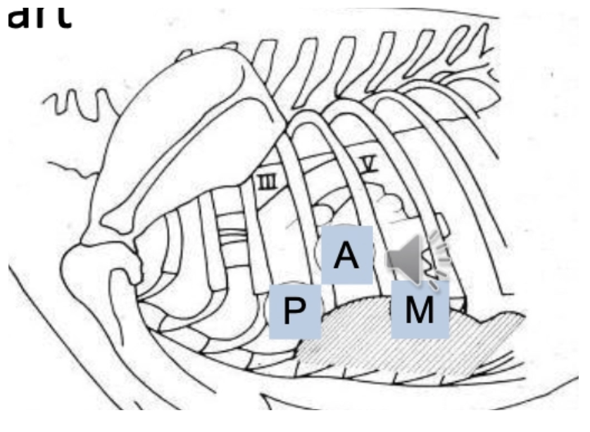
Cardiac Auscultation
4 Ways to classify heart sounds
Diaphragm vs. bell of stethoscope
Approach
Classification:
Frequency (pitch)
Amplitude of vibrations (volume/intensity)
Duration
Quality (timbre) = Affected by heart structures hit by blood
Stethoscope:
Diaphragm = Transmits high frequency and attenuates low frequency
Bell = Transmits high AND low frequency → Ideal for gallop sounds or low-frequency murmurs
Approach:
Have patient standing (avoid displacement of heart) and ensure they are not panting/purring
Palpate for apex beat
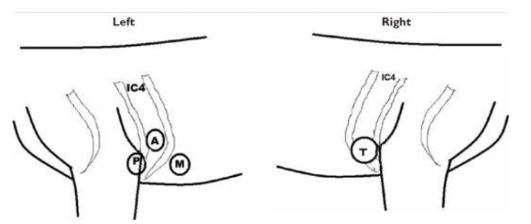
Auscultate ALL valves
Cat: Mid-sternum → Cranially, caudally, left and right
Fill out the following table
Valve | Position | Sound Most Prominent |
|---|---|---|
Mitral | ||
Aortic | ||
Pulmonic | ||
Tricuspid |
Valve | Position | Sound Most Prominent |
|---|---|---|
Mitral | Left 4 - 6th ICS @ costochondral junction | S1 |
Aortic | Move dorsally and cranially from mitral valve (level of the point of the shoulder) | S2 |
Pulmonic | Left 3rd ICS @ sternal border (in axilla) | - |
Tricuspid | Right 3rd - 4th ICS @ level of costochondral junction | - |
Heart Rate and Rhythm
Values for bradycardia/tachycardia of dogs vs. cats
MoA of sinus arrhythmia
Species | Bradycardia | Tachycardia |
|---|---|---|
Dog (Breed-dependent) | < 60 - 70 | > 160 |
Cat | < 120 | > 200 (true tachycardia >160 but stress-tachycardia common in consult) |
Sinus Arrhythmia: Indicates no CHF (good CO and low sympathetic tone)
Inspiration = Expansion of pulmonary vasculature → Decreased SVR
→ Decreased vagal tone → Increased HR
Expiration = Increased vagal tone → Decreased HR
S1 Heart Sound
Definition
Characteristics
Location
4 Causes of increased S1 intensity
4 Causes of decreased S1 intensity
Define split S1 (+ 3 causes)
Definition: “Lub” = Closure of AV valves (mitral and tricuspid)
Coincides with onset of systole and QRS complex (S1 → systole → S2)
Arterial pulse halfway between S1 and S2
Characteristics: Louder, longer and lower-pitched than S2
Location: Loudest over left apex (5th ICS) @ mitral valve
Increased S1 Intensity:
Thin chest wall
Tachycardia/increased SNS
Systemic hypertension
Anaemia
Decreased S1 Intensity:
Obesity/SC emphysema
Pleural/pericardial effusion/diaphragmatic hernia
DCM
Hypovolaemia
Split S1: Mitral and tricuspid valves close at different times (NOT simultaneous) due to
Large dog with slow HR (NORMAL)
Larger heart = AV valves further apart than semilunar valves → Close at slightly different times
Electrical disturbances (eg. ectopic beats or bundle branch block)
Mechanical factors (eg. MV or TV stenosis)

S2 Heart Sound
Definition
Location
Define split S2 (+ 5 causes)
Definition: “Dub” = Closure of semilunar valves (aortic and pulmonic)
Coincides with end of systole and after T wave
Location: Loudest over left heart base
Split S2: Aortic and pulmonic valves close at different times due to
Physiological in healthy, large-breed dogs
Inspiration → increased RV filling → delayed closure of pulmonic valve
Heartworm and pulmonic hypertension
Delayed closure of pulmonic valve: ASD, PS, right bundle branch block, ectopic beats
Premature closure of aortic valve: MS or MR
Delayed closure of aortic valve: AS, left bundle branch block, ectopic beats, systemic hypertension
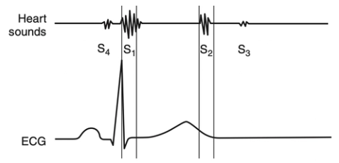
Gallop Sounds
Define S3 (+ DDx)
Define S4 (+ 3 DDx)
Gallop = Low frequency heart sounds during diastole
S3 = Ventricular gallop associated with rapid passive ventricular filling in early diastole
DDx: NOT normal (indicates DCM)
S4 = Atrial contraction in late diastole
DDx: Increased ventricular stiffness and hypertrophy
HCM
Anaemia
Hyperthyroidism
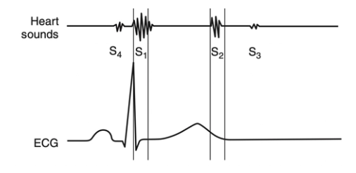
Systolic Click
Definition
DDx
Definition: High frequency sound heard during systole (between S1 and S2)
DDx: Mitral valve disease (eg. dysplasia or MMVD)
Overview of Heart Murmurs
Definition
3 Contributing factors
4 Ways to characterise murmurs
Definition: Caused by turbulent blood flow disrupting laminar flow
Occurs at a high Reynolds number (Re)

Factors Increasing Turbulence:
Big radius change (enlarged chamber)
High velocity
Low blood viscosity (eg. anaemia → haemic murmur)
Characterising Murmurs:
Intensity (grade I - VI)
Character/quality
Timing in the cardiac cycle
Point of maximum intensity (PMI)
Define grade I - VI heart murmur
Grade I | Very soft murmur, barely audible |
Grade II | Soft murmur, but easily heard in one location |
Grade III | Murmur of moderate intensity heard over > 1 location |
Gade IV | Loud murmur and radiates widely (eg. over whole thorax and opposite side) |
Grade V | Loud murmur with precordial thrill
|
Grade VI | VERY loud murmur which can be heard without a stethoscope |
Provide DDx for the following murmur qualities/timing:
Pansystolic, crescendo/decrescendo
Pansystolic, plateau
Diastolic, decrescendo
Continuous, machinery-like
Pansystolic, crescendo/decrescendo = Ejection murmur due to obstruction of ventricular outflow
Subaortic stenosis (SAS)
Pulmonic stenosis (PS)
Pansystolic, Plateau = Holosystolic, regurgitant
Mitral regurgitation (eg. MMVD)
Tricuspid regurgitation
Diastolic, decrescendo
Aortic regurgitation (eg. infective endocarditis)
Pulmonic regurgitation (eg. infective endocarditis)
Continuous, machinery = Patent ductus arteriosus (PDA)

Provide DDx for the following systolic murmurs with PMI at:
Left apex (4)
Left base (4)
Right hemithorax (2)
Left Apex:
MMVD
MV dysplasia
DCM
Endocarditis
Left Base:
SAS
PS
Hypertrophic obstructive cardiomyopathy
Physiological murmur
Right Hemithorax:
VSD
Tricuspid insufficiency
Cat Heart Murmurs
Prevalence
Location description
2 DDx
Prevalence: VERY common (up to 40% of adults)
Location: Left OR right parasternal OR sternal
DDx:
HCM (25%)
Functional/innocent/physiological (75%)
Respiratory Auscultation
Characteristics of pleural effusion
Characteristics of pulmonary oedema
4 Causes of cough with heart disease
Pleural Effusion: Inspiratory AND expiratory dyspnoea with dull respiratory sounds and percussion of fluid line
Pulmonary Oedema: Tachypnoea, dyspnoea and fine crackles at end of inspiration
Cough: Uncommon with heart disease (esp. cats) but caused by
LAE and compression of L mainstem bronchus
Bronchial wall oedema
Pulmonary oedema rarely causes coughing (no cough receptors) BUT severe pulmonary oedema → bronchial wall oedema → coughing
Pulmonary hypertension
Alveolar/bronchiolar fluid accumulation
Thoracic Radiography for Diagnosis of Heart Disease
4 Disadvantages
5 DDx for LAE
4 DDx for R cardiomegaly
DDx for generalised cardiomegaly
-ve:
Patient must be stabilised first (DV superior)
Results affected by treatment
Radiographic signs may lag clinical improvement or deterioration
Cats with HCM have concentric hypertrophy → Normal cardiac silhouette
LAE:
HCM
Early DCM
MMVD
SAS
Systemic hypertension
R Cardiomegaly:
Heartworm
TR
PS
Pulmonary hypertension
Generalised Cardiomegaly:
DCM
Aortic insufficiency
Pericardial effusion
TV dysplasia
Septal defect
PDA
Importance of measuring BP for cardiac disease
Measure baseline prior to ALL therapy
Essential for signs of forward failure (weakness and syncope) → BP to ensure they are not hypotensive
ECG
2 Disadvantages
3 Types of ECG
7 Indications
-ve:
Crude assessment of heart size
Snapshot in time (may miss arrhythmia)
Types:
Conventional = Momentary evaluation of HR and rhythm
Continuous ambulatory Holter monitor = 24hr
Event monitoring = Assess after activation (eg. following collapse)
Indications:
Abnormal HR
Irregular rhythm
Pulse deficit
Variable intensity of heart sounds
Murmur
Split S1/S2
Syncope
3 Features to assess with echocardiography (+ examples)
Structure
Size of chambers and vessels (LA:Ao ratio)
Defects and masses
Pericardial fluid
Valve thickening or stenosis
Blood flow (colour doppler)
Regurgitation
Stenosis
Direction
Speed
Ventricular function
SV
Contractility
Preload
Distensibility
Synergy
CO
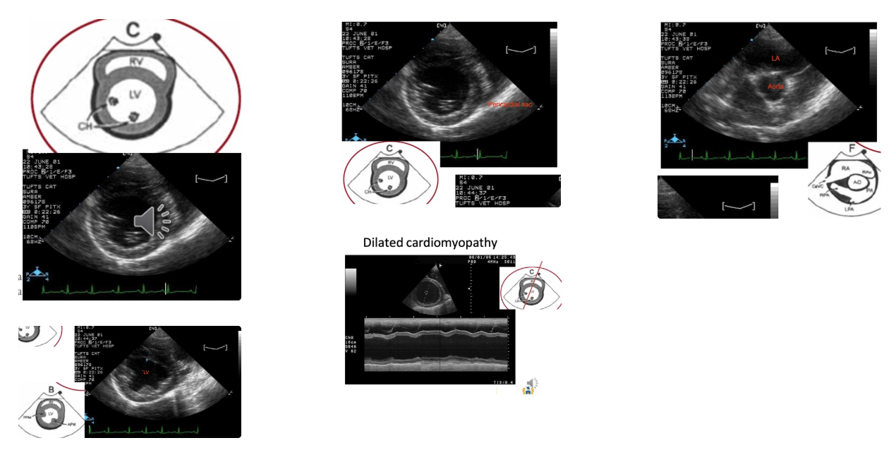
Overview of Blood Testing for Cardiac Disease
3 Functions
3 Tests
Functions:
Diagnose subclinical disease
Differentiate respiratory from cardiac disease
Monitor and prognosis
Tests:
NT-proBNP = N-terminal pro-B-type Natriuretic peptide
Cardiac troponin-1
Serum biochemistry
NT-proBNP
Abnormal elevation
Effect
2 Uses
Abnormal Elevation: Increased secretion from cardiomyocytes with subclinical/clinical heart disease due to wall stress, adrenergic, RAAS concentrations
Effect: Natriuresis (Na+ removal), diuresis and vasodilation
Opposite effect of RAAS
Uses:
Determine likelihood of subclinical DCM or HCM
More specific for disease processes than troponin-1
Differentiating 1˚ respiratory from cardiac disease (when dog presents with respiratory signs)
proBNP low in pulmonary disease and high in cardiac disease
Cardiac Troponin-1
Function
3 Uses
Function: Regulatory protein of contractile apparatus which leaks from myocytes after injury
Uses:
Breed-associated cardiomyopathies (eg. boxer arrhythmogenic right ventricular cardiomyopathy (ARVC)
Right side of heart becomes fatty and fibrotic
Detect myocardial injury (eg. doxorubicin chemotherapy)
With pericardial effusion present: Differentiates haemangiosarcoma (troponin-1 high) from idiopathic effusion
4 Features to assess on serum biochemistry with heart disease
Measures of GFR = Urea, creatinine and SDMA
More useful for LCHF (forward failure)
Crude indicator of renal perfusion (1st major organ supplied by aorta and extremely sensitive to blood flow)
CKD and cardiac disease commonly coexist
Lactate = Insensitive measure of peripheral perfusion
Electrolytes = Essential to measure before and during therapy for CHF
Liver parameters = RCHF monitoring (backward failure
List 9 congenital cardiac diseases
Patent ductus arteriosus (PDA)
Subaortic stenosis (SAS)
Pulmonary stenosis (PS)
Ventricular septal defect (VSD)
Atrial septal defect (ASD)
Tricuspid dysplasia (TD)
Mitral dysplasia (MD)
Tetralogy of fallot (ToF)
Persistent right aortic arch (PRAA)
List 4 compensatory mechanisms for CHF
Increase SNS activation
Increased HR, contractility and SVR
Initially maintains CO and BP
Chronic: Increased myocardial O2 demand, tachyarrhythmia, increased afterload
Activation of RAAS (due to decreased renal perfusion)
Angiotensin II → Vasoconstriction (increased afterload), stimulates aldosterone and ADH
Aldosterone → Increased Na+ and H2O resorption = Increased blood volume and preload
Chronic: Volume overload → Venous congestion and oedema
Release of vasoactive peptides and ADH
Increased water resorption in kidney, vasoconstriction → Increase BP
Chronic: Volume overload and congestion
Myocardial remodelling
Ventricular dilation and compensatory eccentric hypertrophy → Improves SV short-term
Chronic: Dilation prevents normal contraction
Describe the therapeutic approach to CHF (aims and DDx)
Warm vs. col
Dry vs. wet
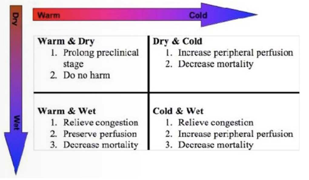
Warm and Dry: Pre-disease (stage A - B)
Dry and Cold: Rare = Poor CO with no CHF
Early DCM → Pimobendan to increase CO
Warm and Wet: Pulmonary oedema and effusion with good CO
Early MMVD and early CHF
Cold and Wet:
DCM and end-stage MMVD
COLD = Poor CO → Improve with pimobendan
WET = CHF → Relieve congestion with diuretics
Causes of Severe CHF
2 Causes of gradual deterioration
4 Causes of acute deterioration
2 Other causes
Gradual Deterioration:
Worsening regurgitation (dilation of annulus)
Myocardial failure due to overload
Acute Deterioration:
Ruptured chordae tendinae
Arrhythmia (supraventricular #1 due to atrial stretching)
LA rupture
Iatrogenic fluid overload
Other Causes:
Pulmonary hypertension
Owner misses doses
Stage A Heart Failure
Definition
4 Treatments
Definition: At high risk for developing heart disease but currently has NO identifiable disorders of the heart (no murmur or structural change)
aka. Pre-disease
eg. Genetic predisposition due to breed (CKCS)
Treatment: Reduce hypertension
Encourage activity to avoid obesity
Avoid excessive salt (increases RAAS which is bad)
Revisit q6 - 12m and monitor SRR
Trend up of SRR → Book consult as may have progressed to CHF
Client education about CHF to reduce risk of developing
No benefit of medications (eg. β-blockers, spironolactone, ACE-i) until onset of CHF
Stage B Heart Failure
Definition
Treatments
Definition: Structural (valve) disease but NO clinical signs
aka. subclinical disease
B1 = No cardiac remodelling/enlargement on imaging (only regurgitation and valve structural changes → murmur)
B2 = Imaging shows evidence of chamber enlargement
Treatments:
B1 = As for stage A
B2 = Pimobendan (must have evidence of cardiac remodelling) + as for stage A
± Monitor with echo
Stage C Heart Failure
Definition
5 Treatments
6 Other drugs to consider
Monitoring
Prognosis
Definition: Clinical signs of CHF with response to standard treatment
Will NOT survive more than a few weeks without treatment
Treatment:
Avoid strenuous exercise
Pimobendan
Low dose frusemide (lowest effective dose)
± ACE-i
Diet = Highly palatable and high energy (reduce weight loss), lower Na+ and increased n-3 PUFA
Drugs to Consider:
± Amlodipine = Vasodilator
± Digoxin = Anti-arrhythmogenic (atrial fibrillation)
Nitrates
Bronchodilators
Cough suppressants
Spironolactone
Monitoring:
SRR, appetite, attitude, ensure drinking
Assess for weakness of syncope
Recheck after 7 - 14 days for urea, creatinine, lytes, Na+ , K+, Cl-
Prognosis: <1yr from onset of CHF with treatment
Stage D Heart Failure
Definition
6 Treatments
Definition: End-stage heart failure, requiring advanced/specialised treatment to remain comfortable
Refractory to treatment
Treatments: Do NOT give fluids (exacerbates pulmonary oedema)
Emergency respiratory distress stabilisation
O2 supplementation
Place IVC
Butorphanol IV = Sedative/anxiolytic
± Thoraco-/abdominocentesis
Max dose frusemide IV until improved RR (4hr) → Stop pulmonary oedema
Refractory → CRI
IV until PO can be given (when RR < 30brpm)
Add ACE-i when no longer acute (not as effective as systemic vasodilation)
Pimobendan IV (when available)
± Other vasodilators if necessary (amlodipine, nitrates, hydralazine)
± Dobutamine with severe oedema
± K+ supplementation (PO or IV) due to hypokalaemia from aggressive diuretic therapy
List 4 common types of presentations of patients with valvular disease
Puppy or kitten
Innocent/physiological murmur vs. congenital disease
Important to have early awareness to prevent disease progression
Annual health check
Asymptomatic detection of murmur or arrhythmia
Mild clinical signs
Cough (cardiac disease may NOT be cause)
Decreased exercise tolerance
Congestive heart failure or low output failure
Esp. large breed dogs
What is the difference between stenosis vs. insufficiency
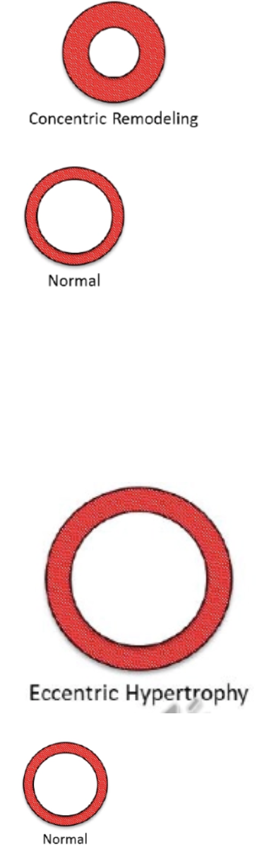
Stenosis = Narrowing of valve orifice while valve is open
Severity of stenosis is related to pressure gradient
Smaller orifice → Larger pressure gradient → Pressure overload
→ Concentric hypertrophy
Insufficiency = Blood flowing through valve in WRONG direction (regurgitation) while valve is “closed”
Severity of insufficiency/regurgitation is related to amount of regurgitant flow
Larger orifice → Greater regurgitant volume → Volume overload
Eccentric hypertrophy
Describe 4 factors which influence severity of valvular disease
Valve affected
R better than L
Tricuspid not as severe as aortic valve
Stenosis OR insufficiency/regurgitation
Severity of impairment
Other concurrent conditions which exacerbate the disease
Cardiomyopathy (eg. DCM → failing myocardium)
Pulmonary/systemic hypertension
CKD
Bacteria-associated disease
Overview of Myxomatous Mitral Valve Disease (aka. Endocardiosis)
Prevalence
Valves affected
Signalment (breed, sex and age)
Aetiology
Pathogenesis
Prevalence: #1 heart disease of dogs (75%)
Common incidental finding at necropsy
Valves: MV only = 60%, MV and TV = 30%, TV only = 10%
Usually MV precedes TV due to larger circulatory bed
Signalment:
Breed = Small - medium dogs
CKCS (>50% by 4yr and almost 100% by 10yr)
Dachshund, poodle, JRT, Yorkie, Chihuahua
Unusual in large breeds (but associated with more rapid CHF)
Sex = Male > Female
Age = Middle-aged to older
Uncommon in cats
Aetiology: Non-inflammatory, degenerative process AND hereditary with complex inheritance pattern
Pathogenesis:
Abnormal collagen degeneration/remodelling (often associated with collapsing airways)
Loose fibroblastic tissue in spongiosa of valve
Deposition of HA and chondroitin sulphate
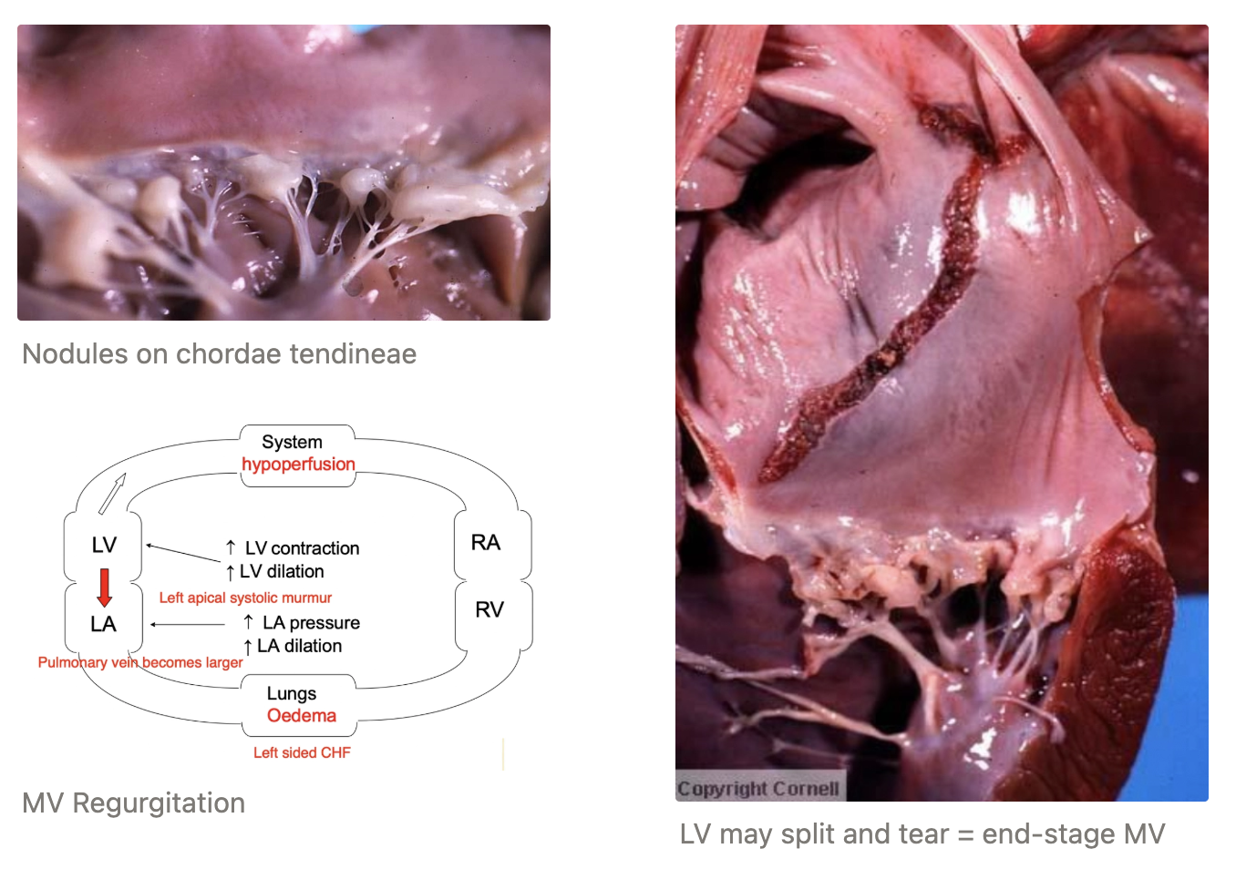
Collagen degeneration of valvular fibrosa and chordae tendinae
Valve laxity and prolapse
Valve becomes thickened and nodular due to scar tissue → Prevents valve from forming tight seal → Regurgitation
Chordae tendinae thicken and may rupture
Cardiac remodelling = Eccentric hypertrophy due to volume overload
Ventricle MAY split
Clinical Signs of MMVD
4 Presentations
4 DDx
Presentations: LCHF #1
Asymptomatic with holosystolic apical murmur on routine examination
Insidious onset of clinical signs
Acute respiratory distress (pulmonary oedema)
Acute decompensation (myocardial failure, tear in LA or ruptured chordae tendinae)
DDx:
Pulmonary disease (eg. chronic bronchitis or bronchopneumonia)
Differentiate with proBNP
If dog with mitral murmur and cough has NORMAL HR, it does NOT have CHF
Tracheobronchial malacia
Bacterial endocarditis
Congenital heart disease
Describe 7 diagnostic tools for MMVD
Advantages
Disavantages
Results
Thoracic Radiography
+ve:
Reliable method to evaluate LA size
2nd best method to monitor response to treatment
Results:
Mitral Regurgitation
LAE = Dorsal displacement of trachea and divergence of mainstem bronchi
LVE = No cardiac waist and bulge
Pulmonary venous distension
Pulmonary oedema = Interstitial → Alveolar
Tricuspid Regurgitation
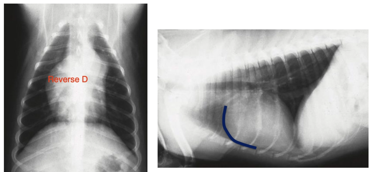
Right-sided enlargement = Reverse D and increased cardiosternal contact
Distended CdVC
Hepatomegaly/splenomegaly
Loss of serosal detail due to ascites
Blood Pressure
Echocardiography
+ve:
Assess anatomy of valves and size of chambers
Confirm diagnosis and determine prognosis
Prognostic Indicators:
LA:Ao > 1.6 = LAE
End-systolic diameter/volume = #1 indicator of volume overload
Small dogs usually normal to increased LV function
FS > 50% due to increased diastolic diameter
Large dogs = Decreased in advanced cases (echo more indicated in large dogs)
FS 20 - 40%
± Cardiac Biomarkers (NT-proBNP)
± Airway Sampling
± MDB
CBC = Rule out anaemia as exacerbating cause
Biochemistry = Kidney parameters and electrolytes
UA = Cardio-renal syndrome (difficult to manage as kidneys want to be wet and lungs want to be dry)
± ECG
+ve: May be indicated when arrhythmia suspected (i.e. heavily disease ventricle or atrium)
-ve: Echo better to assess for atrial and ventricular enlargement
Overview of Endocarditis
Dog vs. cat presentation
Pathogenesis
Risk of individual valve infection
4 Agents
2 Sources
7 Risks
Presentation: Rare and life-threatening disease
Dog = Acute
Cat = Chronic
Pathogenesis:
Bacteraemia results in colonisation of the free margins of the heart valves
Large friable/vegetative mass develops
Destruction of valves → Regurgitation
Destruction of adjacent endocardium
Thromboembolism in kidneys and spleen
Valves: Mitral > Aortic »» Tricuspid > Pulmonic
Proportional to pressure on each valve
Agents: G+ aerobic #1
Staph. aureus
β-haemolytic Streptococci
E. coli (acute)
Bartonella (25% in USA)
Sources:
Discospondylitis
Dental disease
Risks:
Large dog
Immunosuppressive agents or disease
Inappropriate/inadequate antimicrobial therapy
Indwelling large IV or urinary catheter
Prior valvular disease (eg. subaortic stenosis due to jet lesions)
Bacteraemia
Periodontal disease?
Diagnosis of Endocarditis
5 Clinical signs
4 Diagnostic tools
Clinical Signs:
Fever
Vomiting
Lameness (IMPA and discospondylitis)
New heart murmur (74% systolic and 26% diastolic)
Diagnosis:
CBC and biochem (azotaemia/kidney failure)
Blood cultures
Treat based on most likely infection while awaiting cultures
Urine cultures
Echocardiography to differentiate between endocardiosis vs. endocarditis
Shows: Vegetative and destructive lesions
Difficult to distinguish with U/S but use younger signalment
Treatment of Endocarditis
2 Treatments
Prognosis
Treatment:
AB q4 - 6hr for 4 - 6w until resolution of lesion
Parenteral → PO
Based on culture and sensitivity
CHF therapy
Prognosis: Poor (20%)
G- infection worse than G+ infection
Aortic valve worse than mitral valve
Overview of Subaortic Stenosis
Prevalence
Sites of stenosis
Signalment (breed)
Aetiology
Pathogenesis
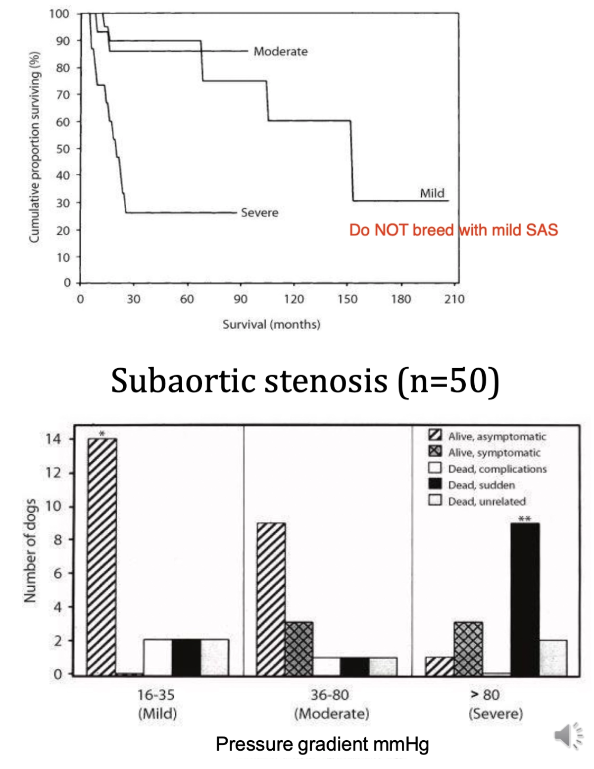
Prevalence: #1 congenital cardiac defect in NZ dogs
Sites: Subvalvular (>95%), valvular (5%), supravalvular (rare)
Signalment: Large breeds (boxer, GSD, GR, GSP, GD, Newfoundland, Rottweiler, bulldog, Labrador, bullmastiff, samoyed)
Rare in cats
Aetiology: Congenital valvular disease
Can by dynamic (narrowing changes overtime)
Pathogenesis:
Fibrous subaortic ring develops which reduces left ventricular outflow
LVE hypertrophy to pump blood through congenital narrowing stenotic region (pressure overload → concentric hypertrophy)
HR increases to maximise CO → Reduced myocardial perfusion with increased O2 demand
Underperfused myocardium → Ventricular arrhythmia

Diagnosis of Subaortic Stenosis
5 History findings
2 PE findings
3 Diagnostic tools (+ results)
History:
Progressive (silent < 3 months)
Exercise intolerance (low CO)
Syncope (low CO)
Sudden death (ventricular arrhythmias ~70% <3yr)
LCHF with MR = Dyspnoea (esp. cats)
PE:
Systolic crescendo-decrescendo left basilar murmur
Slow-rising hypokinetic pulse
Diagnosis:
Radiography = Aortic bulge and L-sided cardiomegaly
ECG = Exercise-precipitated ventricular arrhythmia
Echocardiography = Confirm diagnosis and determine severity of lesion


Treatment of Subaortic Stenosis
3 Treatments
Prognosis
Treatments:
Balloon dilation of stenotic valve (NOT often successful vs. PS)
β-1 blockers (eg. atenolol)
Reduce HR in moderate to severe cases
Prevent arrhythmic effect of catecholamines in body and reduce myocardial O2 demand
Increased coronary perfusion
Exercise restriction
Prognosis: Poor if severe
Overview of Pulmonic Stenosis
Prevalence
Site of stenosis
Signalment (breed)
Pathogenesis
Prevalence: Common congenital valvular defect in dogs
Sites: Valvular #1 (> subvalvular > supravalvular)
Signalment: Small breed dogs (beagle, Newfoundland, samoyed, Chihuahua, English bulldog, GR, Chow Chow, mini schnauzer)
Normally >1yr
Pathogenesis:
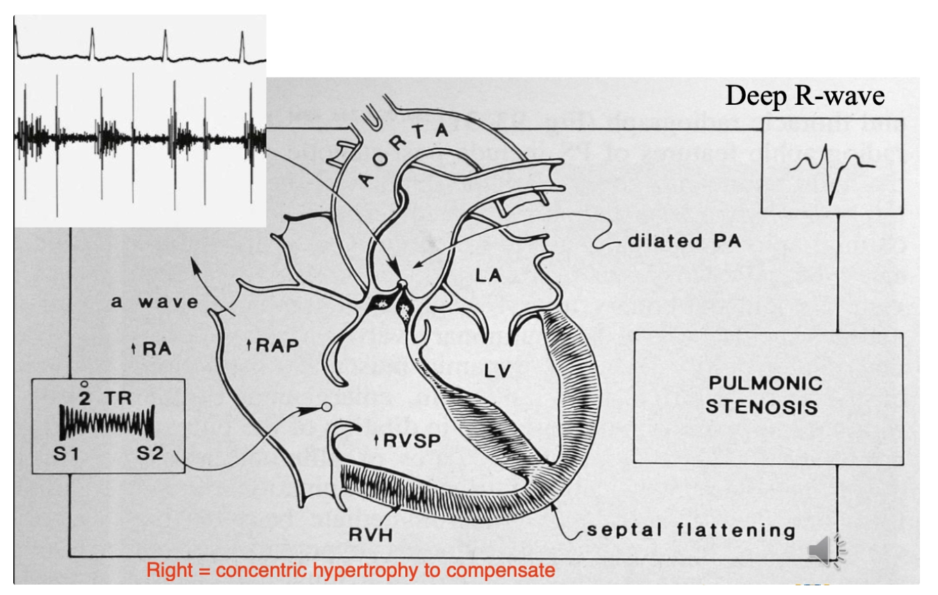
Congenital narrowing of pulmonic valve → Obstruction to RV outflow
Concentric hypertrophy of RV
± RCHF
Diagnosis of Pulmonic Stenosis
3 History findings
5 PE findings
4 Diagnostic tools (+ results)
History:
Usually asymptomatic
Exercise intolerance and syncope
RCHF
PE:
Left basilar crescendo-decrescendo systolic murmur and precordial thrill
Jugular pulse
Arrhythmia and sudden death
Ascites and hepatomegaly
Diagnosis:
Radiograph = RVE, pulmonary artery bulge, hypoperfusion of lungs
ECG = Exercise-precipitated ventricular arrhythmia and R-sided cardiomegaly
Echocardiography = Confirm diagnosis and determine severity of lesion
Treatment of Pulmonic Stenosis
3 Treatments
Prognosis
Treatments:
Balloon dilation OR surgery (good efficacy)
Exercise restriction
Treat CHF
Prognosis: Very good (even without treatment)
AV Valve Dysplasia
2 Types (+ signalment)
Clinical signs
Types:
Mitral Dysplasia = #1 congenital defect in cats (also large dogs)
Tricuspid Dysplasia = Male large dogs
Clinical Signs: Related to valvular insufficiency/regurgitation
Lesions tolerated for many years (acute or chronic)
Classification of Cardiomyopathies
Aetiology
3 Types
Aetiology: 1˚ OR 2˚
Types:
Dilated cardiomyopathy (DCM) = Systolic pump failure
Hypertrophic cardiomyopathy (HCM) = Diastolic compliance failure
Restrictive cardiomyopathy (RCM) = Diastolic compliance failure with some systolic dysfunction
Overview of Dilated Cardiomyopathy (DCM)
Definition
5 Aetiologies
Pathogenesis
Signalment (breed, age and sex)
Side of heart affected
Definition: Eccentric hypertrophy of 2 OR 4 heart chambers (LV #1) → Myocardial dysfunction
Characterised by SYSTOLIC dysfunction = Low %Fractional shortening (FS)

Aetiologies:
Idiopathic (90%)
Genetic predisposition
Taurine deficiency
MoA: Unknown, but concentrates inside excitable tissues eg. myocardium
Diets: Lamb meal, rice, grain-free diets, low S-amino acids (Met/Cys) necessary to synthesise Tau
Also carnitine deficiency in boxers → Poor mitochondrial function
Immune-mediated
Infectious agents (2˚ to myocardial damage)
Doxorubicin toxicity
Pathogenesis:
Progressive decrease in contractility → Decreased SV and increased eSV
Increased RAAS and SNS in attempts to maintain BP
Dysfunctional eccentric hypertrophic compensation due to volume overload → Increased wall stress ± mitral regurgitation (stretching of the LV damages the MV which causes insufficiency and exacerbates LCHF)
LCHF ± atrial arrhythmia due to reduced coronary filling AND cardiac remodelling (fibrosis)
Signalment:
Breed = Giant
Doberman, boxer, Irish Wolfhound, Great Dane, St Bernard, Newfoundland, English sheepdog, Labrador, golden retriever, huntaway
X-linked in Great Danes
Age = 6m - 14yr (~5yr)
Sex = Male > Female
Side: L, R or biventricular failure
Clinical Signs of Dilated Cardiomyopathy
3 Presentations
5 PE findings
Presentations:
Long subclinical phase (due to compensation) → Short clinical phase
LCHF or RCHF (better prognosis)
Tachyarrhythmia (eg. a fib) → Syncope or sudden death
Often 1st clinical signs in boxers and dobermans
PE:
Irregularly irregular heart beat (atrial fibrillation #1)
VPC or VT for boxers and dobermans
Mitral valve murmur due to stretching of annulus (lower grade than MMVD)
Gallop rhythm (S3)
LCHF or RCHF
Low CO signs
Diagnosis of Dilated Cardiomyopathy
7 Diagnostic tools (+ results)
5 Major criteria for DCM
3 Minor criteria for DCM
Prognosis
Diagnosis:
Radiography = Presumptive diagnosis of DCM (generalised cardiomegaly in young, large breed dog with NO prior clinical signs)
CHF mimics end-stage MMVD (BUT latter typical of older, small breed dogs and preceded by clinical signs)
Results:
Generalised cardiomegaly
Interstitial/alveolar lung pattern
± Pleural effusion/ascites
Echocardiography = Differentiate between DCM vs. MMVD as cause of MR
DCM = Annulus stretched and pulled away from septum vs. MMVD = Valve degeneration and thickening
Results:
Increased LV eSD and eDD (heart stays large during systole due to failed contraction)
FS% < 25% (reduced contractility)
Increased LA:Ao ratio (2˚ to MR)
Reduced wall thickness
Blood Pressure
± ECG = Atrial fibrillation/ventricular arrhythmia
MDB = Monitoring for treatment
± proBNP = Elevated
± Taurine assay to seek for 1˚ cause (esp. with atypical breeds)
Major Criteria:
Increased LV eSD
Increased LV eDD
Reduced FS%
More spherical LV
Elevated NT-proBNP
Minor Criteria:
Left and bi-atrial enlargement
Increased mitral valve E point of septal separation (MV pulled apart)
Arrhythmia
Prognosis: <6m when CHF develops

Dobermans with DCM
Age of onset of clinical signs
3 Diagnostic tools (+ results)
Prognosis
Age: ~6.5yr
Long pre-clinical phase (“occult DCM”)
Clinical phase = CHF (75%) and forward failure (25%) ± paradoxical bradycardia
Diagnosis:
Radiography = L-sided cardiomegaly
24hr Holter monitoring = Ventricular arrhythmia (>100 VPCsin 24hr)
Atrial fibrillation = Worse prognosis
Echocardiography
Prognosis: <3m after onset
Diastolic LVID (LV internal diameter) negatively correlates with prognosis
Larger the internal diameter of LV at diastole = worst prognosis
Pimobendan improves survival (~ 1yr) prior to CHF
Sudden death common due to arrhythmia
Cocker Spaniels with DCM
Age
Radiographic findings
Prognosis
Age: 5 - 6yr
Radiography: Generalised cardiomegaly (often with concurrent MMVD)
Prognosis: Better than other breeds with DCM
Often asymptomatic for long time
Cats with DCM
Aetiology
Pathogenesis
2 Treatments
Aetiology: Idiopathic OR taurine deficiency
Potentially reversible with taurine supplementation
Pathogenesis: Systolic myocardial dysfunction of ALL heart chambers → Pleural and pericardial effusion (LCHF)
Treatments:
CHF management
Digoxin
Arrhythmogenic Right Ventricular Cardiomyopathy ARVC)
Signalment
3 Diagnostic tools (+ results)
3 Categories (+ prognosis)
Treatment of arrhythmia
Signalment: 1 - 11yr (~6yr) boxers
Diagnosis:
Radiography = NORMAL
RV becomes fibrotic and fatty
± Mild eccentric hypertrophy of LV due to volume overload
24hr Holter monitoring = Ventricular arrhythmia (>50 VPC in 24hr)
Positive trace on lead II indicates origin from R side of heart
Echocardiography = Mild eccentric LV hypertrophy
Categories:
Survival | ||
I (concealed) | Asymptomatic with arrhythmias | 2yr |
II (overt) | Arrhythmias with syncope and prone to sudden death | 1 - 2 yr |
III (myocardial failure) | Myocardial failure with CHF and arrhythmias | < 6 months |
Treatment of Arrhythmia: Sotalol (β-blocker) → Treat ventricular arrhythmia
Feline Myocardial Diseases
2 1˚ disorders
5 2˚ disorders
1˚:
Feline HCM
Feline RCM
2˚: Restrict left ventricular function = pressure overload of ventricle = maladaptive concentric hypertrophy
Hyperthyoidism
Systemic arterial hypertension
Acromegaly
Aortic stenosis
Taurine deficiency
Overview of Feline Hypertrophic Cardiomyopathy (HCM)
Definition
Prevalence
5 Aetiologies (1˚)
Signalment (breed, sex and age)
3 Gross pathology findings
3 Histological findings
Definition: 1˚ disease of the LV free wall and intraventricular septum → Diastolic failure due to concentric hypertrophy → Reduced ventricular compliance and myocardial relaxation
± Obstructive outflow from LV (associated with SAM)
MV becomes stuck
Prevalence: 15% of cats (increases with age)
Very rare in dogs (except compensatory concentric hypertrophy 2˚ to SAS)
Aetiologies:
Genetic (breed disposition with mutation in myosin-binding protein C)
Autosomal dominant train in Maine Coons and American Shorthairs?
Idiopathic #1
Viral???
Immune-mediated???
Toxic???
Signalment:
Breed = Maine Coon and American Shorthair (large cats)
Sex = Male > Female
Age = 5m - 17yr (~4.8 - 7yr)
PM:
Concentric hypertrophy of LV
Papillary muscle hypertrophy
Increased heart weight : BWT ratio
Histology:
Myocardial fibre disarray
Increased fibrous connective tissue
Arteriosclerosis and vascular dysplasia
Pathology of Feline Hypertrophic Cardiomyopathy (HCM)
Pathogenesis
Systemic anterior motion of the mitral valve (SAM)
Definition
3 Causes
Diagnosis
Treatment
Prognosis
Feline aortic thromboembolism (FATE)
Cause
5 Clinical signs
Pathogenesis: Diastolic dysfunction
Abnormal concentric hypertrophy of LV → Thin lumen with reduced eDV → Small SV
Low CO
Tachycardia to increase CO (exacerbates issue due to reduced diastolic filling time)
RAAS activation → Increased end-diastolic pressure
Cardiac distortion causes MV leaflet prolapse → LVOT obstruction (exacerbates disease)
LAE → Thromboembolism
LCHF
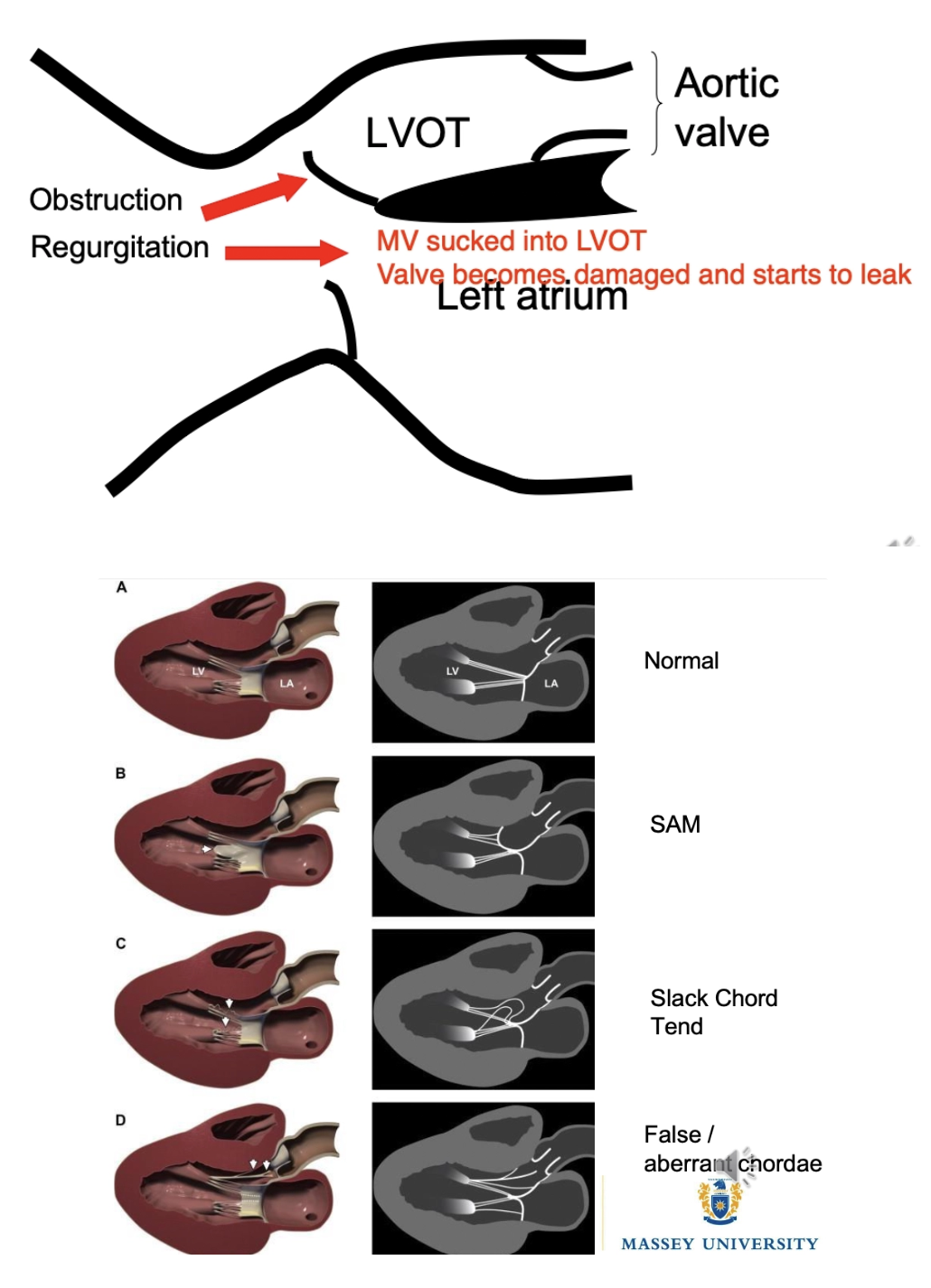
SAM: MV pulled out of normal position and sucked into LVOT → Dynamic obstruction
Causes:
Position of papillary muscles
Slack chordae tendinae not pulling valve closed
“False/abberent” chordae
Diagnosis: Echocardiography
Treatment: β-blocker
Prognosis: Same as no obstruction
FATE: Clots form in LAE (stagnant blood) and break off to lodge anywhere in body leading to
Paralysis (esp. HL due to lodging in caudal bifurcation fo Ao)
Pain
Pulselessness
Pallor
Poikilothermia

Clinical Signs of Feline Hypertrophic Cardiomyopathy (HCM)
2 Signs of subclinical disease
4 Signs of clinical disease
Subclinical Disease:
Left parasternal murmur (20 - 60%)
Gallop sounds (S4)
Clinical Disease: Often present very late in course of cardiac disease due to propensity for low activity
Non-specific clinical signs (depression, anorexia and inactivity)
LCHF
Can be peracute onset (silent periods with no clinical signs)
May be preceded by stressful event or IV fluid administration
Respiratory distress (cough rare)
Arterial thromboembolism = Posterior paresis (bifurcation of caudal aorta)
Also: Haemorrhagic diarrhoea (GIT), azotaemia (kidney) and seizures (brain)
Arrhythmias
Diagnosis of Feline Hypertrophic Cardiomyopathy (HCM)
6 Diagnostic tools (+ results)
MDB = Rule out 2˚ HCM
Hyperthyroidism (T4)
Hypertension
Aortic stenosis
Acromegaly
NT-proBNP = Differentiate from 1˚ pulmonary disease when presenting in respiratory distress
Radiography
Pulmonary oedema = Patchy, multifocal interstitial to alveolar lung pattern
Pleural effusion (2˚ to pulmonary hypertension and biventricular failure)
Valentine-shaped heart = Bi-atrial enlargement
Distended pulmonary arteries and vein (pulmonary hypertension
± ECG = Left anterior fascicular block, VPC or atrial fibrillation
Echocardiography = Diagnosis and assess LVOT
LV hypertrophy (free wall and intraventricular septum = LVWd)
LAE
Normal to increased contractility (reduced diastolic measures)
Narrowed LVOT
SAM
Blood pressure

Treatment of Feline Hypertrophic Cardiomyopathy (HCM)
6 Treatments
Follow-up
Cat with pulmonary oedema on frusemide
Cat with pleural effusion
Cat with FATE but no CHF
5 Principles of FATE treatment
4 Negative prognostic indicators
Treatment:
Frusemide (lower dose than dogs as cats more sensitivity)
± Pimobendan if frusemide insufficient
-ve: Can make LVOT obstruction worse by increasing contractility of heart → Prolapse of mitral valve
± Antiarrhythmic (β-blocker) to slow HR and reduce effect of SAM
ACE-i
± Thoracocentesis (more predisposed to pleural effusion than dogs)
FATE treatment
Follow-Up:
Pulmonary oedema on frusemide
Check bloods 1 - 2w after discharge → Adjust dose based on SAP, biochemistry and PE
Recheck q3m with ECG
Recheck q6 - 12m with radiography
Pleural effusion = Check q3m
FATE
Check q1m for 3m → q3m if stable
Consider annual echo and radiography q6 - 12m
FATE:
Analgesia = Opioids
Induce hypocoagulable state to reduce further thrombus formation
Clopidogrel = Inhibit platelet aggregation
± Heparin, aspirin and vasodilators (no evidence of efficacy)
Heparin = Antithrombin III co-factor which neutralises clotting factors
Do NOT use thrombolytic therapy
eg. Plasminogen activator (rHU t-PA) which converts fibrin bound plasminogen to plasmin
-ve: Low efficacy and $
eg. streptokinase/urokinase which converts blood plasminogen to plasmin
-ve: Risk of hyperkalaemia, metabolic acidosis, haemorrhage and $
Improve blood flow to infarcted arterial bed by avoiding hypotension and dehydration
Treat concurrent CHF if present
Provide supportive care = Cage rest and nutritional support
Negative Prognostic Indicators:
CHF
Severe LAW
Thromboembolism
Older age
Describe the classifications of hypertension
Hypertension = Increased SAP (MAP superior assessment)
Classification based on risk of target organ damage (TOD) and present TOD
Use breed-specific RR (eg. sighthounds have normally higher BP)
SAP (mmHg) | Substage | Risk of TOD |
<140 | Normotensive | Minimal |
140 - 159 | Pre-hypertensive | Low |
160 - 179 | Hypertensive | Moderate |
≥180 | Severely hypertensive | High |
Hypertension SAP > 160mmHg (180mmHg in-clinic due to physiological hypertension)
3 Types of hypertension
Situational Hypertension (aka. “white coat” hypertension)
Normal patient with hypertension due to SNS stimulation caused by excitement/anxiety/pain
No treatment necessary as resolves with removal of stimulus
Idiopathic Hypertension (aka. “primary malignant hypertension” or “essential hypertension”)
Sustained hypertension with no identifiable cause
20% of hypertensive cats
Secondary Hypertension
Treatment of 1˚ condition MAY resolve hypertension
List 9 causes of secondary hypertension (most to least common)
CKD (bidirectional)
Hyperthyroidism
Hyperadrenocorticism
Diabetes mellitus
Chronic hepatic disease
Pheochromocytoma (adrenal gland neoplasia → Secrete excess adrenaline/noradrenaline)
Chronic anaemia
Hyperaldosteronism
Polycythaemia
Cardiovascular disease does NOT cause hypertension (vs. humans)
4 Target organs damaged by hypertension (+ clinical signs)
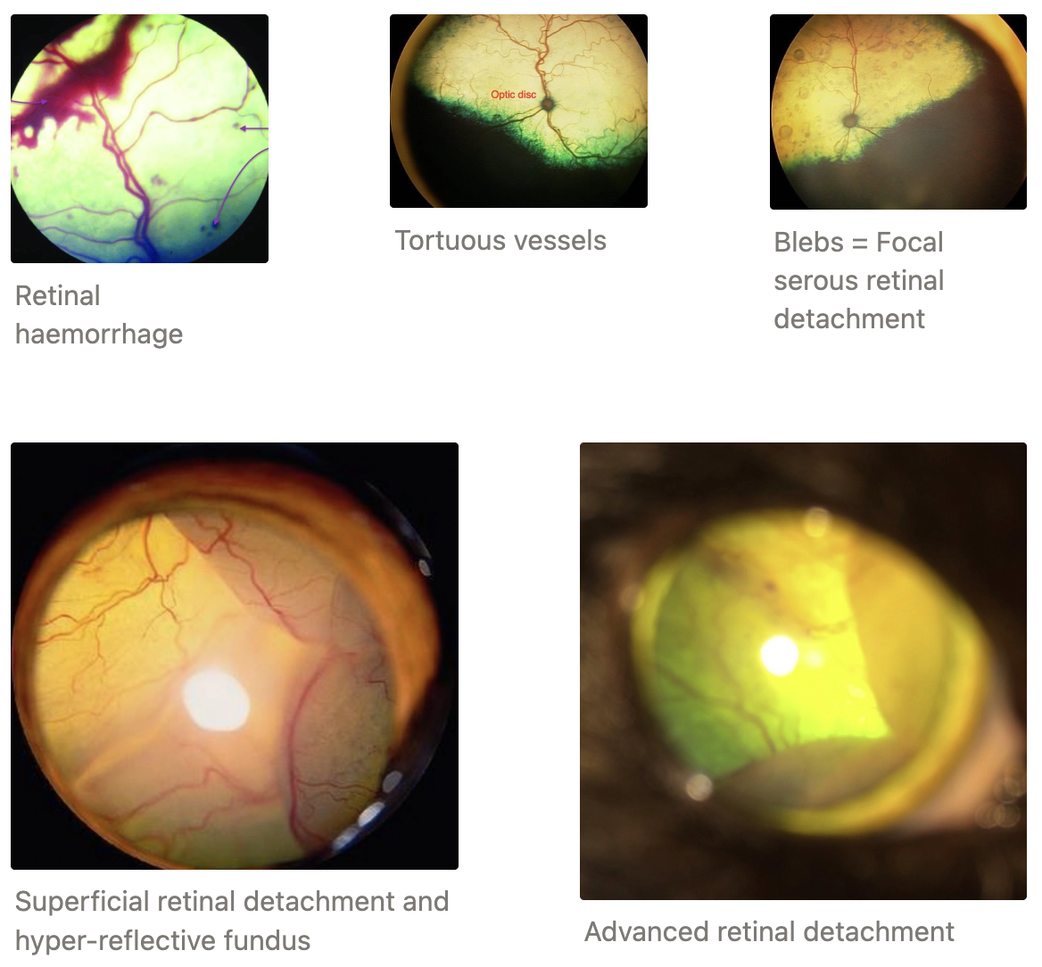
EYES #1
Sudden blindness (often reversible)
Retinal oedema
Dilated and tortuous retinal vessels
Hyphaema
Retinal detachment
Dilated, hyper-reflective fundus
Scleral/conjunctival hyperaemia/iris aneurism
Slow PLR
Secondary glaucoma
KIDNEYS = CKD and proteinuria
BRAIN = High ICP
Cerebrovascular accidents (stroke)
Depression
Seizure
Ataxia
Focal brain and spinal cord lesions (aneurism)
HEART AND BLOOD VESSELS
Gallop sounds
Murmurs
LV concentric hypertrophy (pressure overload due to high systemic BP)
LCHF
Arrhythmia
Bleeding due to blood vessel changes (epistaxis, stroke, aortic rupture)
Vague Clinical Signs of Hypertension:
Restlessness
Anxiety
Panting
Behaviour changes
3 Indicators for blood pressure measurements
Dogs and cats ≥9yr
Diagnosis of condition associated with 2˚ hypertension
Evaluate patient with evidence of TOD

Label the following probe/doppler positions for measuring blood pressure
Forelimb
Digital artery (2) = Palmar metacarpals distal to carpal pad
Cuff between carpus and elbow (1)
Hindlimb
Dorsal pedal artery (6) = DM tarsus OR caudal branch of saphenous artery (5) = Plantar metatarsal proximal to metatarsal pad
Cuff just above or below tarsus (3 or 4)
Tail
Coccygeal artery (7) = Midline of tail
Cuff at base of tail
12 Standardised protocols for BP measurement (+ 3 things to avoid)
Quiet environment
Owner present (MAY make worse)
Allow 5 - 10 minutes for patient to acclimatise to environment
Gentle restraint in R lateral recumbency (cats can be in sternal to reduce stress)
Ensure cuff and probe are level with the heart
Cuff >10cm below RA → Falsely elevated BP
Formula to correct: Subtract 0.8mmHg for every 1cm cuff sits below the RA (when > 10cm)
Cuff width 30 - 40% limb/tail circumference
Too small/tight = False elevation of BP
Too large/loose = False lowering of BP
Always discharge 1st value
Record 5 - 7 consecutive and consistent values (<20% variability) → Take average
Record all parameters (position, site of measurement, environment, operator and cuff size)
Same operator each time
Use U/S gel to improve contact between Doppler probe and skin
Use headphones with doppler to reduced noise disturbance for patient AND ease of hearing
Avoid:
Avoid sedation (lower BP)
Do NOT clip hair to make detection of blood flow with Doppler easier
Consider noisy clippers = stress
Moisten hair with alcohol and liberally apply U/S gel instead (esp. cats)
Avoid taping cuff to secure in place
Taping can increase occlusion pressure (esp. when 360˚)
Use small pieces if necessary
When should hypertension treatment be initiated?
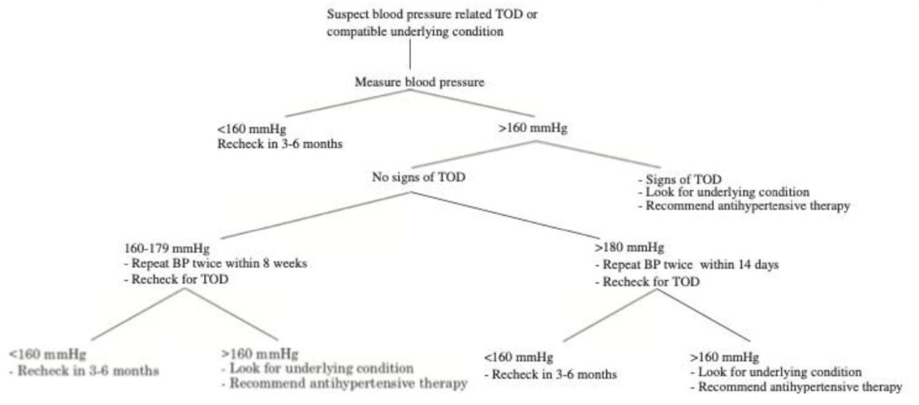
Begin Treatment When:
Single record of hypertension WITH EOD (end-organ damage = fundic examination)
Hypertension without EOD confirmed with ≥ 2 measurements on separate occasions (160 - 179mmHg) within 8 weeks
Two instances may be a few days apart
Hypertension without EOD confirmed with ≥ 2 measurements on separate occasions > 180mmHg within 14 days
7 Diagnostic tests to perform with a hypertensive patient
Fundic examination
MDB
Urine protein:creatinine ratio
Total T4 (hyperthyroidism)
Abdominal U/S
+ Echocardiography
± Brain MRI
Hypertension Treatment
Aim of treatment
3 Management recommendations
Dog treatment progression
Cat treatment progression
Aim: SAP < 160mmHg (ideally 120 - 140mmHg)
In-clinic values (avoid hypotension when home)
ALSO no hypotensive signs (weakness and tachycardia)
Do NOT start medication in unstable or dehydrated patients as it may drop GFR
Management:
Avoid excess Na+ in diet
Ensure stable and hydrated
Drugs to GRADUALLY reduce BP to avoid side effects
Dogs: Progress to next step when refractory
ACE-i (eg. Benazepril)
ACE-i x2
ACE-i + Amlodipine
ACE-i + Amlodipine + Telmisartan ± Hydralazine
Cat:
Amlodipine #1 OR Telmisartan
Increase Amlodipine
Amlodipine + Telmisartan
Short-term and long-term monitoring of hypertension treatment
Short-Term:
Monitor BP 7 - 10d after treatment (1 - 3d in emergency setting)
Monitor treatment with clinical signs and renal values (markers of GFR)
Increased potassium and creatinine with benazepril
Accept small increase in creatinine and SMDA due to medication
Hypotension with excessive amlodipine
Long-Term:
Recheck q3m for PE and eye exam
Recheck q6m for renal value assessment
Overview of Pericardial Effusion
Define pericardial effusion vs. cardiac tamponade
3 Types of pericardial effusion (+ causes)
Pathogenesis
Pericardial Effusion: Excessive/abnormal fluid accumulation in the pericardial sac
Normal <2mL for lubrication
Cardiac Tamponade: Impaired ventricular filling due to increased intrapericardial pressure from
Pericardial effusion
Masses
Organs (eg. diaphragmatic hernia)
Types of Pericardial Effusion:
Blood
Idiopathic #1 (up to 75%)
Viral
Immune-mediated
Neoplasia (HSA, chemodectoma, lymphoma)
Chemodectoma = Pressure tumour of carotid body sinuses
Ruptured LA (eg. MMVD)
Trauma (eg. HCM)
Coagulopathy (eg. rat bait toxicity)
Transudate
RCHF
Hypoalbuminaemia
Exudate
Virus (eg. FIP = low cellularity exudate)
Bacteria
Pathogenesis:
Pericardial effusion → Increased intrapericardial pressure
Results in diastolic collapse of RA
Right side before left side as RA = lowest pressure chamber
RV tamponade if severe
Impaired ventricular filling
Reduced CO (RV pumps less blood to left side of heart)
Hypotension
Increased venous pressure in vena cava
Blood backs up
Diagnosis of Pericardial Effusion
5 History findings
5 Clinical signs
4 Diagnostic tools (+ results)
History:
Sudden onset
Signs of RCHF (slow onset)
Generalised weakness
Collapse
Sudden death
Clinical Signs:
Jugular vein: Distension, jugular pulses and hepatojugular reflux
Muffled heart sounds with normal lung sounds
Cannot detect apex beat
Tachycardia
Hypokinetic pulse or pulsus paradoxus
ALSO bounding hyperkinetic pulse due to increased difference between SAP and DAP
Poor peripheral perfusion
Cold extremities
Extended CRT and pale MM
Diagnosis:
T-fast
Radiography → Globose heart
± ECG → Electrical alternans = Variation in QRS height due to heart swinging in fluid of pericardium = different position in relation to leads
Echocardiography = Definitive diagnosis

5 Differences between pericardial effusion and DCM
Feature | Pericardial Effusion | DCM |
|---|---|---|
Heart sounds | Muffled (effusion) | Loud |
ECG | Electric alternans | NO electrical alternans |
Cardiac silhouette | Globose and sharp edged | Blurred edges due to movement with heart beat |
Pulmonary oedema | Less/absent | Prominent feature |
Pulsus paradoxus | Common | Rare |
List 2 treatments of pericardial effusion
Pericardiocentesis = Drain fluid
Pericardiectomy = Removal of part of pericardium to allow bleeding into pleural space
Pericardiocentesis
Patient positioning
Surgical preparation
3 Pieces of equipment
Location of catheter placement
Monitoring
4 Laboratory features to evaluate
Positioning: LEFT lateral recumbency
Surgical Preparation: Ribs 3 - 8 on right side from sternum to mid thorax
Apply local anaesthetic (lignocaine) from skin to pleural surface
Equipment:
12 - 16G catheter with added side holes
Side holes prevent occlusion of catheter
Too many = weaker catheter and allows air to enter space
Extension set + 3-way tap
Stops movements
50mL syringe
Location: RIGHT side with large cardiac notch
Cardiac notch = Area where heart touches chest wall without touching lung
4th - 6th ICS
Monitoring: Ventricular tachycardia on ECG = Hit ventricle
Laboratory Assessment:
Assess clotting of fluid
Blood from pericardial space should NOT clot (defibringinated blood via normal fibrinolysis cascade)
Clot = Accidental aspiration from RV or actively bleeding into pericardium
HCT usually lower than peripheral blood BUT can be much higher
Cytology
pH of fluid? NOT useful

Pericardiectomy
2 Advantages
4 Indications
3 Methods
+ve:
Blood resorbed by lymphatics
Larger space → Less pressure build up than pericardial space
Indications:
Idiopathic haemorrhagic pericardial effusion
50% recur after pericardiocentesis
≥ 3 pericardiocentesis → Pericardectomy
Curative!
Septic pericarditis
Left atrial tear
Neoplasia (palliative and NOT curative)
Methods:
Open thoracotomy
Fluoroscopy to remove bottom half
Pericardial window (make space in pericardial tissue)
7 Principles for treating sick cardiac patients
“SOCROTH”
Minimise chronic STRETCH
Increases O2 demand
Myocyte injury and arrhythmia
Remove pulmonary OEDEMA
Lung heavy, wet and stiff → V:Q mismatch
Fatigue on respiratory muscles
Increases O2 demand
Improve CO
Increase HR or contractility
Reduce regurgitation
Normalise heart RATE and RHYTHM
Tachycardia → Reduces myocardial perfusion and increases demand
bradycardia → MAY reduce CO
Arrhythmia → MAY be fatal
Improve OXYGENATION of blood
Minimise THROMBOEMBOLISM
Ischaemic infarct and organ damage
Esp. kidneys, brain, heart and saddle embolism
Treat HEARTWORM
Occludes pulmonary arteries
Cardiology Drugs
5 Diuretics
5 Vasodilators
3 Positive inotropes
2 Negative inotropes (reduce CO for severe LVOT obstruction)
3 Antithrombotics
Diuretics:
Frusemide
Thiazide
Spironolactone
Amiloride
ACE-i
Vasodilators:
Pimobendan
Nitroglycerine
Sodium nitroprusside
Amlodipine
Hydralazine (arterial)
Positive Inotropes:
Pimobendan
Digoxin
Dobutamine
Negative Inotropes:
Diltiazem
β-blocker (atenolol, sotalol, propanolol)
Antithrombotics:
Aspirin
Heparin
Clopidogrel
Anti-Arrhythmogenic Drugs/Treatments
AV block (2)
Sinus arrest (2)
Atrial standstill (2)
Atrial fibrillation (3)
Supraventricular tachycardia (3)
Ventricular tachycardia (2)
AV Block:
Atropine
Pacemaker
Sinus Arrest:
Atropine
Pacemaker
Atrial Standstill:
Treat 1˚ cause of hyperkalaemia
Pacemaker
Atrial Fibrillation:
Digoxin
Diltiazem
β-blocker (atenolol, sotalol, propanolol)
Supraventricular Tachycardia:
Diltiazem
β-blocker (atenolol, sotalol, propanolol)
Digoxin
Ventricular Tachycardia:
Acute = Lignocaine
Chronic = Sotalol
Frusemide
Indication
MoA
Onset
Peak
Duration
Routes
4 Side effects
Dosing protocol
Indication: Acute, fulminant CHF with pulmonary oedema
Fastest way to turn ECF into urine → Reduce volume overload
Vasodilator when IV
MoA: Loop diuretic
Inhibits active absorption of Cl- in ascending loop of Henle → Reduced resorption of Na+, K+ and Cl- in the DCT → Increased water loss in DCT
Onset: 5 minutes (60 minutes PO)
Peak: 30 minutes (1 - 2hr PO)
Duration: 2 - 3hr (6hr PO)
Route: IV (lowest effective dose PO at home for management)
Do NOT use with digoxin → Exacerbates toxicity
Side Effects:
Hypokalaemia #1 and electrolyte depletion
Hypotension
Pre-renal azotaemia
Antitussive (BAD if productive cough)
Dosing: Proportional to severity of CHF
IV bolus q1 - 2hr → CRI if refractory
Monitor RR q15 - 20 minutes
Maximum effective dose = 12mg/kg/d

Thiazides
2 Examples
2 Indications
MoA
Advantage
Examples:
Hydrochlorothiazide
Chlorothiazide
Indications:
Combined with frusemide if refractory and severe CHF
Long-term diuretic to manage CHF alone
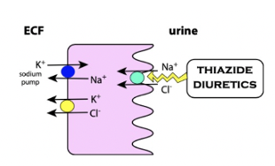
MoA: Decrease membrane permeability to Na+ and Cl- in the DCT
+ve: Less K+ washout than frusemide but more potent diuretic than spironolactone
Spironolactone
Indication
Advantage
MoA
Indication:
Combined with frusemide if refractory and severe CHF
Long-term management of CHF (mild diuretic)
+ve: No K+ washout
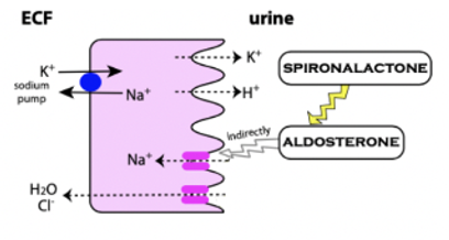
MoA: Competitive aldosterone antagonist → Prevents binding in collecting duct → Increased Na+ elimination (and Cl-)
ACE-i
3 Examples
Indication
MoA (+ 2 effects)
Caution
Examples:
Benazepril
Enalapril
Ramipril
Indication: #1 long-term management of mild CHF (stage C)
NOT for acute CHF (slow onset of action)
No benefit during pre-clinical stage
MoA: Inhibits function of angiotensin converting enzyme (ACE) which converts ATI → ATII
Vasodilation (ATII = Vasoconstrictor)
Moderate diuresis (ATII stimulates aldosterone secretion)
Caution: Ensure well-hydrated, normal renal parameters and no emesis
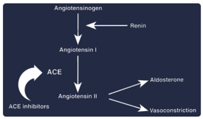
Telmisartan
Indication
MoA
Indication: Hypertension treatment
3rd line therapy for dogs and 1st line therapy for cats
MoA: Angiotensin receptor blocker
Inhibits angiotensin II from binding receptor in vascular smooth muscle → Vasodilation
Digoxin
Indication
2 Effects
Disadvantage
Indication: Atrial fibrillation ± other supraventricular tachyarrhythmias (eg. dogs with DCM)
Aim to get HR < 140bpm
Effects:
Positive inotrope = Increase myocardial contractility
Negative chronotrope = Decrease HR
-ve: Narrow TI (must use with good awareness of drug)