PSYCH 230 - Exam 2
1/119
There's no tags or description
Looks like no tags are added yet.
Name | Mastery | Learn | Test | Matching | Spaced |
|---|
No study sessions yet.
120 Terms
glutamate
most common neurotransmitter in the nervous system, found throughout the brain
excitatory
induces EPSPs
receptors induce depolarization following NT binding
GABA (gamma amino butyric acid)
most common inhibitory neurotransmitter
causes IPSPs
causes the postsynaptic neuron to become hyperpolarized
acetylcholine (ACh)
usually EPSPs
in both the brain and the PNS
in brain - sensation, action, learning
in PNS - peripheral motor neurons, parasympathetic system
the neurotransmitter at the neuromuscular junctions
spread out across the muscle fiber, causing the muscle to contract
neuromuscular junctions
synapses b/w motor neurons and muscle fiber
dopamine
movement, reward-seeking, motivation
produced in substantia nigra (movement), ventral tegmental area (VTA)
serotonin
known as “happiness NT”, but is also involved in sleep and appetite
antidepressant drugs increase serotonin
produced in the Raphe nuclei
mixed EPSPs and IPSPs
opioids
endorphin and enkephalin
natural morphine
pain reduction, reward, euphoria
mixed EPSPs and IPSPs
bind to opioid receptors
synthesized following pain, exercise, laughter
agonists
turn on NT system
EPSP/IPSP
presynaptic - release NT
postsynaptic - activate receptors
inverse agonists
postsynaptic binds to receptors but induces opposite effect
presynaptic agonists
release NT
L-Dopa, cocaine, amphetamine, Adderall, SSRIs (ex. Prozac)
postsynaptic agonists
activate receptors
synthetic opioids, benzodiazepines
L-Dopa
a dopamine precursor used to medicate Parkinson’s Disease (reduces dopamine levels)
cocaine
inhibits reuptake of dopamine by blocking dopamine transporter
increases dopamine levels
amphetamine
blocks and reverses dopamine transporter
increases levels of dopamine and norepinephrine
stimulation, euphoria, wakefulness, improved cognitive control
Adderall
prescribed combo of amphetamine and dextroamphetamine
used in the treatment of ADHD and narcolepsy
SSRIs (selective serotonin reuptake inhibitors)
block reuptake of serotonin
synthetic opioids
Fentanil - 100x more potent than morphine
Carfentanil - 100x more potent than fentanil
pain reduction, tranquillizer darts
overdose inhibits brainstem breathing circuits
benzodiazepines
Xanax, Valium
sedative, hypnotic, anxiolytic (anti-anxiety), anti-epileptic, muscle relaxant
bind to GABA receptors and facilitate GABA effects
antagonists
turn off NT system
presynaptic - prevent release
postsynaptic - block receptors
presynaptic antagonists
prevent release
postsynaptic antagonists
block receptors
typical antipsychotics
antipsychotic drugs for schizophrenia - pimozide, haloperidol
block D2 dopamine receptors
prevent dopamine from activating
used to treat schizophrenia
atypical antipsychotics
block D2 dopamine receptors
block serotonin receptors
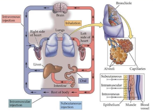
route of drug administration
oral ingestion
injection (subcutaneous, intramuscular, intravenous)
intramuscular - flu shot
inhalation
long-term effects
receptor down-regulation
neural sensitization
neurotoxicity
receptor down-regulation
tolerance to drug - homeostatic regulation in postsynaptic cell causes receptor degradation
withdrawal in absence - normal NT gives low signal
neural sensitization
hyper-responsive to drug
dopamine sensitization and addiction - wanting vs. liking
neurotoxicity
amphetamine kills dopamine neurons
toxic at 10x street dose
techniques for measuring action potentials
electrophysiological methods
optical methods
electrophysiological methods
intracellular recording
extracellular recording
intracellular recording
when electrode tip is inside neuron
senses positive action
whether action potential happens or not
sometimes, electrode tip can interfere w/ the action potential
electrode entering the cell can damage it
harder to puncture a neuron
only recording from 1 neuron
extracellular recording
when electrode tip is outside neuron
sodium rushes in, so positive ions are going away from the electrode tip
senses negative action
less invasive for cell
easier to carry out
not limited to recording only from 1 neuron at a time bc axons are right next to each other
cannot record IPSPs and EPSPs
good enough to record spikes
like an echo of the action potential
neurons represent locations
advantages of electrophysiological methods
actual spikes
fast
disadvantages of electrophysiological methods
intracellular recording can harm the cell
optical methods (calcium imaging)
calcium-sensitive dyes - molecules that become fluorescent in presence of calcium
report how much calcium is inside the neuron
fluoresce more when there is more calcium (reports spiking)
can record from many neurons through calcium-imaging
advantages of optical methods
indirect measurement of spikes
seeing the exact location of neuron (see and measure)
allows us to rerecord the same neuron
disadvantages of optical methods
slower
recording same neurons across days
cell-type specificity
overexciting the laser can harm the cell
sensation
the activation of sensory brain pathways by a physical stimulus
perception
the extraction of a mental representation from sensation
psychophysics
how the quantitative aspects of physical stimuli correlate w/ the perceptions they evoke
psychometric curve
x-axis - stimulus intensity
y-axis - some aspect of stimulus perception
in psychophysics - stimulus detection (%)
in sensory coding - neural activity (spikes per second)
sensory coding/processing
how the quantitative aspects of physical stimuli correlate w/ the neural activity they evoke
receptor cells
specialized cells that respond to physical sensory stimuli
respond electrochemically
convert sensory stimuli into neural signals
sensory stimulus triggers receptor cells
spontaneous firing
a sensory neuron occasionally fires spikes w/ no (obvious) relation to any sensory stimulus
trial-to-trial variability
Raster Plot
peri-stimulus time histogram (PSTH)
Raster Plot
x-axis - time
includes time of stimulus
y-axis - trials
peri-stimulus time histogram (PSTH)
time profile of firing
avg of the trials in the Raster plot
shows on avg what the neuron does in response to the specific stimulus
x-axis - time
y-axis - spike rate (spikes/sec)
receptive field
the region of sensory space in which a stimulus will modify the firing rate of that neuron
rate coding
a model of neuronal communication where the intensity of a stimulus is encoded by the frequency of a neuron’s action potentials (spikes)
temporal coding
when info is encoded in the precise timing of neuronal spikes, rather than just the rate of firing
cortical maps/topography
touch information from adjacent parts of the body are represented in adjacent parts in the cortex
homunculus
“tiny man”
refers to an orderly representation of the body in the brain
interesting disproportions

Mach Bands illusion
each vertical stripe has exactly the same luminescence, but higher and medium stations in our visual pathway make it appear as though the left side of each stripe is a bit lighter than the right side of each stripe
retinal ganglion cells (RGC)
receive signals from photoreceptors transmitted through horizontal, bipolar, and amacrine cells and send info up the visual pathways
amacrine and horizontal cells
lateral interactions w/in the retina
bipolar cells
carry info from photoreceptors to retinal ganglion cells
photoreceptors
transduce light signals (rods and cones)
rods
highly sensitive to light
ideal for vision in dim environments
respond similarly to diff light wavelengths
sensitive black and white sensors
similarly activated for colors
cones
less sensitive/need more light to be activated
come in 3 types that are each sensitive to either red, green, or blue wavelengths
phototransduction
the process of how we transform light into a neural signal
light hits the photoreceptors in our eyes and has passed through the layers of the lens
light strikes a light-absorbing pigment molecule (rhodopsin) in disc of photoreceptor
rhodopsin breaks into retinal and opsin
opsin closes Na+ gates, hyperpolarizing the photoreceptors
stops glutamate release
in the dark, photoreceptors constantly release a little bit of the glutamate
light hitting the rhodopsin causes a chain reaction that closes sodium channels and ultimately stops glutamate release
color blindness
a lack in one or more of the cone pigment
fovea
sharpest vision
corresponds to center of gaze
non-photoreceptor cells are pushed aside, which increases our sensitivity
highest density of cones, few rods
our focus of gaze is optimized for day
at night, we are nearly blind bc of this part
receptive field of RGCs
on-center off-surround
off-center on-surround
on-center off-surround
light in center excites (fire more)
light in surround inhibits
when an “on bipolar cell” receives less glutamate, it releases more glutamate
ex. if a small bright spot of light lands in the center of the cell’s receptive field, the cell fires a lot, but if the whole area is evenly lit, the cell barely responds
off-center on-surround
light in center inhibits
light in surround excites
when an “off bipolar cell” receives less glutamate, it releases less glutamate
lateral inhibition
capacity of an excited neuron to reduce the activity of its neighbors
in the retina-building intuition
edge detection
types of receptive fields enhance sensitivity to edges of images
on-center off-surround
off-center on-surround
bionic retina
camera captures image and sends info to the microprocessor
microprocessor converts data to an electronic signal and transmits it to receiver
receiver send signals through a tiny cable to an electrode panel implanted by doctors on back wall of eye (retina)
retinal implant emits pulses which travel through the optic nerve to the brain
brain perceives patterns of light and dark which correspond to the electrodes stimulated on the retinal implant
allows for partial vision, even when eyes are closed
implant is tacked onto retina
blind spot
there can be no photoreceptors where the nerve starts
bc of lack of machinery to detect light in the specific area
nasal hemiretina
closer to the nose, info coming through this side crosses over to the opp hemisphere of the brain
temporal hemiretina
closer to the temples, info coming through this side stays on the same hemisphere of the brain
lateral geniculate nucleus (LGN)
bundle of axons from thalamus to primary visual cortex (V1) is called optic radiation
axons travel through the optic radiation to V1
maintains retinotopic organization
primary visual cortex (V1)
leads to higher-level visual processing
neurons respond to oriented lines
retinotopic organization
each V1 neuron responds to a stimulus in a small area in the field of view
neighboring V1 neurons respond to stimuli in nearby locations in the visual field
hierarchical processing
a cognitive process in psychology where information is processed in a structured, top-down manner (higher-level concepts to lower-level details)
blindsight
when humans are forced to guess/use vision, they do well (unconscious vision)
visual perception
the process by which the brain interprets and organizes visual information from the environment, allowing us to make sense of what we see
ventral “what” stream
LGN → V1 → V2 → V4 → inferior temporal lobe (IT)
object recognition, feature conjunctions, feature recognitions
responses to increasingly complex stimuli
V4 responds to complex geometric shapes
ex. shapes like triangles
IT responds to visual objects in a position-invariant and size-invariant manner
ex. objects like cars, faces
fusiform face area (FFA)
has neurons that respond to faces
in the IT
prosopagnosia
face blindness
lesion in FFA - can be specific difficulty in recognizing faces
dorsal “where” stream
LGN → V1 → V2 → V5 (MT) → parietal cortex
spatial attention - guiding our view to points of interests
using vision for guidance of actions
detecting and analyzing movements
change blindness occurs bc we can’t pay attention to the full field of view simultaneously
critical for guiding visual attention
change blindness
occurs bc we can’t pay attention to the full field of view simultaneously
saccades
fast eye movements that focus out fovea on a small area or interest at any time
parietal cortex
important for eye movement and perception
integrates sensory info (gaze and spatial attention)
guides voluntary eye movement
neglect syndrome
can be caused by a unilateral lesion of the parietal lobe
visual system neglects one side of visual field
sounds frequency
determines our sense of pitch
measure - cycles/sec, Hz
sound amplitude
determines our sense of loudness
measure - decibel, dB
pure tone
a sound w/ a sinusoidal waveform
complex sounds
sounds that are not pure tones
most sounds
Fourier Transform
decomp of a sound (or other signal) to the frequencies that make it up
sum of 2 frequencies split into those 2 frequencies
spectrogram
shows the frequency of composition of sounds
frequency domain corresponds to power spectrum
how many of each frequency shows up in the time vs. amplitude graph
total frequency composition of the original sound
pitch perception
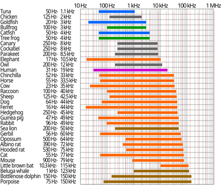
humans - 20-20,000 Hz
tympanic membrane
is struck by sound air pressure waves and forwards the vibration to the inner ear via the bones in the middle ear
cochlea
a coiled tube containing the basilar membrane
basilar membrane
vibrates w/ the sound wave
high frequency sounds cause the basal end to vibrate
basal end - higher frequencies
low frequency sounds cause the apical end to vibrate
apical end - lower frequencies
hair cell stereocilia
convert sounds to electrical signals
vibration of the basilar membrane causes movement
auditory nerve
sends signals from hair cells to the cochlear nucleus in the brainstem
hearing aid
a small electronic device that amplifies sounds
3 basic parts - microphone, amplifier, speaker
pick up sounds and makes them louder in the speaker
allows you to regain the ability of the hair cells you have lost
cochlear implant
used in complete or near-complete deafness
bypasses/replaces hair cells
directly stimulates the auditory nerve
electrical stimulation
coiled electrode array
via surgery
cochlear nucleus
receives signals from hair cells sent by the auditory nerve and sends info to the superior olivary nucleus
sound localization
sound location has to be computed, it is NOT encoded in the peripheral receptors
needs to compare b/w the ears
having 2 ears provides cues to localize sounds