August 2019
1/45
There's no tags or description
Looks like no tags are added yet.
Name | Mastery | Learn | Test | Matching | Spaced | Call with Kai | Chat |
|---|
No analytics yet
Send a link to your students to track their progress
46 Terms
A1) Name two types of anchoring junctions between cells?
Adherens junctions (zonula adherens)
Desmosomes (macula adherens)
A2) What is the major collagen type found in an epithelial cell's basement membrane?
Type IV collagen
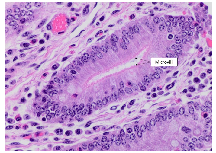
A3) What is the typical (average) length of microvilli?
Ca. 1 µm (typisk 1–2 µm)
A4) What is the main cytoskeletal protein found in microvilli?
Actin (mikrofilamenter)
A5) What is the main function of microvilli on the surface of these epithelial cells?
At øge celleoverfladens areal, så absorption (og i mindre grad sekretion) bliver mere effektiv.
Billede af mikrovilli
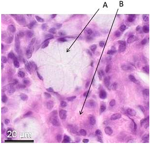
A6) Specify the type of secretory end piece indicated by A.
A peger på et mucous acinus (mucous secretory end piece).
Begrundelse: Cellerne er lyse/skummende, fordi mucin farves dårligt med H&E, og lumen virker relativt stor.

A7) Specify the type of secretory end piece indicated by B.
B peger på et serous acinus (serous secretory end piece).
Begrundelse: Cellerne er mørkere farvede, mere basofile og har et mere granulært cytoplasma.
A8) Indicate which statement about loose connective tissue is not correct.
A. Inflammation processes can occur in loose connective tissue.
B. Loose connective tissue is a cell rich connective tissue.
C. Loose connective tissue is rich in vessels and nerves.
D. Loose connective tissue fills gaps in the organs.
E. The majority of tendons and ligaments typically consist of loose connective tissue.
E. The majority of tendons and ligaments typically consist of loose connective tissue.
Denne er forkert.
Sener og ligamenter består hovedsageligt af dense regular connective tissue, ikke loose connective tissue.
A9) Indicate which statement about reticulum cells is correct.
A. Reticulum cells typically occur in epithelium.
B. Reticulum cells form reticular fibers in lymphoid tissues.
C. Reticulum cells are formed from leucocytes migrating from the peripheral blood.
D. Reticulum cells have a horseshoe-shaped nucleus
E. Reticulum cells secrete lysosomal enzymes.
✅ B. Reticulum cells form reticular fibers in lymphoid tissues.
Hvorfor?
Reticulumceller er specialiserede fibroblastlignende celler i lymfoide organer.
De producerer retikulære fibre (type III kollagen), som danner det støttende netværk i fx lymfeknuder og milt.
Hvorfor er de andre forkerte?
A: Findes ikke typisk i epitel.
C: De stammer ikke fra leukocytter.
D: Hesteskoformet kerne er typisk for nogle monocytter/makrofager.
E: Lysosomale enzymer er ikke deres karakteristiske funktion.
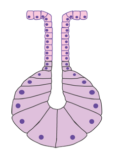
A10) Below is a cartoon of a multicellular gland. What is the general classification for this type of gland?
Simple acinar gland
Hvordan klassificeres glands
A11) Where is the nucleus placed (relative to whole cell) in a multilocular adipocyte?
Centrally placed nucleus
Husk: Multilokulære (brune) adipocytter har mange små fedtdråber og en relativt central kerne.
Unilokulære (hvide) adipocytter har en perifert forskudt kerne.
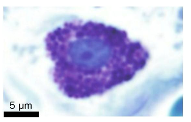
A12) What is the name of the cell type?
Mast cell
Begrundelse: Toluidinblåt farver de mange granula metakromatisk (lilla/violette granula), hvilket er karakteristisk for mastceller.
A13) Name two substances found in the granules of the cell.
✅ Histamine
✅ Heparin
(Andre mulige svar kunne være proteaser som tryptase eller chymase.)
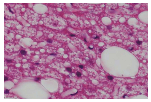
A14) What is the main classification of tissue (be specific) that can be observed in this specimen?
Billedet viser mange små lipidvakuoler i cellerne → multilocular adipose tissue (brown adipose tissue).
✅ Brown adipose tissue (multilocular adipose tissue)
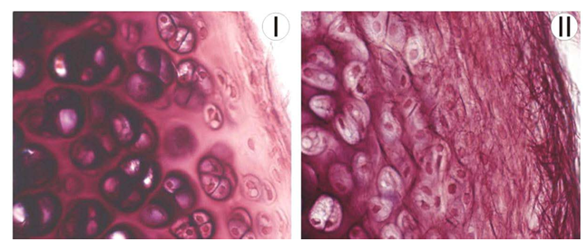
Specify the type of tissue seen in the two pictures
A15) Tissue I
I væv I ses chondrocytter i lakuner og en homogen matrix uden tydelige elastiske fibre.
✅ Hyaline cartilage

Specify the type of tissue seen in the two pictures
A16) Tissue II
I væv II ses chondrocytter i lakuner samt et tæt netværk af mørke elastiske fibre.
✅ Elastic cartilage
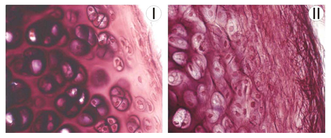
A17) Specify the combination of extracellular matrix fibres that are the typical components of tissues I and II
A. Tissue I: elastic fibers; tissue II: type IV collagen.
B. Tissue I: reticular fibers; tissue II: type IV collagen.
C. Tissue I: Type I collagen and elastic fibers; Tissue II: Reticular fibers.
D. Tissue I: Type II collagen; tissue II: type II collagen and elastic fibers.
E. Tissue I: Type II collagen; Tissue II: Reticular fibers.
Hyalin brusk (I): Type II kollagen
Elastisk brusk (II): Type II kollagen + elastiske fibre
✅ D. Tissue I: Type II collagen; tissue II: type II collagen and elastic fibers.
A18) Specify which statement is correct.
A. The preparation shows articular cartilage
B. The preparation shows elastic cartilage
C. The tissue contains hydroxyapatite
D. The tissue is surrounded by a perichondrium
E. The cells seen secrete mainly collagen type I
Below is a tissue preparation that is stained with Masson-Goldner-Trichrome.
✅ A. The preparation shows articular cartilage
Vi kan se ledbrusk, som er hyalin brusk. (ik al hyalin brusk er ledbrusk)
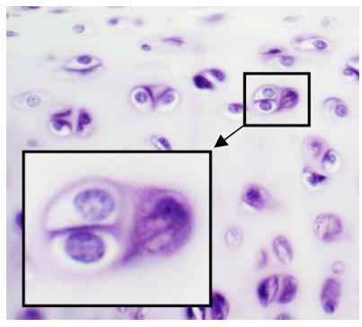
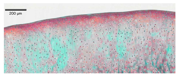
A19) What is the name of the region of cells that is highlighted in the box and arises during interstitial growth of cartilage?
Den markerede struktur viser to chondrocytter, som er opstået ved deling af én chondrocyt.
✅ Isogenous group (isogenous cell nest)
Dette er karakteristisk for interstitial growth af brusk.
•Isogen gruppe (isogenous groups) = opstået fra 1 enkelt chondrocyt, der har undergået mitotiske delinger
A20) What is the name of the structure in which the osteocytes' extensions are located?
✅ Canaliculi
(Osteocytterne ligger i lakuner, mens deres udløbere løber gennem canaliculi.)

A21) Specify the colors of tissue type 1 and tissue type 2 in a fresh unfixed preparation (in vivo).
Væv 1 = hvidt fedtvæv → ser gult ud in vivo pga. carotenoider opløst i fedtet.
Væv 2 = rød knoglemarv → ser rød ud pga. det høje indhold af blodkar og hæmatopoietiske celler.
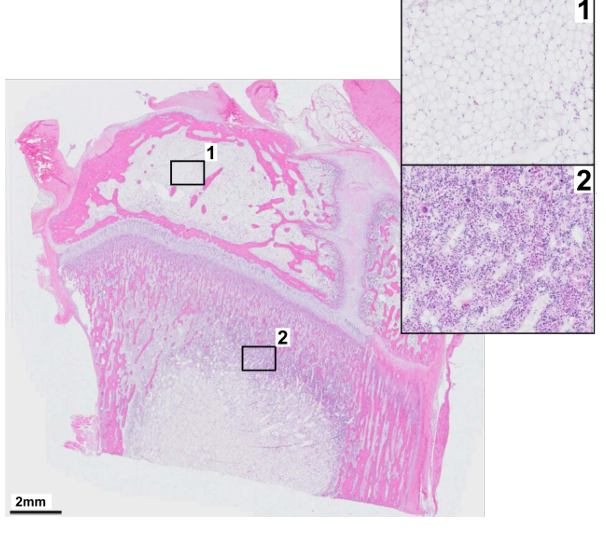
A22) Name the main function of tissue type 2.
✅ Hematopoiesis (blood cell production)
Væv 2 = rød knoglemarv

A23) What is the major cell type found in tissue type 1?
✅ Adipocytes (unilocular adipocytes)
Væv 1 = hvidt fedtvæv
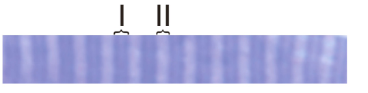
A24) Below is a tissue preparation of skeletal muscle stained with toluidine blue.
Which of the two marked structures (I or II) will become shorter during contraction?
✅ Svar: I
Hvorfor?
Struktur I er I-båndet, som bliver kortere under kontraktion. Struktur II er A-båndet, som ikke ændrer længde.
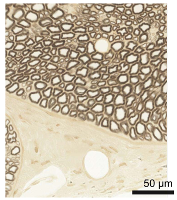
A25) Below is a preparation that is stained with Osmium. Name the cell type that forms the dark brown "circular" structures in the upper part of the image.
✅ Svar: Schwann cell
Hvorfor?
Osmium farver myelin mørkebrunt. De mørke ringe er tværsnit af myeliniserede nervefibre, hvor myelinen dannes af Schwann-celler i det perifere nervesystem.
B1) Give two examples of a so-called second messenger and its signaling receptor.
Second messenger | Receptor/effektor |
|---|---|
cAMP | PKA (Protein Kinase A) |
cGMP | PKG (Protein Kinase G) |
Ca²⁺ | Calmodulin |
IP₃ | IP₃-receptor (på ER) |
DAG | PKC (Protein Kinase C) |
B2) Briefly describe the function/role of scaffold proteins in cell signaling.
Scaffoldproteiner samler flere signalproteiner i et kompleks, så signaloverførsel bliver hurtigere, mere effektiv og mere specifik.
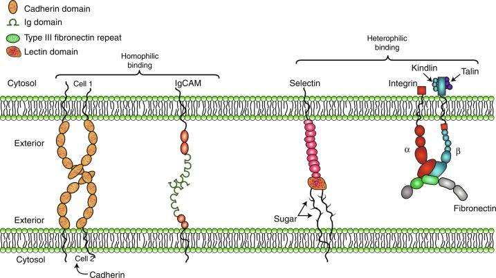
B3) Give an example of the subcellular localization of integrin receptors.
Focal adhesions: Integriner binder ECM-proteiner (fx fibronectin) til aktin-cytoskelettet.
Hemidesmosomes: Integriner binder basallamina (laminin) til keratinfilamenter.
Basolateral plasmamembran: Integriner forankrer epitelceller til ECM i basalmembranen.
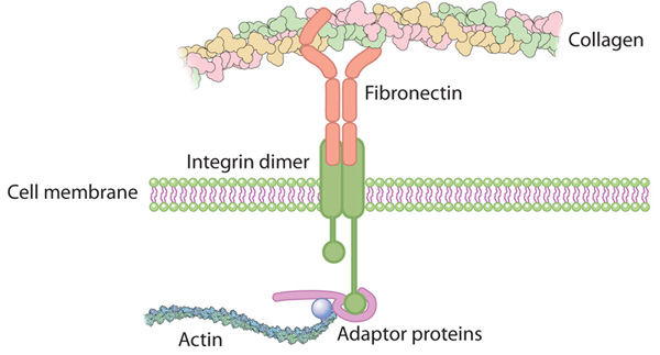
B4) Describe how integrin receptors connect the extracellular matrix with the cytoskeleton.
Integriner binder ECM-proteiner (fx fibronectin) extracellulært og er intracellulært forbundet til aktin-cytoskelettet via adaptorproteiner som talin og vinculin.

B5) Describe the principle of the dynamic role of integrin receptors in cell migration.
Integriner danner nye adhæsioner ved cellens forende og nedbryder adhæsioner ved bagenden, så cellen kan bevæge sig fremad.
B6) Give an example of how cadherin adhesion receptors differ from integrin receptors.
Cadheriner medierer primært celle-celle-adhæsion, mens integriner medierer celle-ECM-adhæsion.
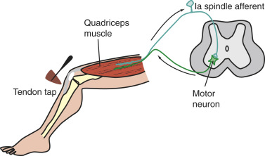
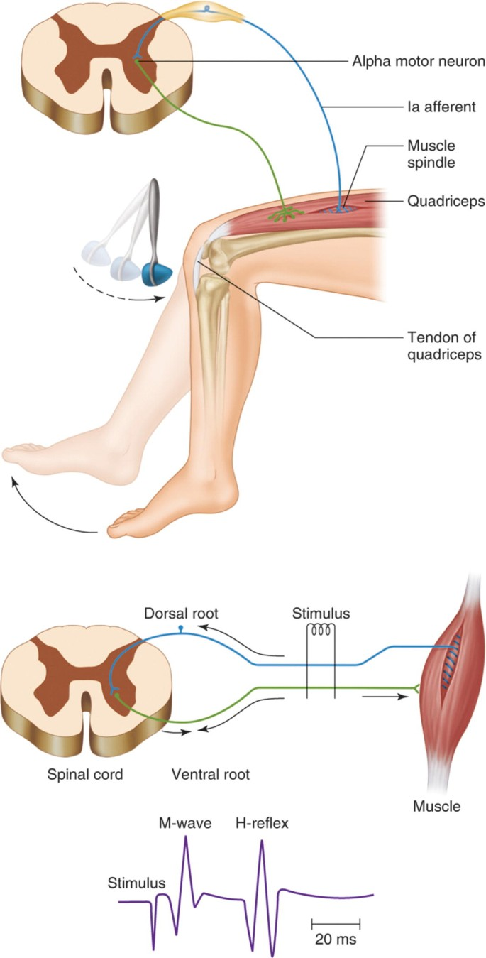
C1) Describe the circuit diagram for the T-reflex (stretch reflex).
Stræk af musklen aktiverer muskeltenens Ia-afferente neuron, som laver en monosynaptisk excitatorisk synapse på et α-motorneuron i rygmarven, hvilket får den samme muskel til at kontrahere.
C2) Explain how the T-reflex differs from the H-response (H-reflex) in terms of the circuit diagram and the methods of induction and recording.
Begge bruger hovedsageligt den samme Ia-afferent → α-motorneuron refleksbane. T-refleksen udløses ved et mekanisk stræk (reflekshammer), mens H-refleksen udløses ved elektrisk stimulation af nerven. Begge registreres typisk med EMG.

C3) Explain briefly the advantage of the H-response compared to the T-reflex in clinical testing.
H-refleksen kan fremkaldes mere kontrolleret og reproducerbart, fordi stimuleringen er elektrisk og kan standardiseres.
C4) Explain how the eye changes its focal power by accommodation.
Ved akkommodation kontraherer m. ciliaris, zonulatrådene aflastes, og linsen bliver mere rund, hvilket øger dens brydningsstyrke.
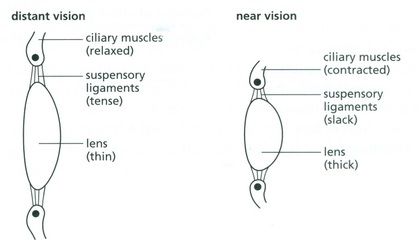
C5) Describe how and why the ability to accommodate changes with age.
Akkommodationsevnen falder med alderen (presbyopi), fordi linsen bliver mindre elastisk og derfor har sværere ved at ændre form.
C6) What type of lenses are suitable to correct the above age-related changes?
Akkommodationsevnen falder med alderen (presbyopi), fordi linsen bliver mindre elastisk og derfor har sværere ved at ændre form.
Konvekse (plus-)linser anvendes til at korrigere presbyopi. De øger øjets brydningsstyrke og kompenserer for, at den aldersstive linse ikke længere kan blive tilstrækkeligt rund til at fokusere lys fra nærliggende objekter på nethinden.
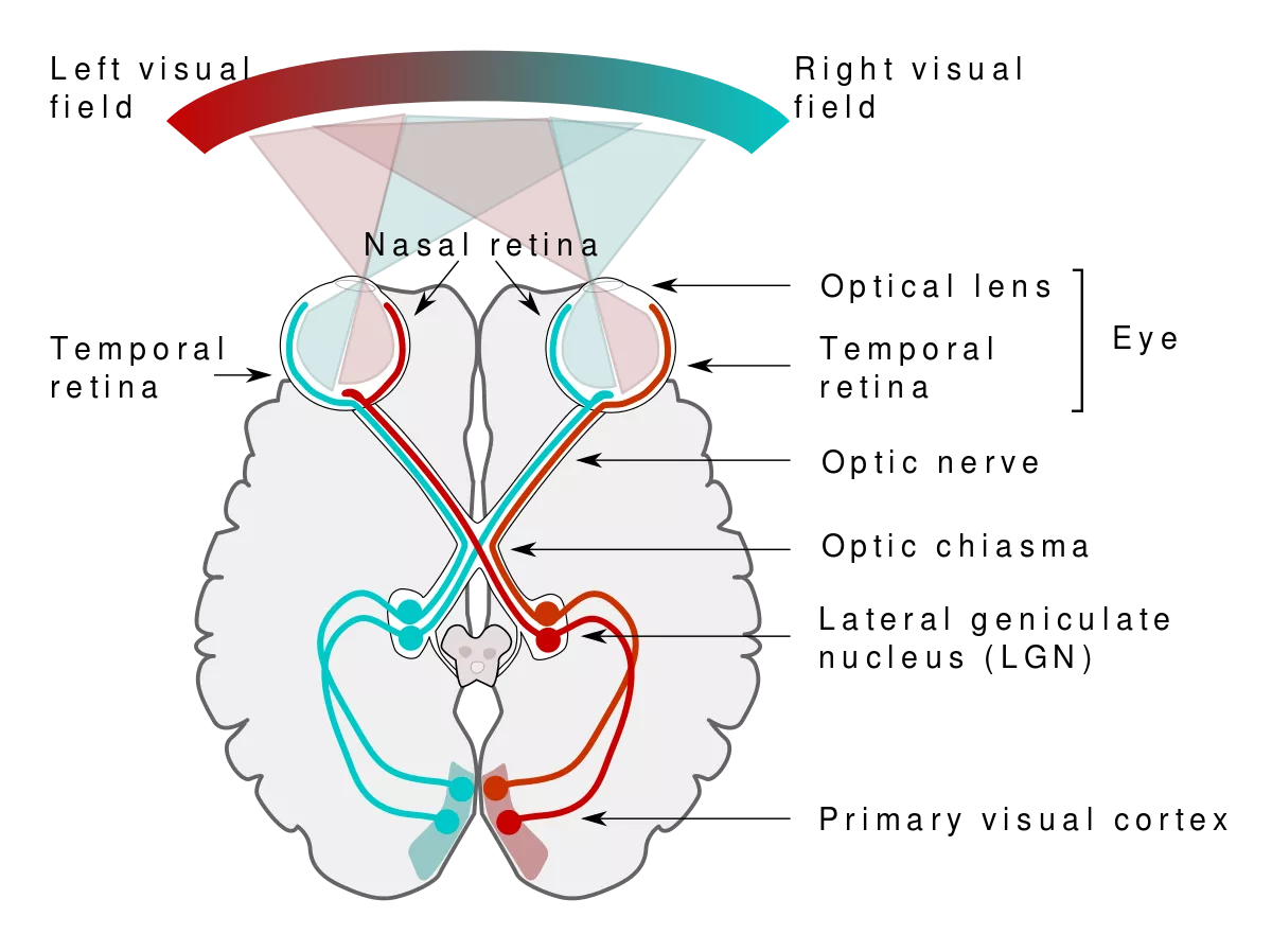
D1) What would happen if you lesioned the left LGN?
(a) You would not be able to see the right visual field.
(b) You would not be able to see the left visual field.
(c) You would not receive any information from your right eye (functionally blind in the right eye).
(d) You would not receive any information from your left eye (functionally blind in the left eye).
✅ Svar: (a) You would not be able to see the right visual field.
Struktur | Udfald |
|---|---|
Left LGN | Tab af højre synsfelt (right homonymous hemianopia) |
Right LGN | Tab af venstre synsfelt (left homonymous hemianopia) |
D2) What is the difference between ocular dominance columns and orientation columns?
Ocular dominance columns: Neuroner responderer primært på input fra enten højre eller venstre øje.
Orientation columns: Neuroner responderer bedst på en bestemt orientering af en kant eller streg.
D3) Where does binocular input first occur in the visual system and why does it emerge at this location?
Binokulært input opstår først i primær visuel cortex (V1).
I LGN holdes input fra de to øjne adskilt, mens neuroner i V1 modtager input fra begge øjne og kan sammenligne dem for dybdesyn (stereopsis).
D4) Describe briefly the main functions of cerebellum and a characteristic deficit after damage to this brain structure?
Cerebellum koordinerer bevægelser, balance, timing og motorisk læring.
Skade giver typisk ataxi (ukoordinerede bevægelser).
D5) Describe the morphology of the principal nerve cell in the cerebellar cortex.
Den vigtigste nervecelle er Purkinje-cellen, som har en stor cellekrop og et meget forgrenet, fladt dendrittræ orienteret i ét plan.

D6) Describe the afferent connection to the principal nerve cell in the cerebellar cortex and indicate their origin.
Climbing fibers: Kommer fra inferior olive og danner stærke synapser direkte på Purkinje-cellens dendritter.
Mossy fibers: Kommer fra mange kilder (bl.a. rygmarv, vestibulære kerner og pons) og påvirker Purkinje-celler indirekte via granule cells og parallel fibers.
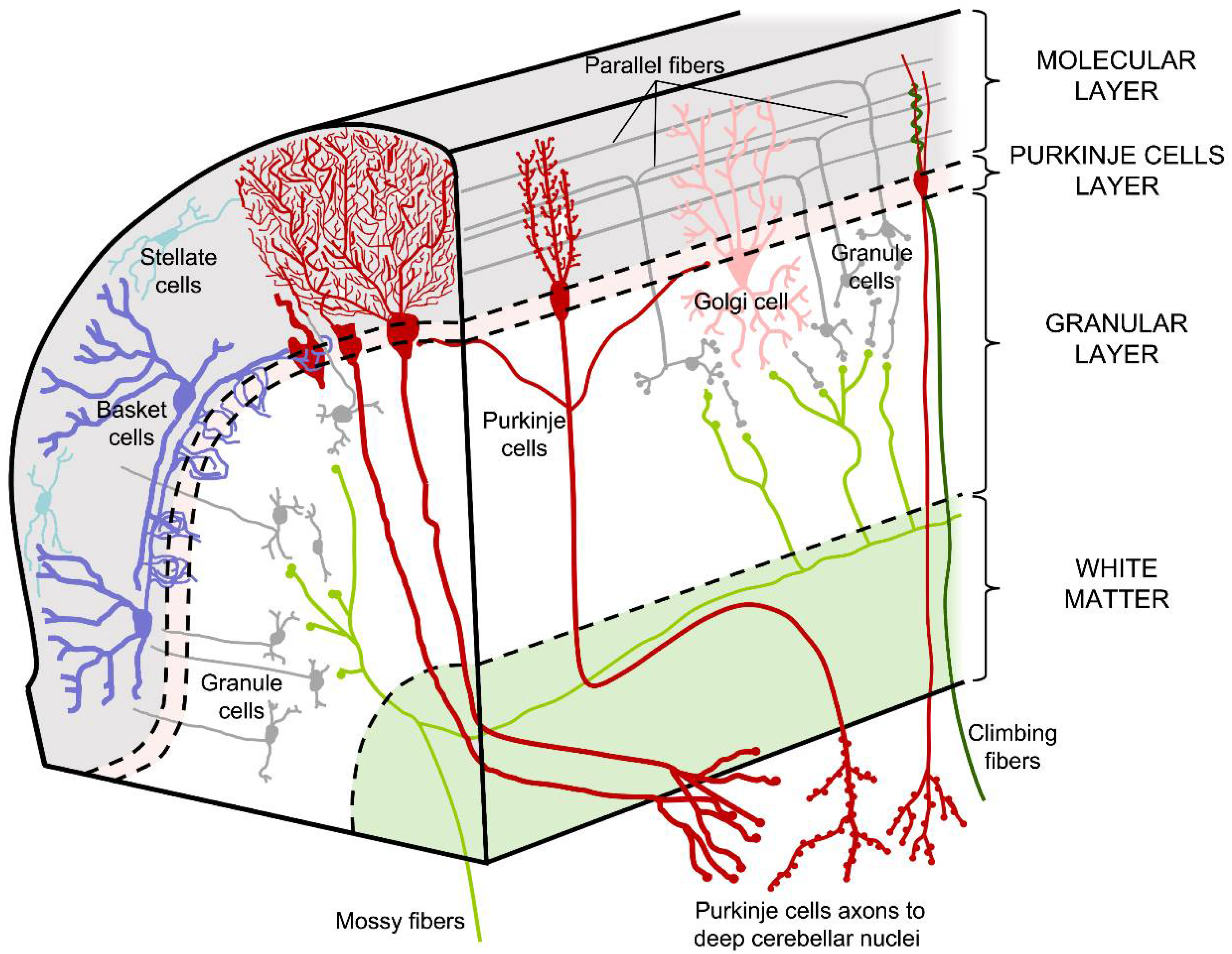
D7) Discuss briefly the functions of the afferent connection to the principal nerve cell in the cerebellar cortex.
Climbing fibers: Fejl- og læringssignal, vigtigt for motorisk læring.
Mossy fibers/parallel fibers: Formidler sensorisk og motorisk information om den aktuelle bevægelse.