Anatomy and Physiology Skull Bones
1/81
There's no tags or description
Looks like no tags are added yet.
Name | Mastery | Learn | Test | Matching | Spaced |
|---|
No study sessions yet.
82 Terms
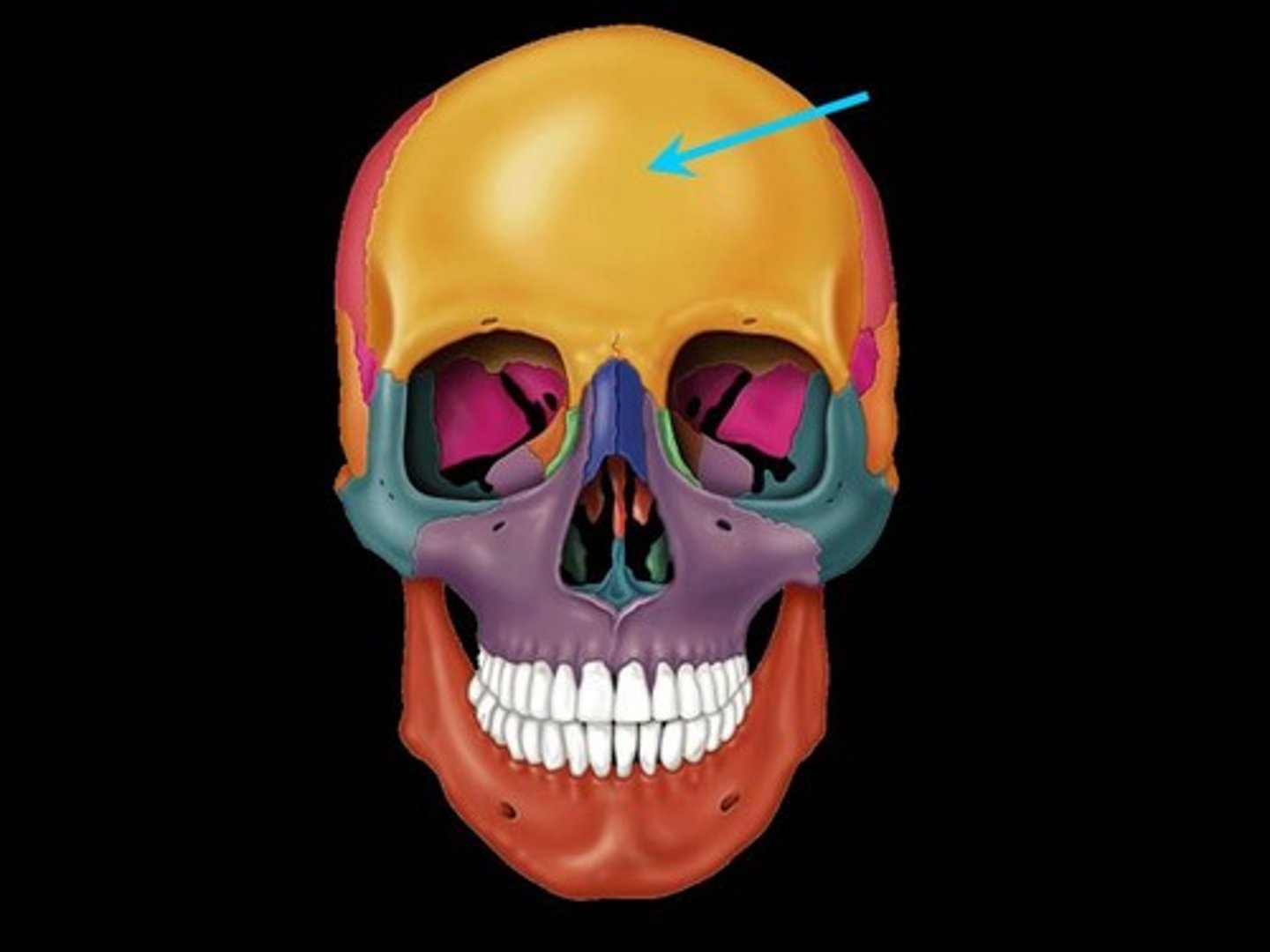
Frontal Bone (unpaired)
forehead
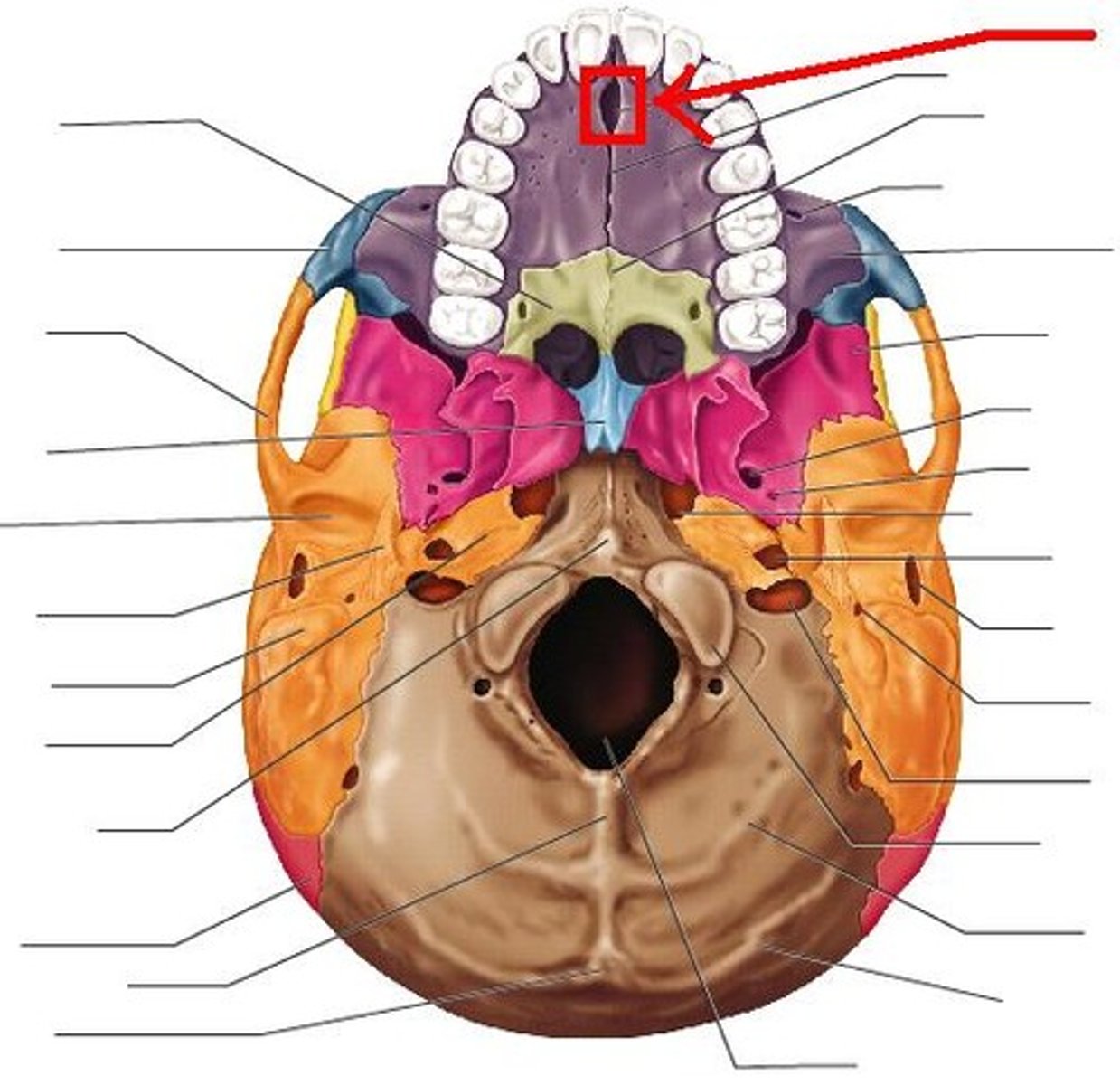
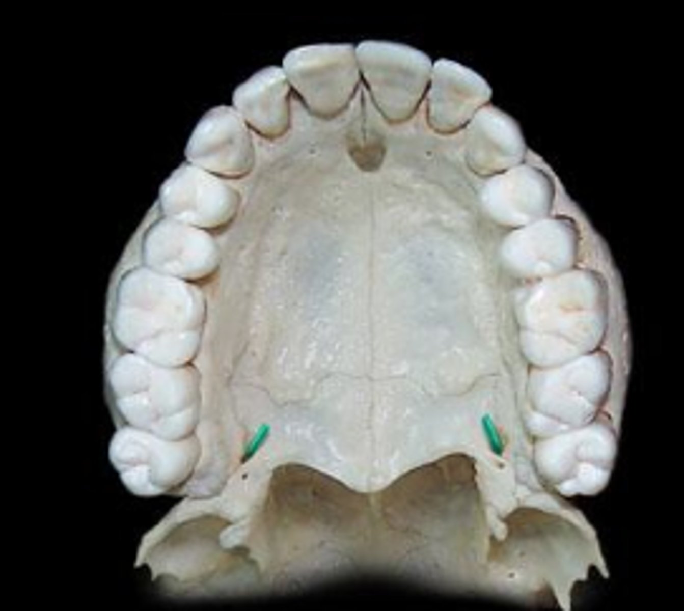
incisive foramen
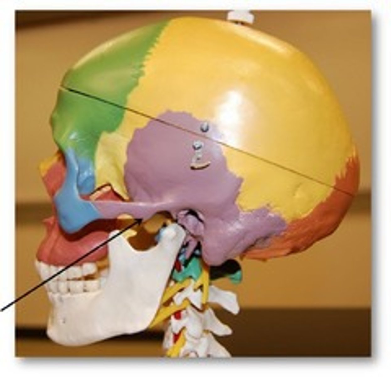
articular eminence
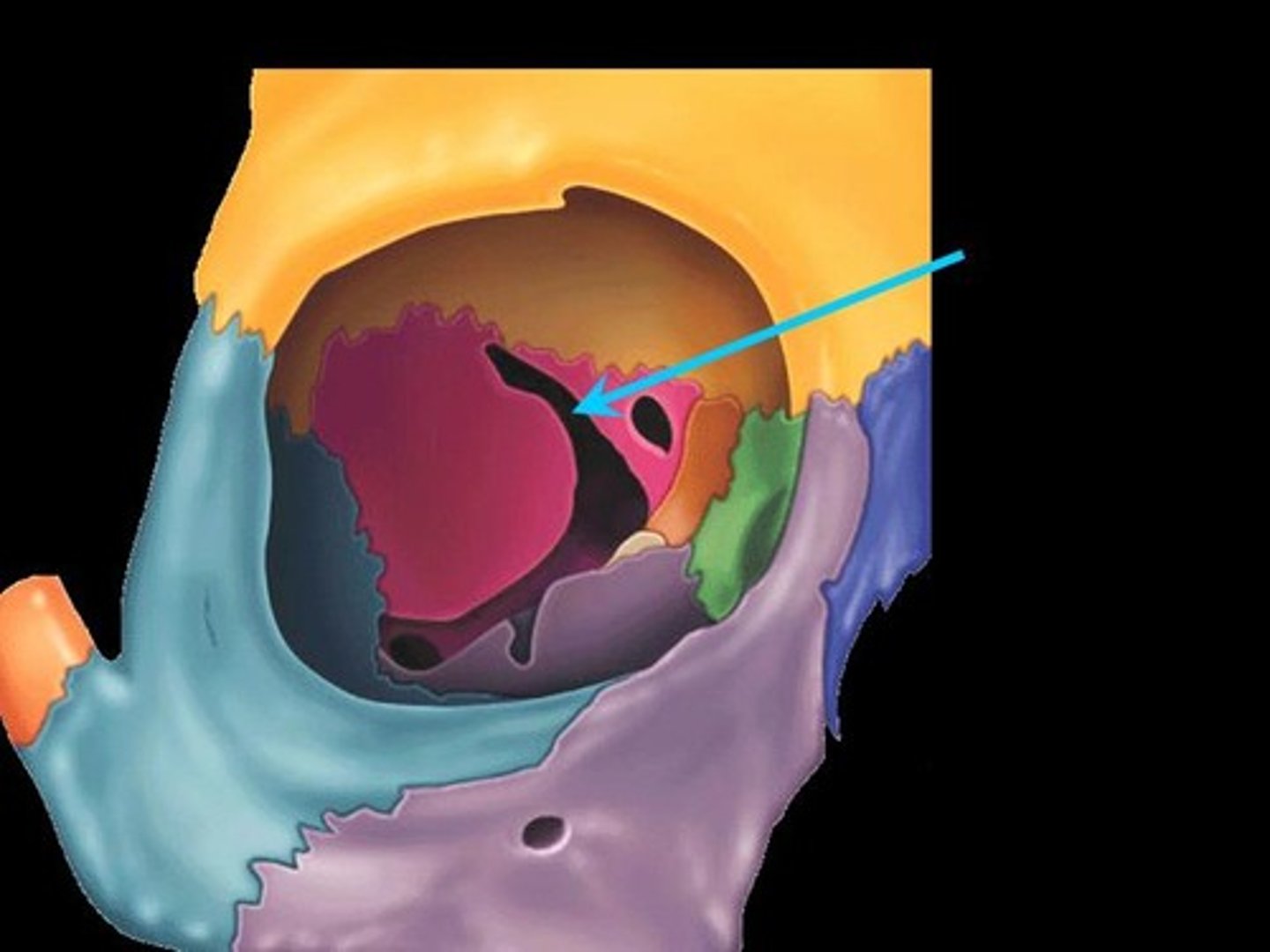
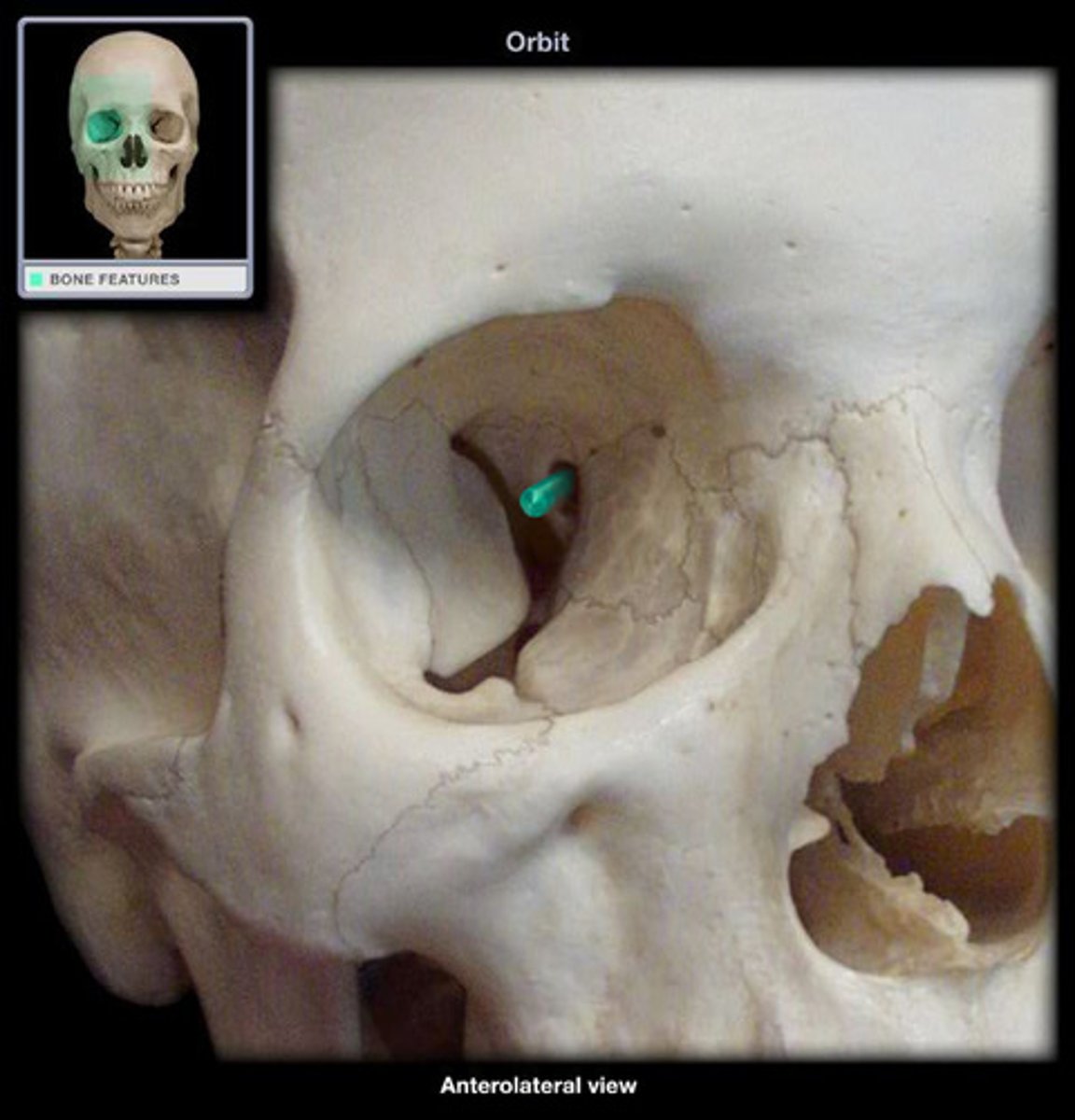
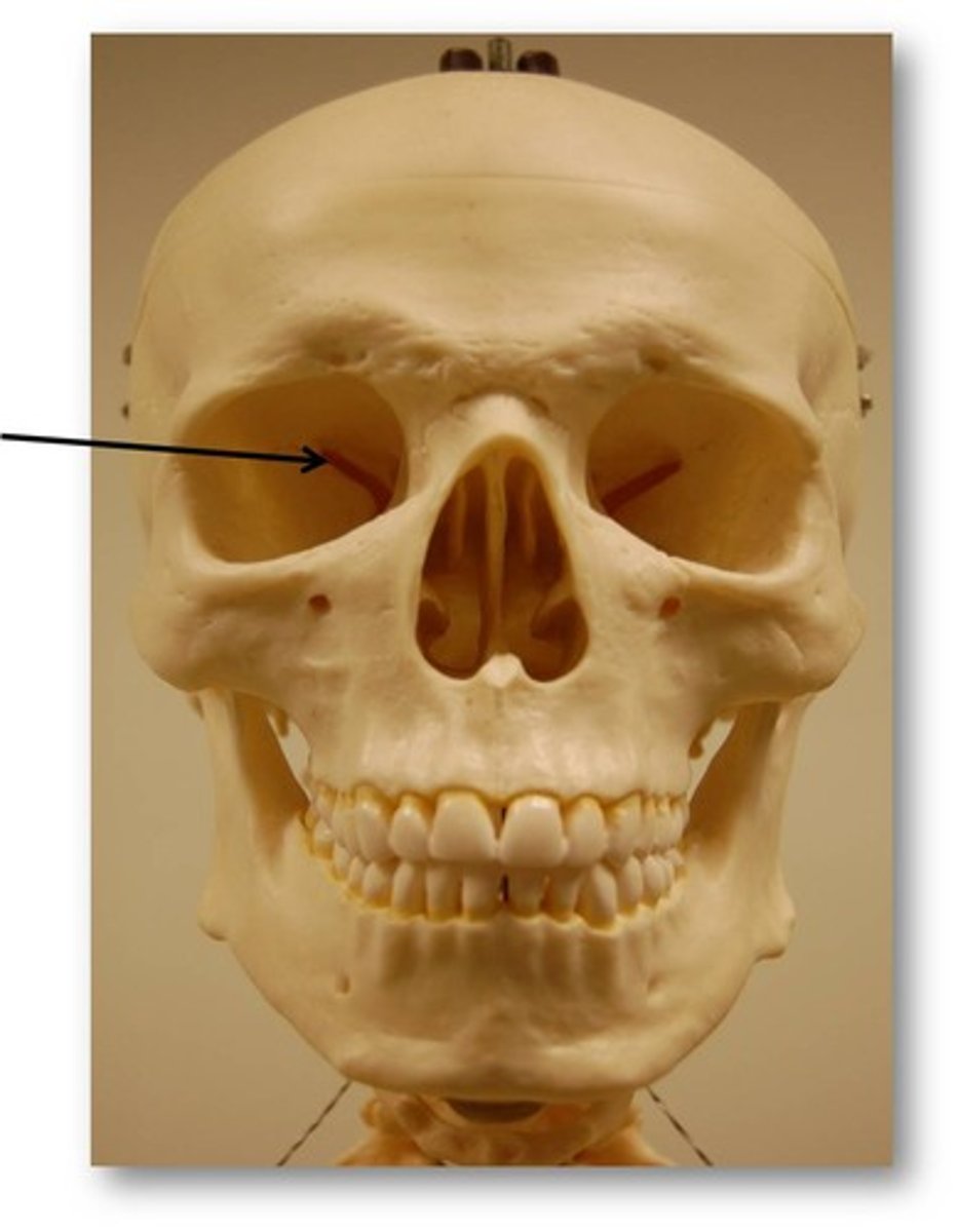
Superior Orbital Fissure
sphenoid
Optic Canal
optic nerve
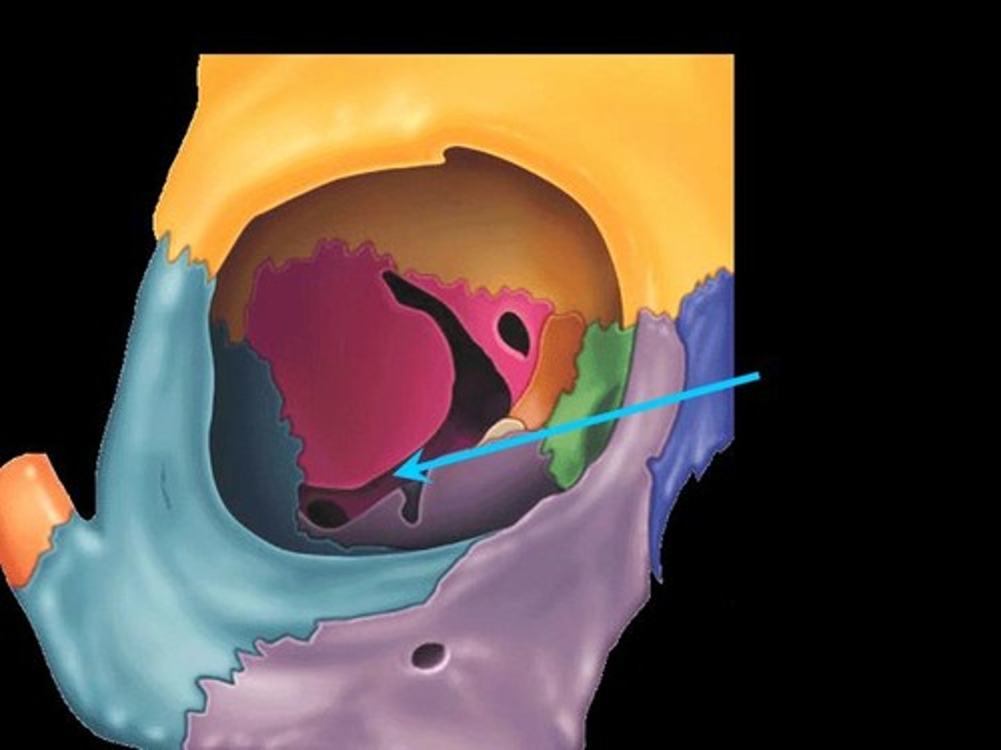
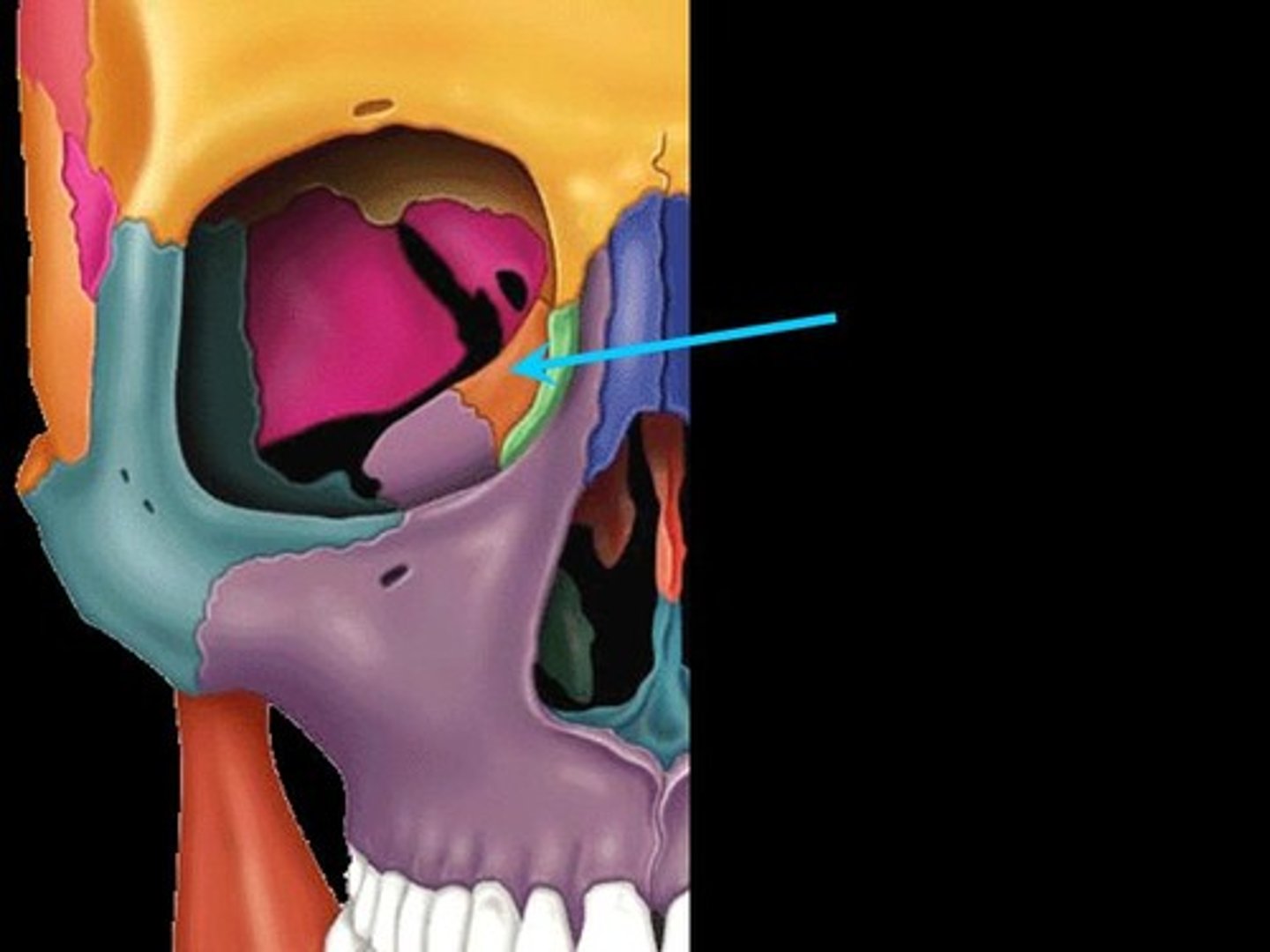
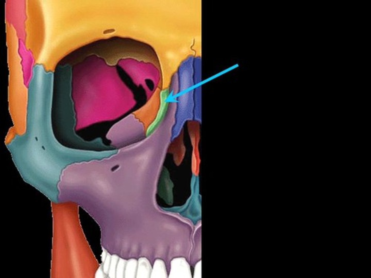
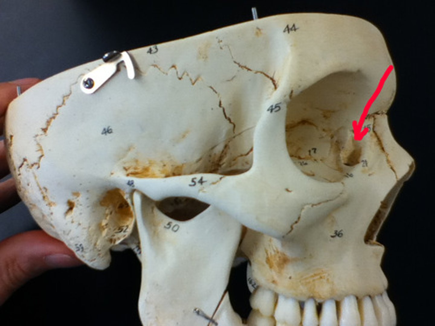
inferior orbital fissure
fissure in the orbit floor between maxilla and greater wing of sphenoid
Middle Nasal Concha
the middle thin, spongy, bony plate with curved margins, part of the ethmoidal labyrinth, projecting from the lateral wall of the nasal cavity and separating the superior meatus from the middle meatus;
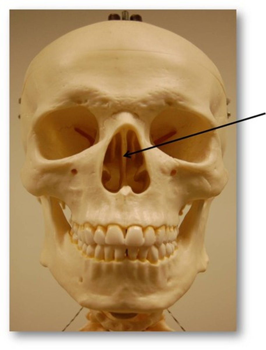
Perpendicular Plate
forms superior part of nasal septum
Inferior Nasal Concha
located on each side of the nasal septum, attached to the lateral wall of the nasal cavity; increase epithelial surface area and create turbulence in the inspired air
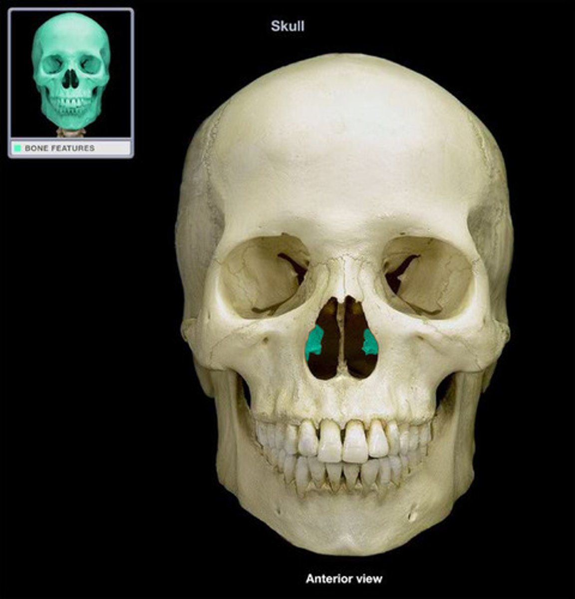
Vomer (unpaired)
nasal septum
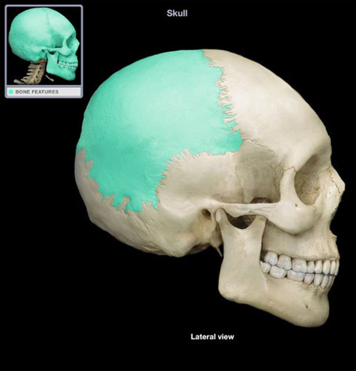
parietal bone (paired)
a bone forming the central side and upper back part of each side of the skull.
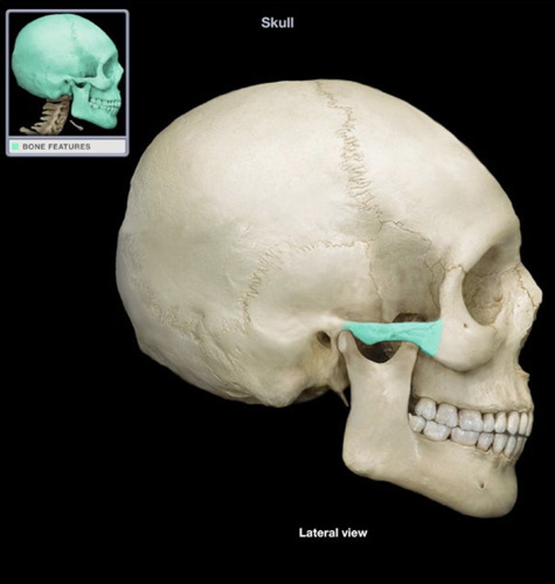
Sphenoid Bone (unpaired)
forms part of the base of the skull and parts of the floor and sides of the orbit
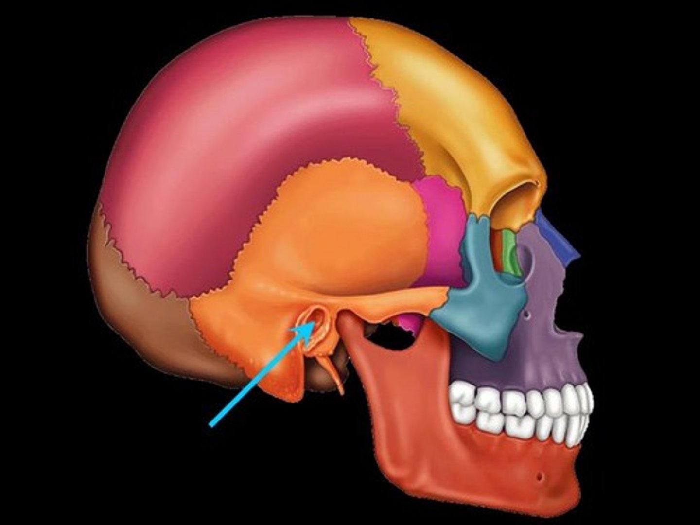
Temporal Bone (paired)
bone that forms parts of the side of the skull and floor of the cranial activity. There is a right and left temporal bone.

Ethmoid Bone (unpaired)
Light spongy bone between the eye sockets; forms part of the nasal cavities.
Lacrimal Bone (paired)
small fragile bone making up part of the front inner walls of each eye socket and providing room for the passage of the lacrimal ducts
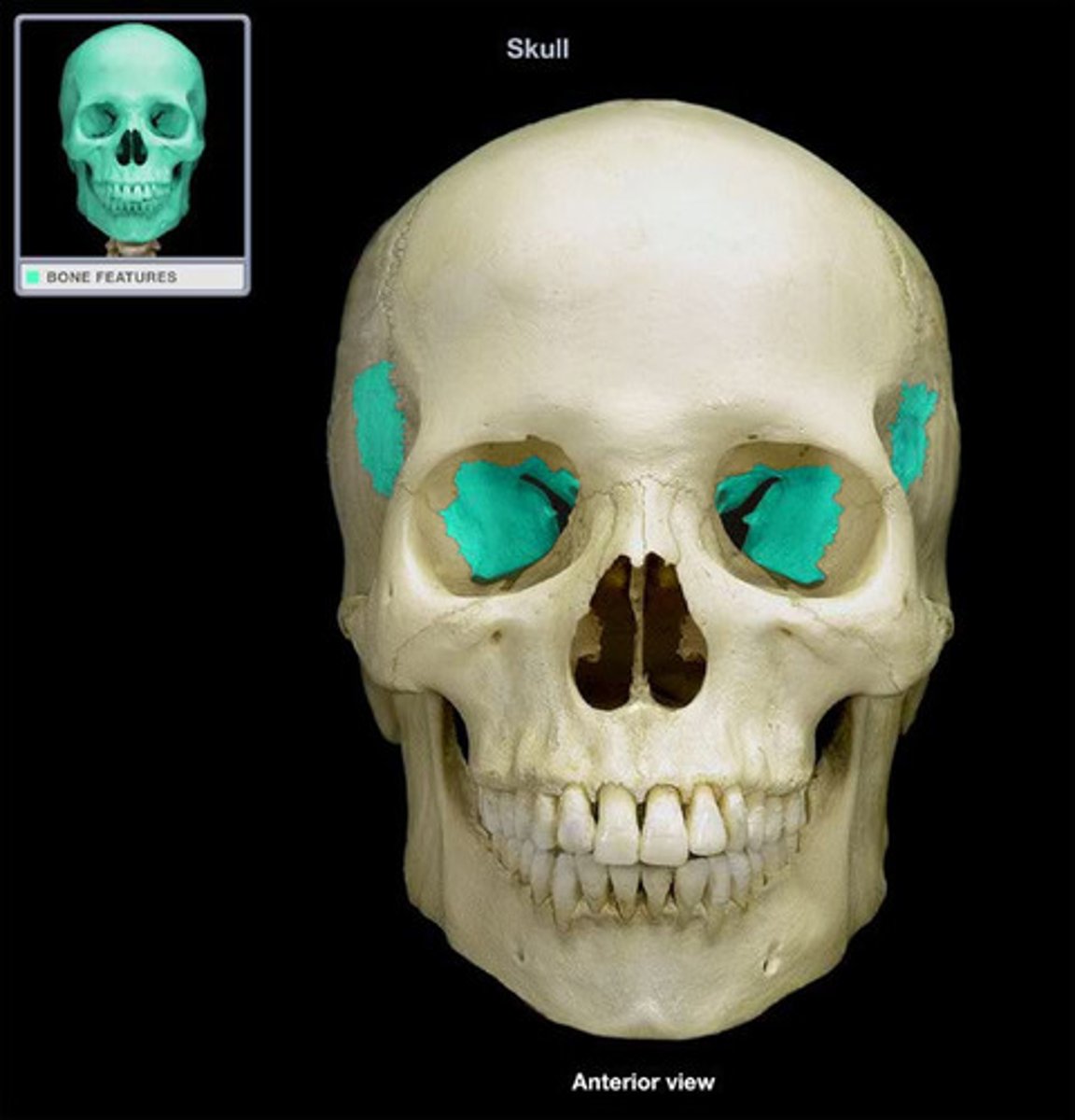
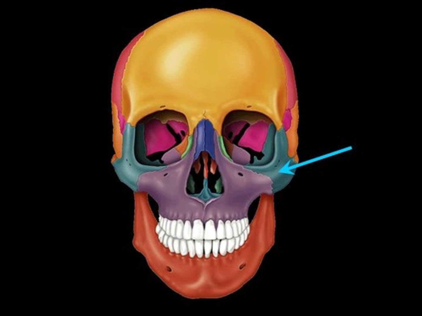
Zygomatic Bone (paired)
cheek bone
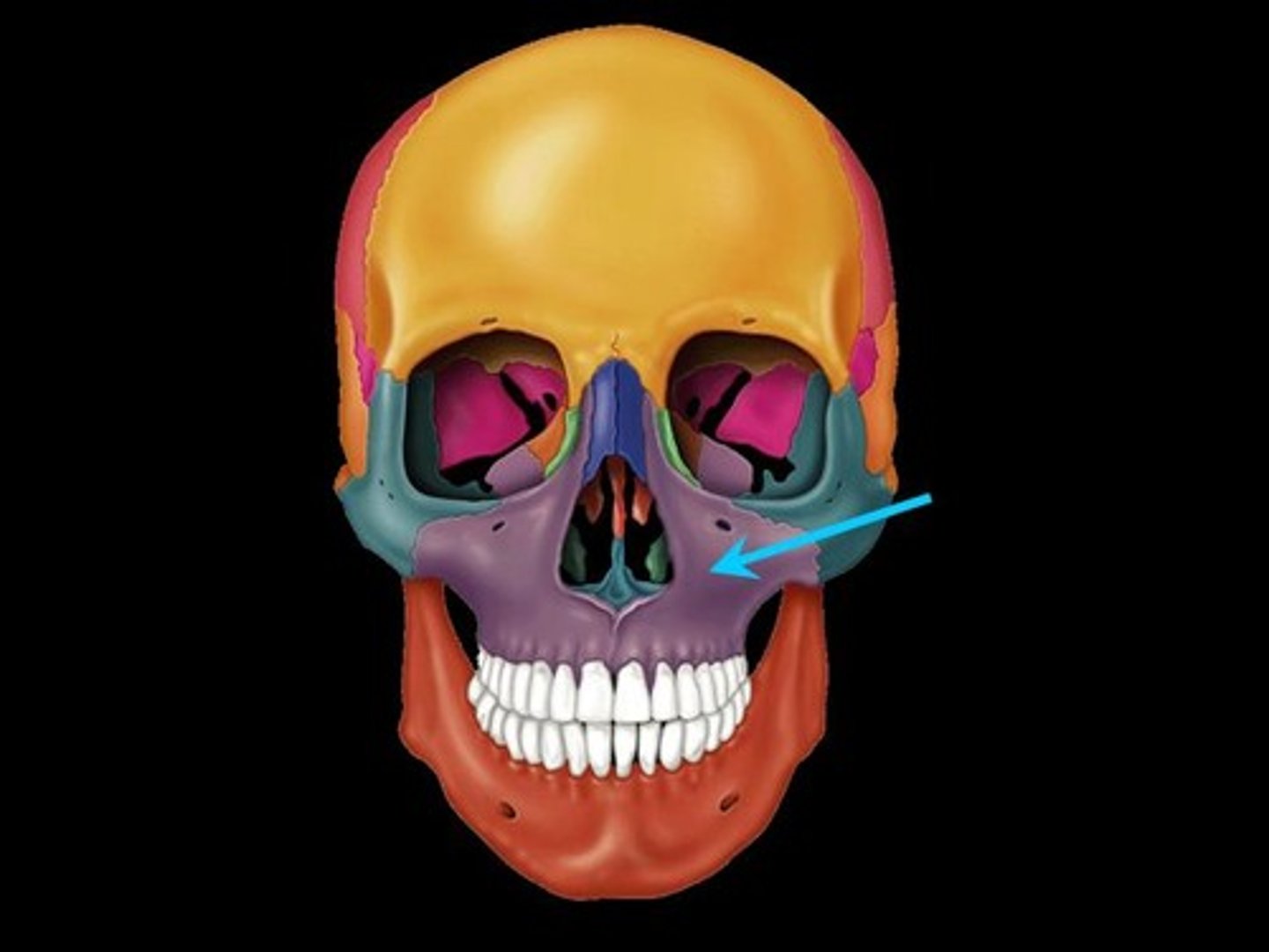
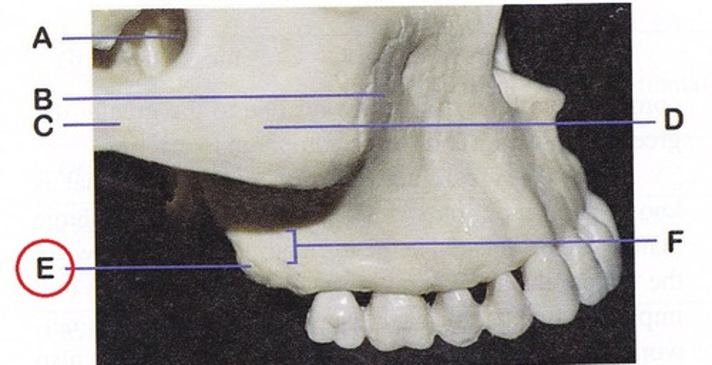
Maxilla (paired)
upper jaw
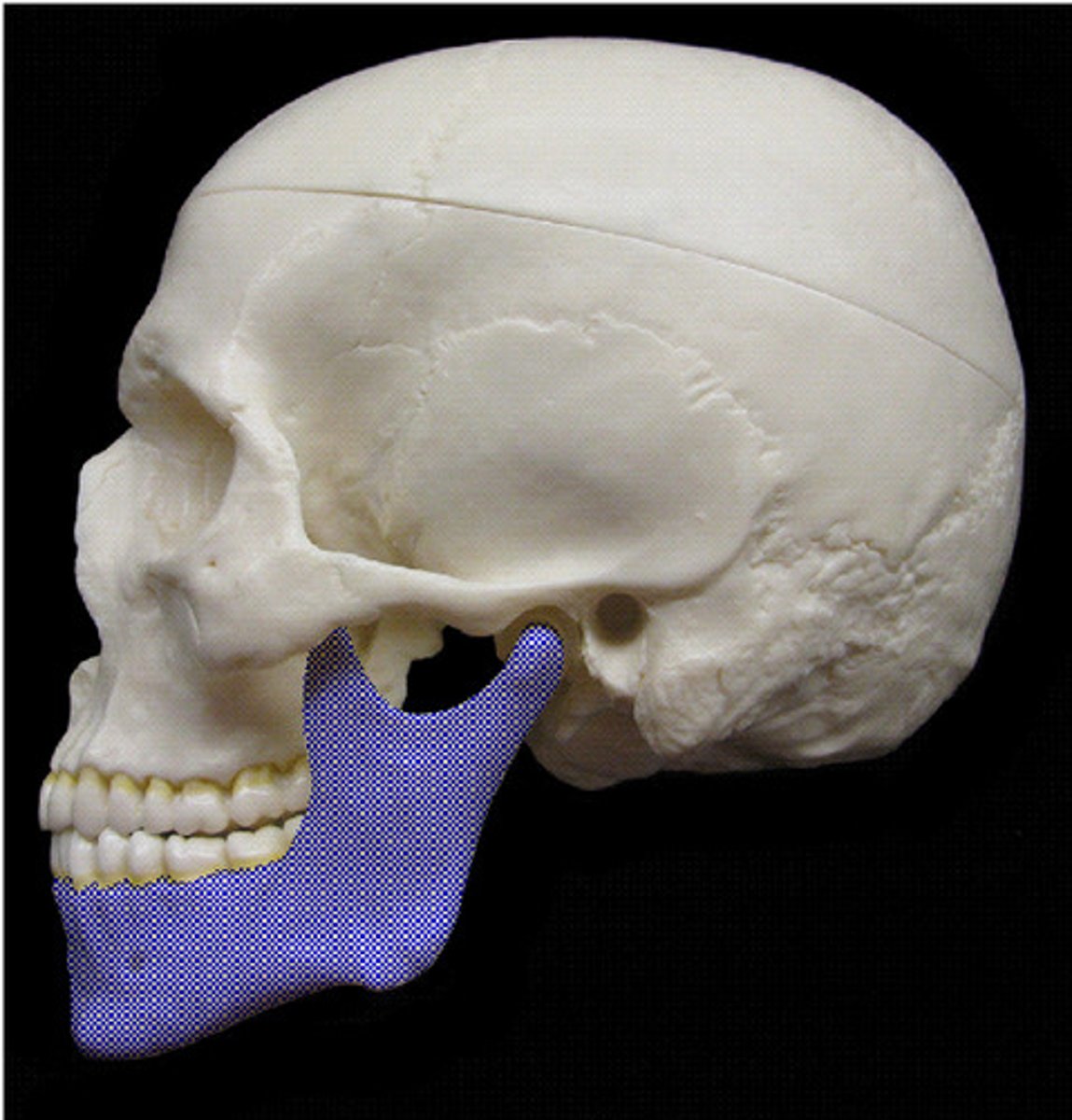
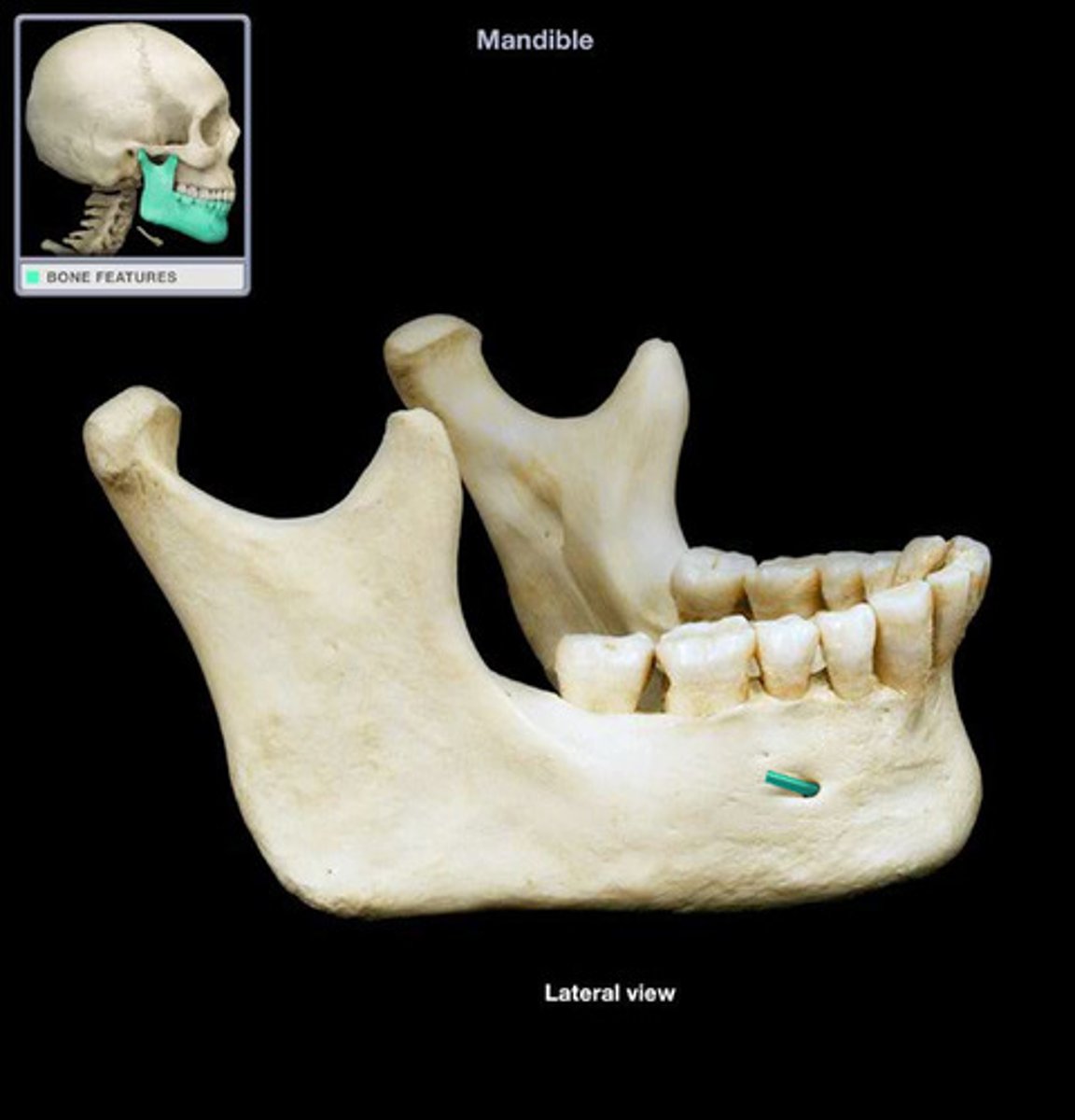
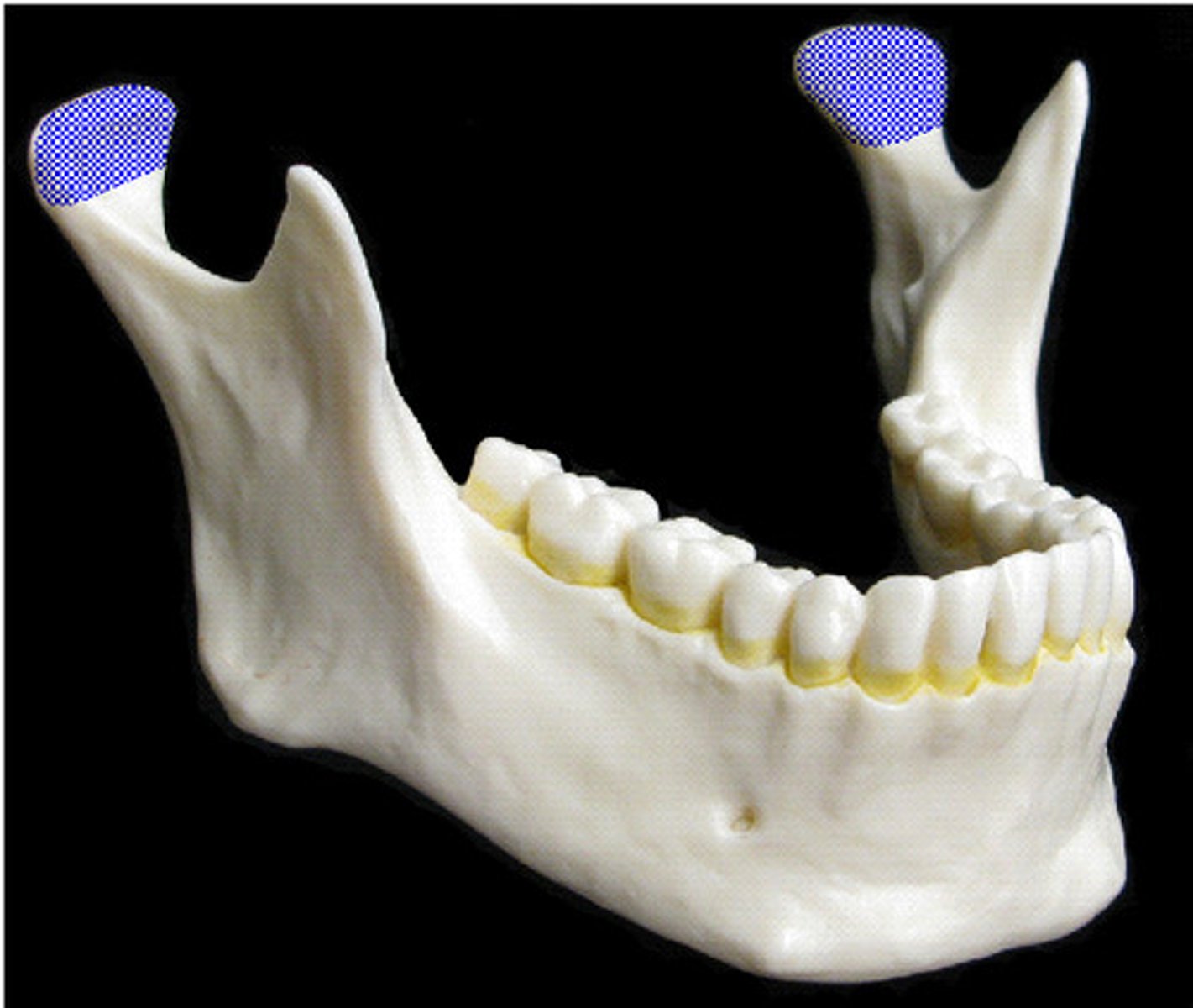
Mandible (unpaired)
lower jaw
Mental Foramen
opening in the mandible for passage of chin artery/nerve
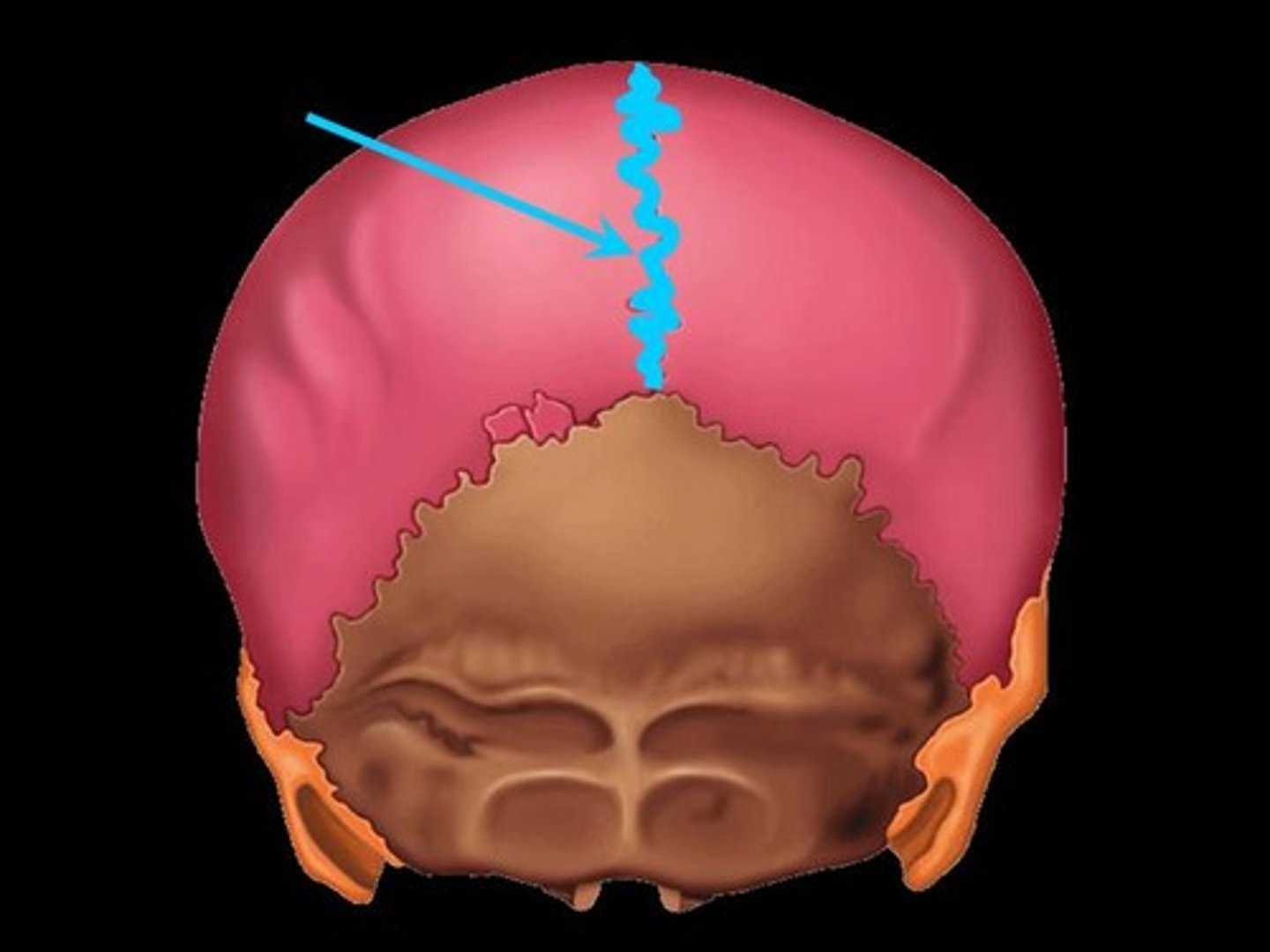
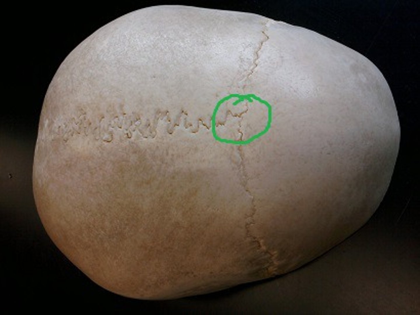
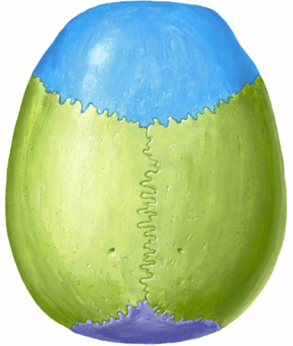
Sagittal Suture
between the two parietal bones
Sutural Bone
tiny bones between cranial bones
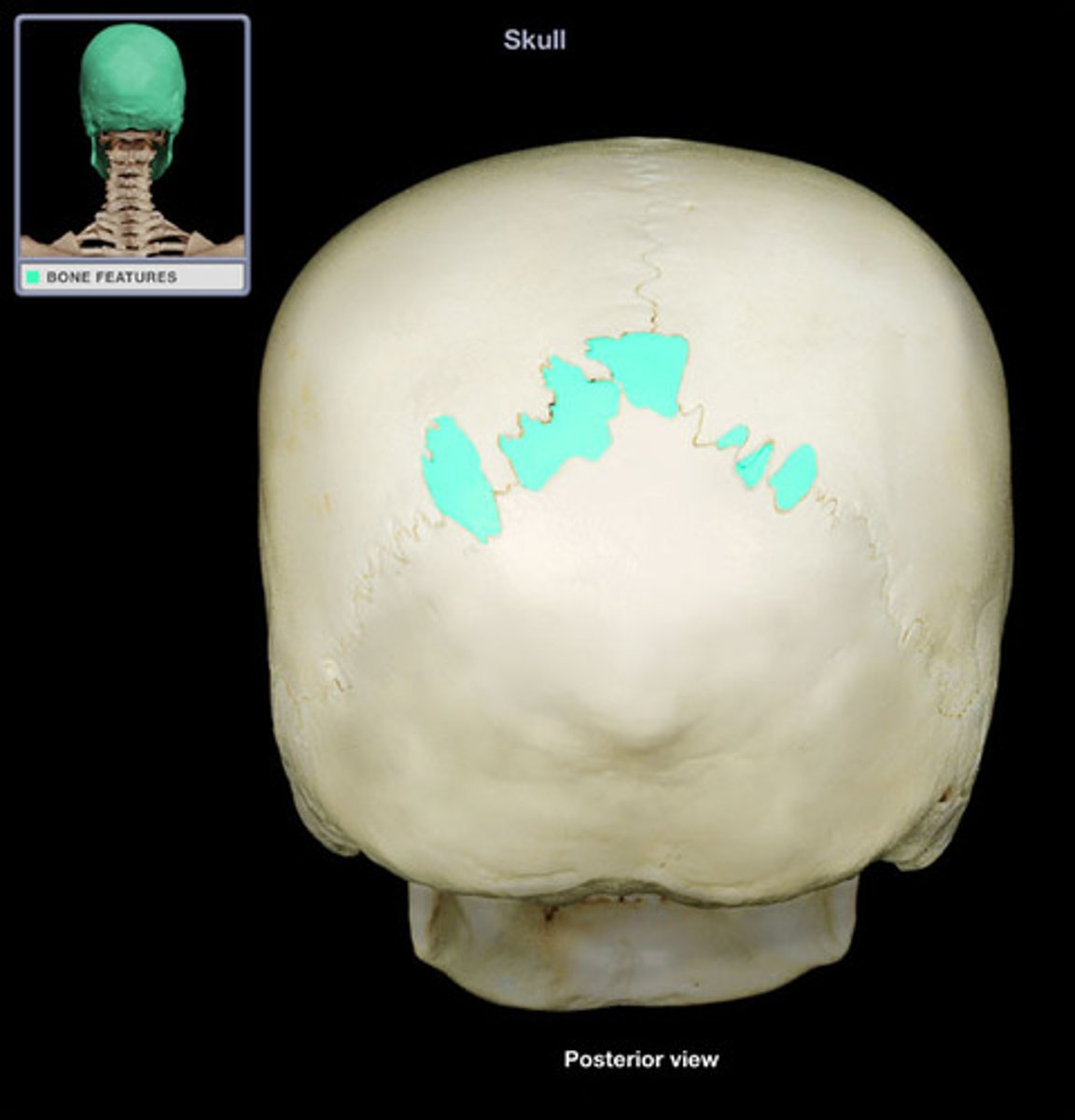
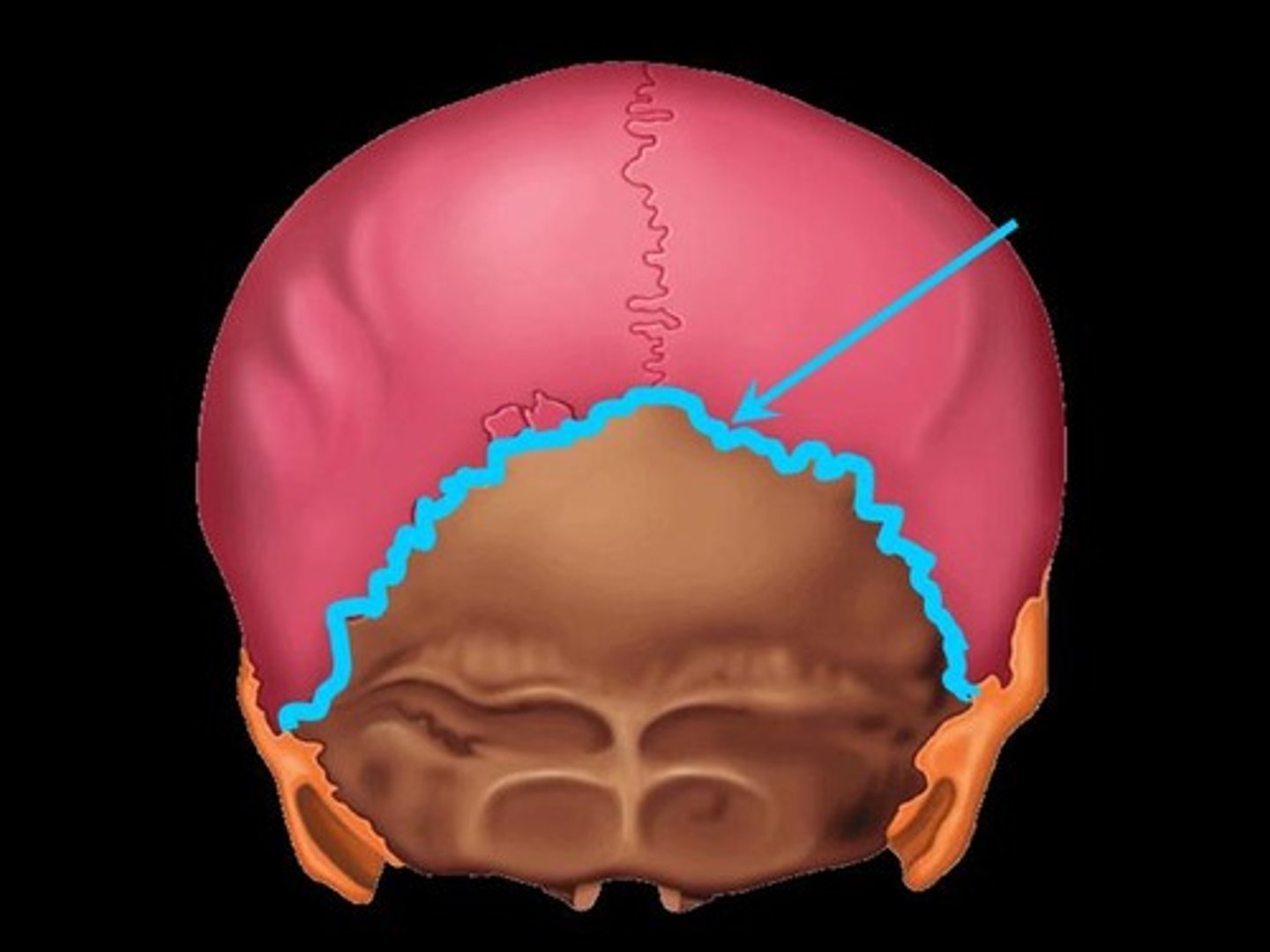
Lambdoid Suture
between parietal bones and occipital bone

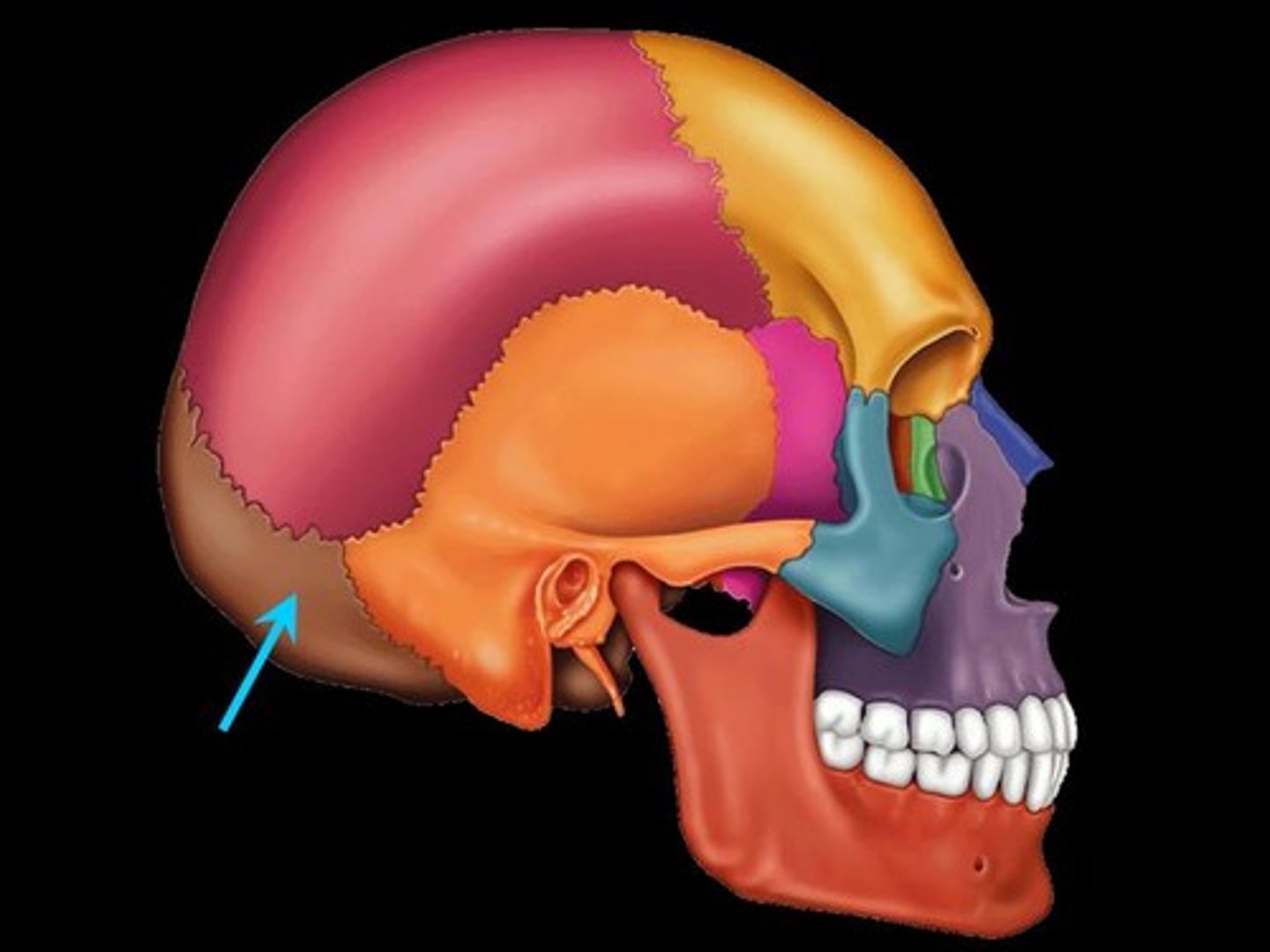
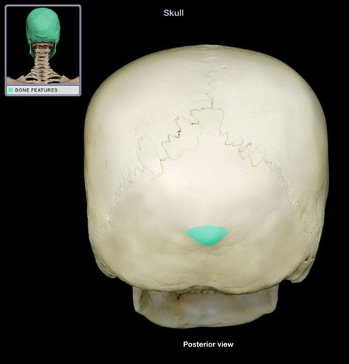
Occipital Bone (unpaired)
back of head
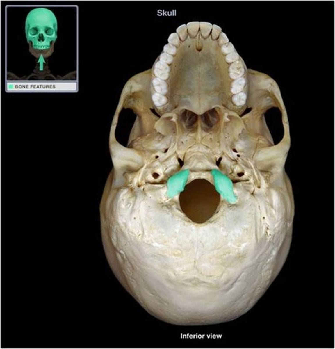
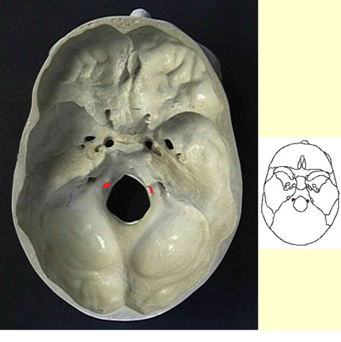
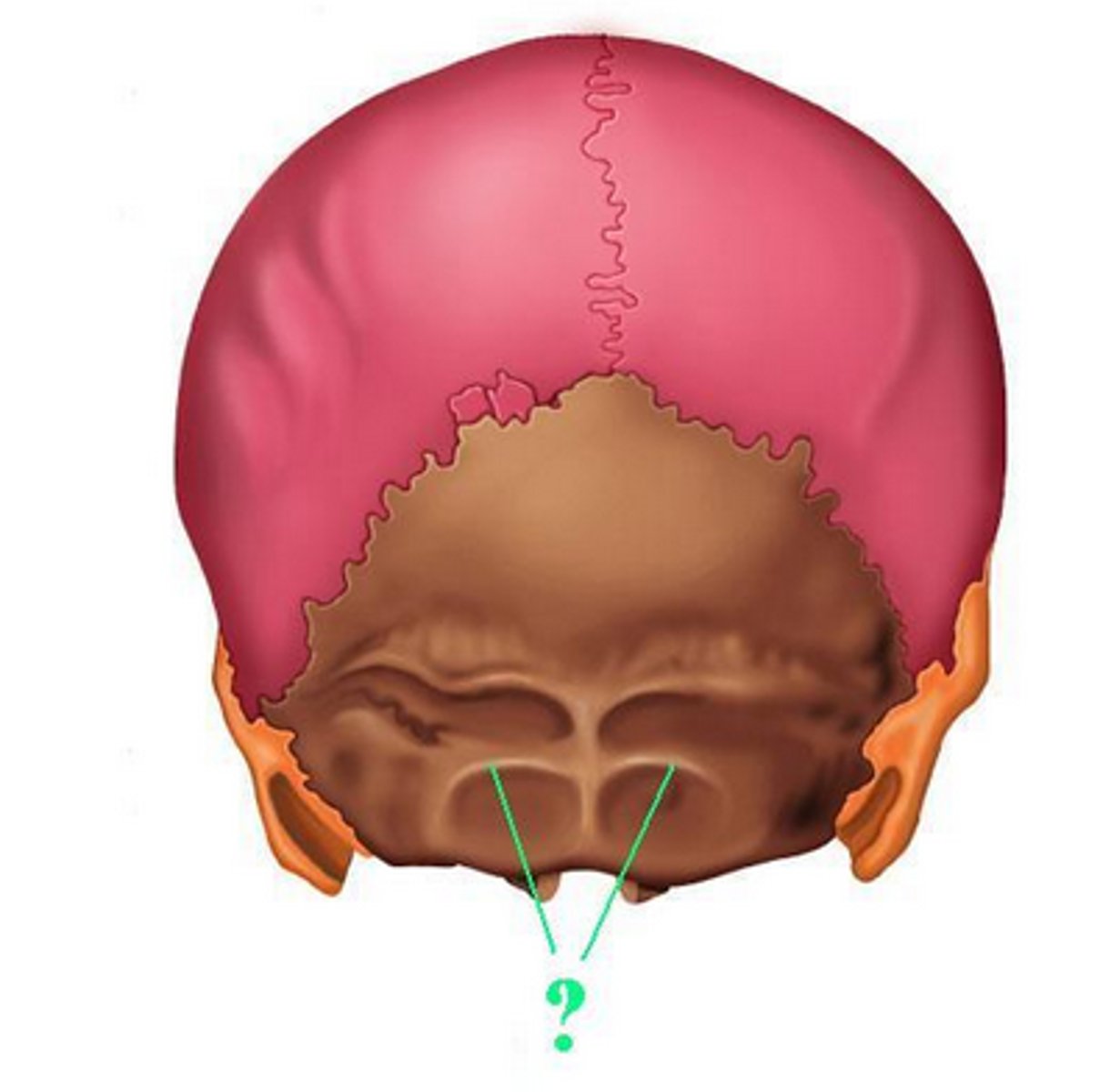
Occipital Condyle
ridges on left and right of foramen magnum
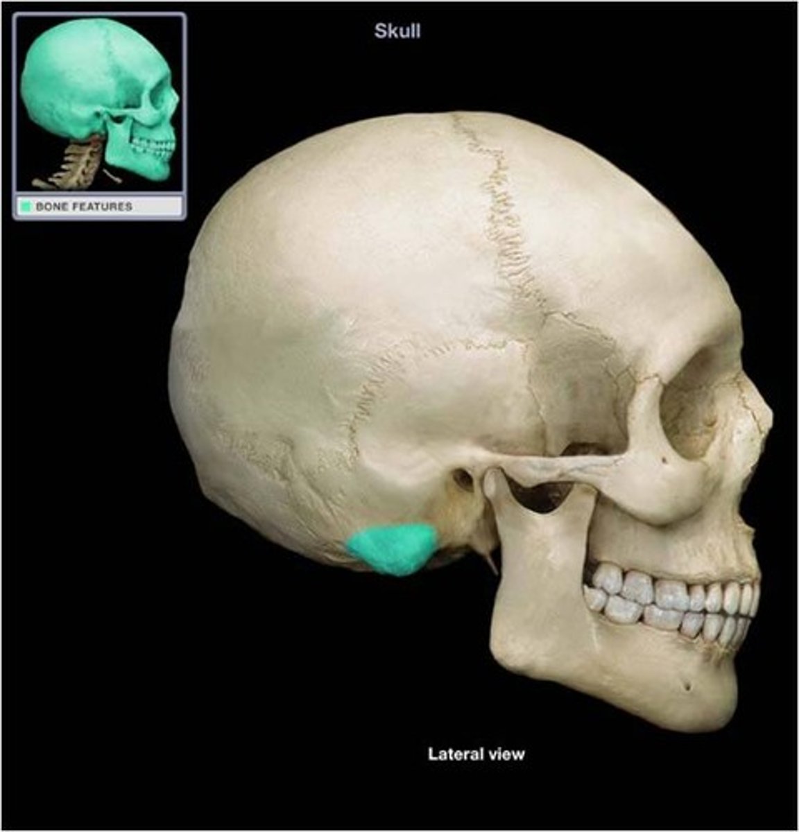
Mastoid process of temporal bone
round projection on the temporal bone behind the ear
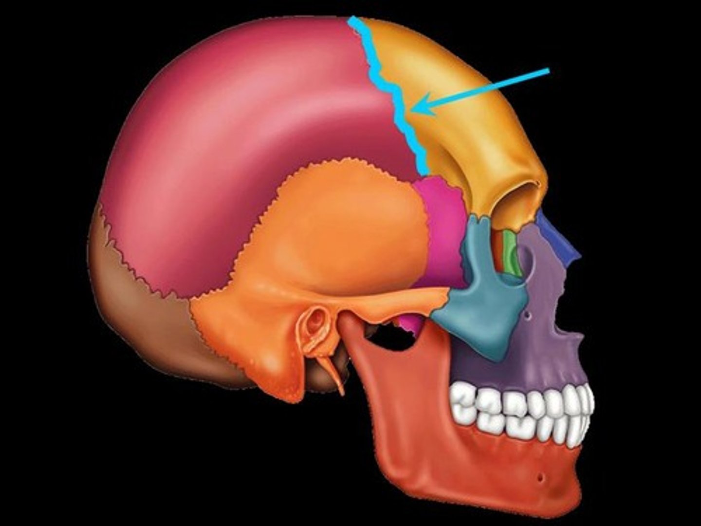
Coronal Suture
the suture between the parietal and frontal bones of the skull
Lacrimal Fossa
Anterior and lateral depression of roof of orbit which accommodates the lacrimal gland
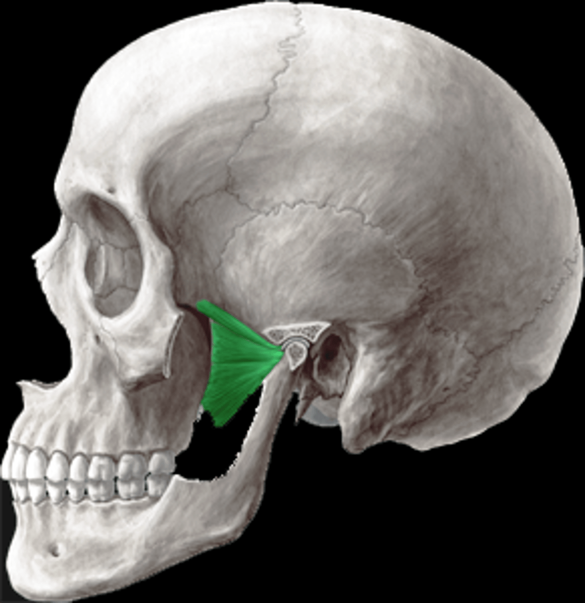
Coronoid process
"crown-shaped" insertion point for the large temporalis muscle that elevates the lower jaw during chewing
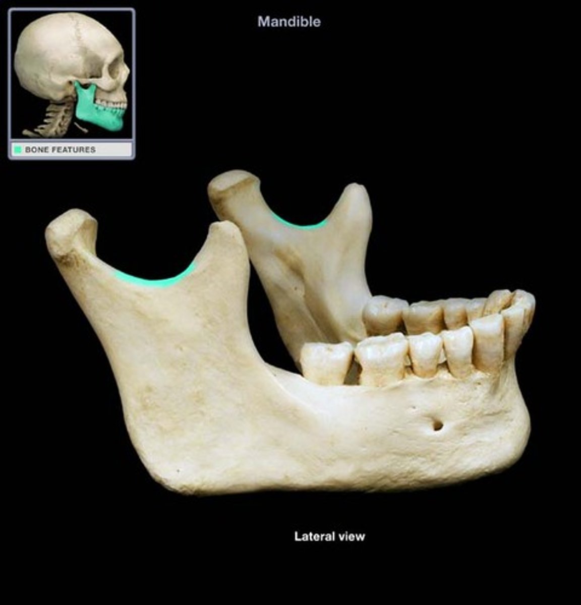
coronoid notch
mandibular notch
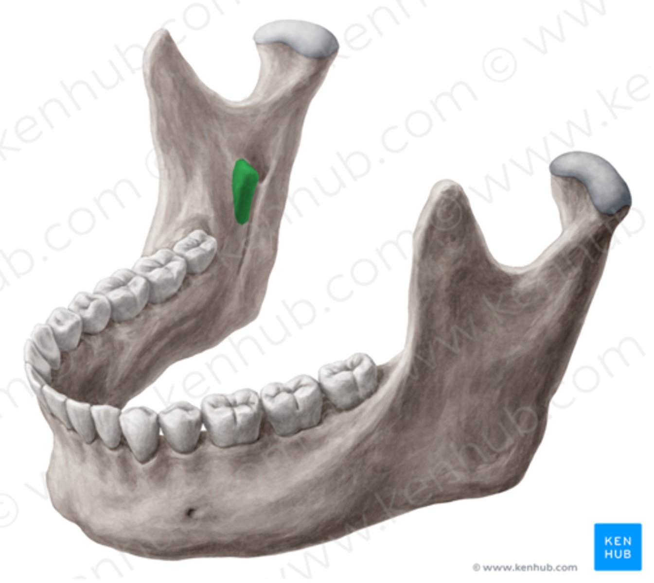
Lingula
mylohyoid line

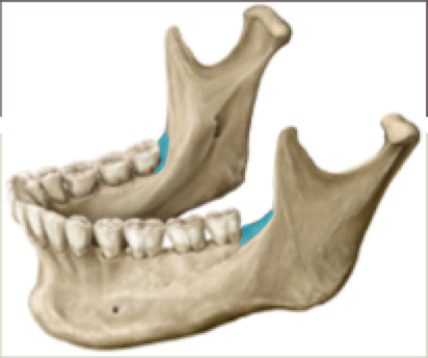
retromolar triangle
Mandibular angle
the posterior, inferior corner of the lower jaw
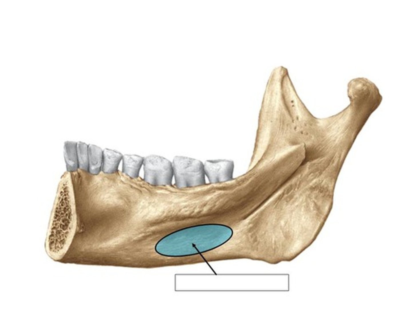
sublingual fossa
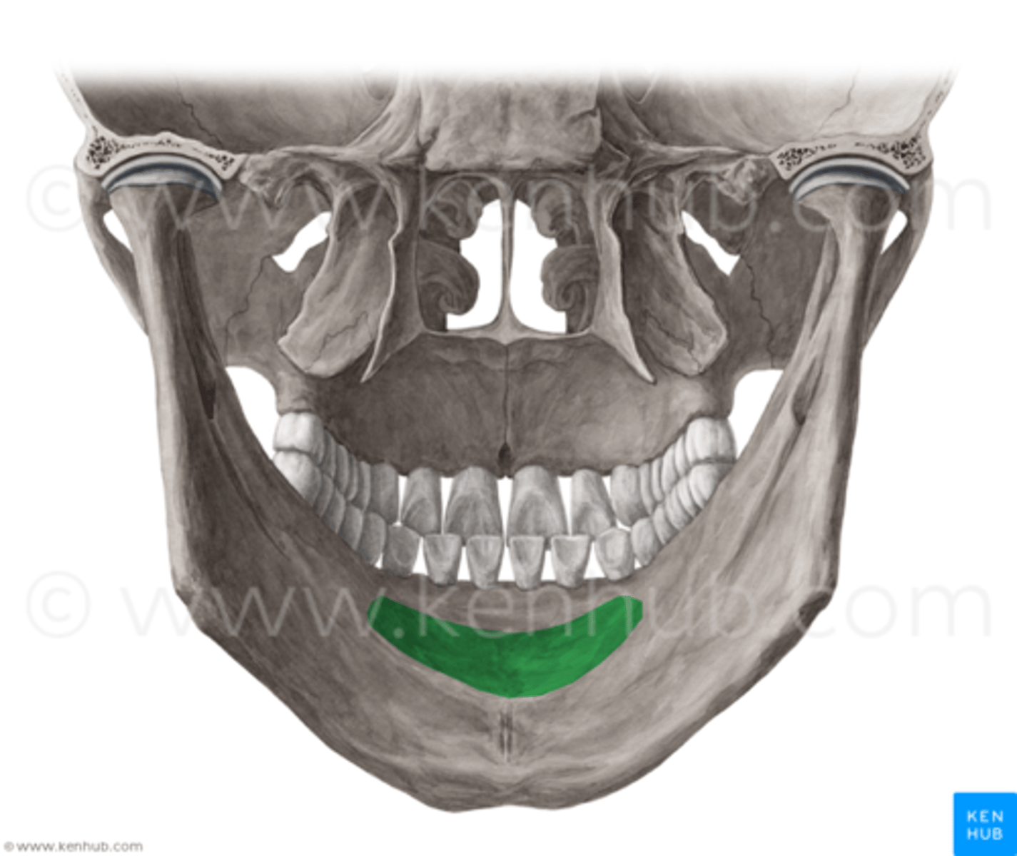
submandibular fossa
Mastoid process
round projection on the temporal bone behind the ear
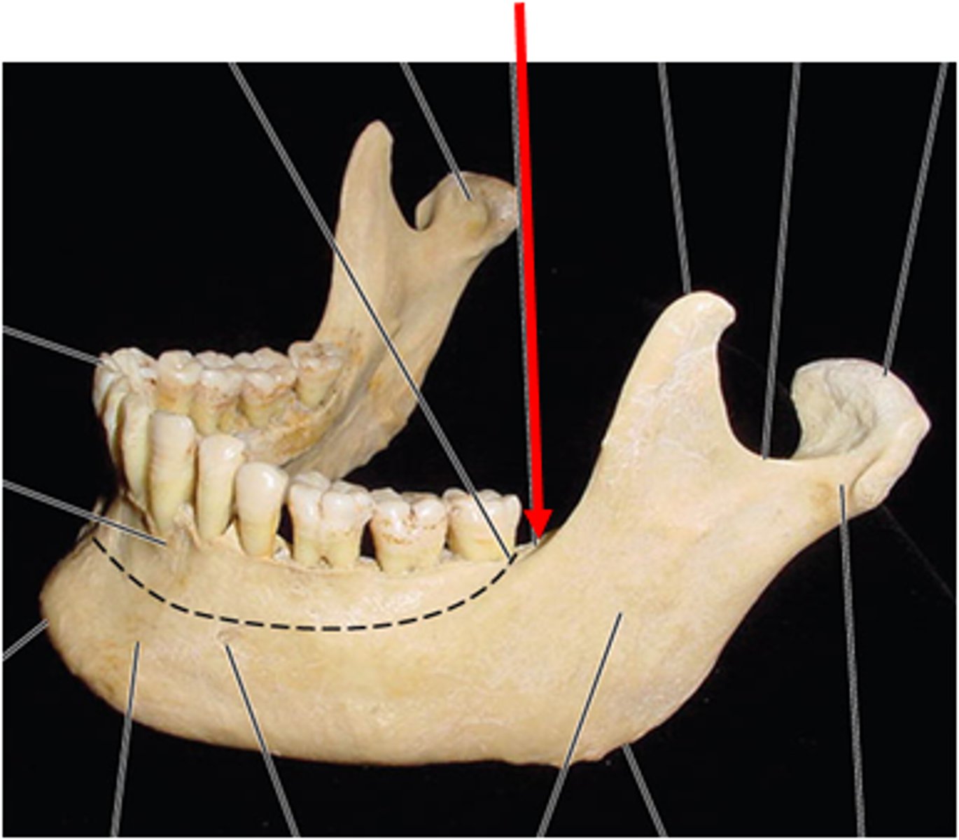
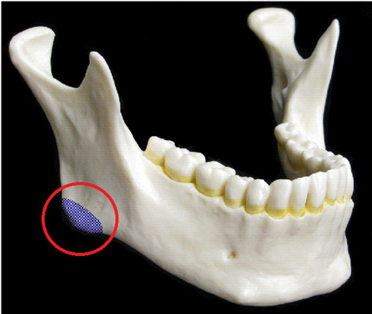
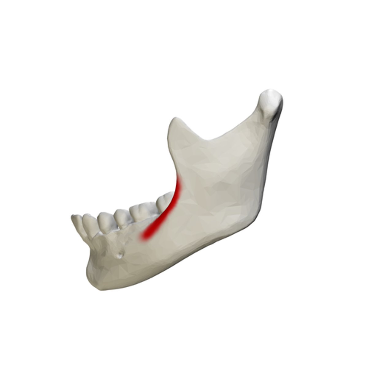
external oblique line
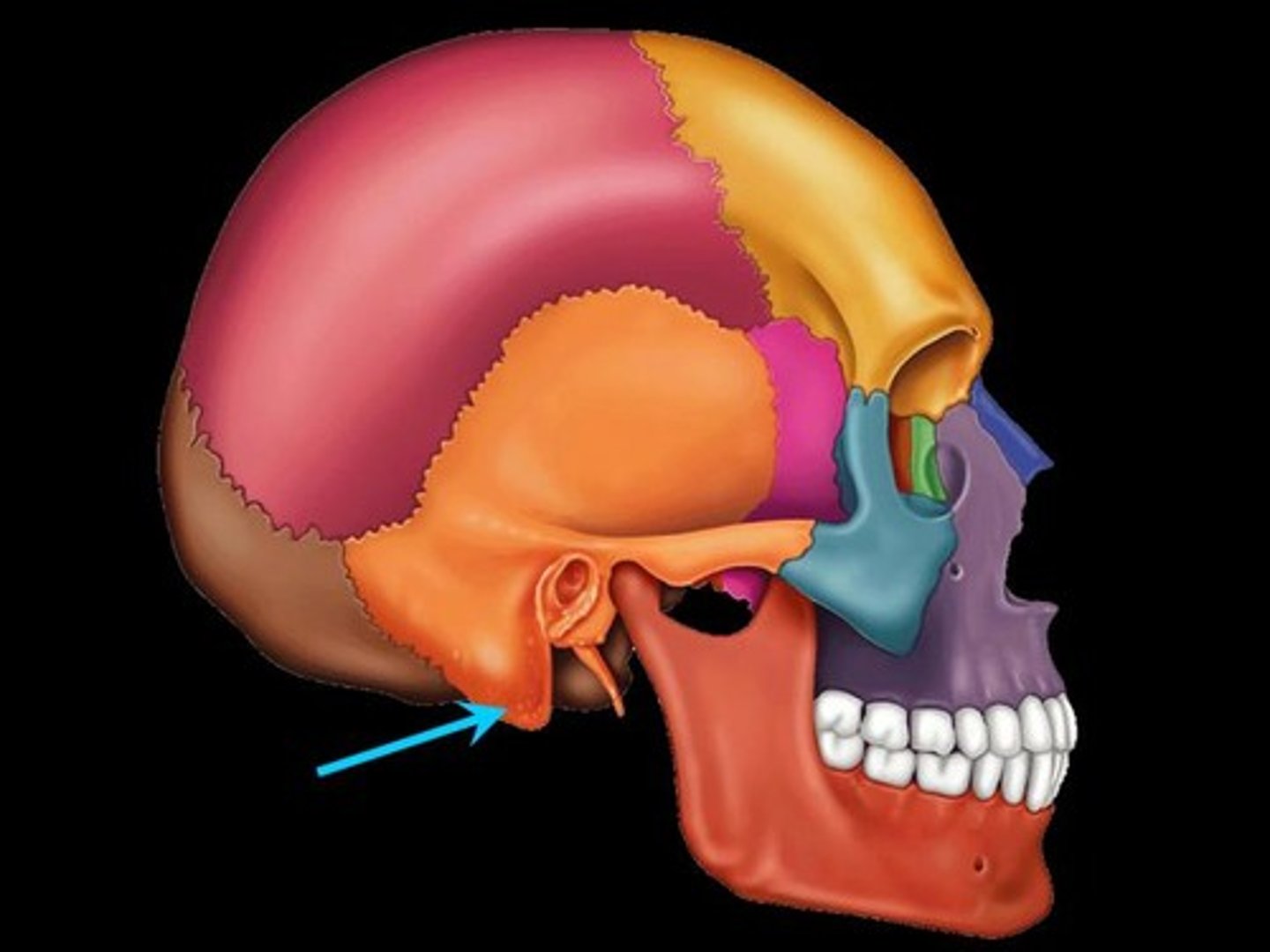
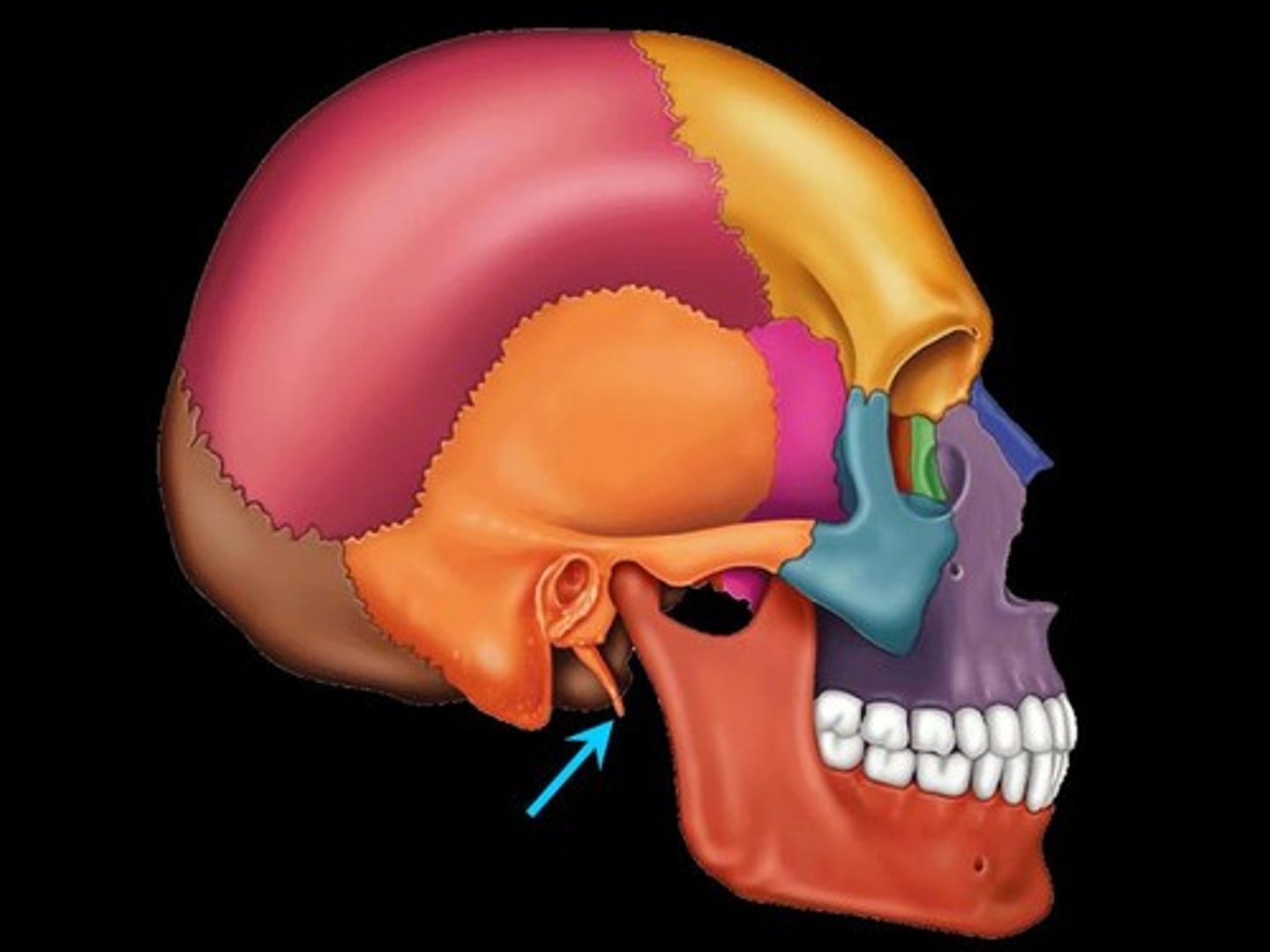
Styloid process
pole-like process extending downward from the temporal bone on each side of the skull
external occipital protuberances
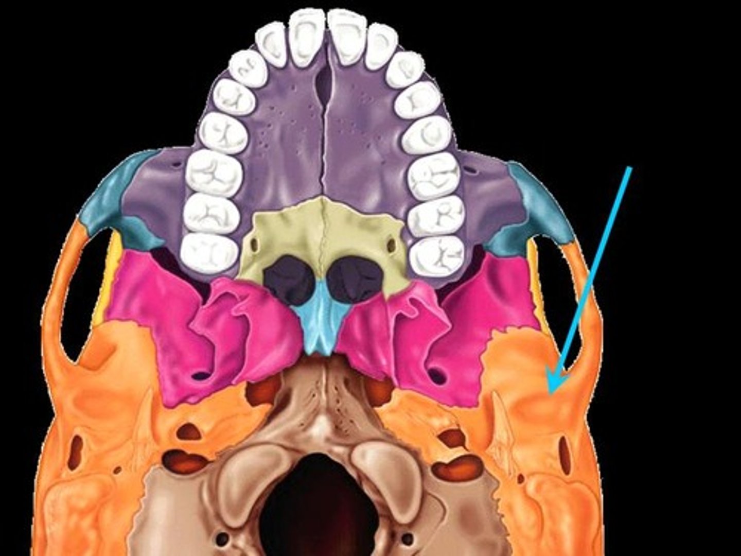
maxillary tuberosity
External acoustic meatus
ear canal
Zygomatic process
cheek bone
Lambdoid suture
between parietal bones and occipital bone
greater palatine foramen
Squamous Suture
Between parietal and temporal bones

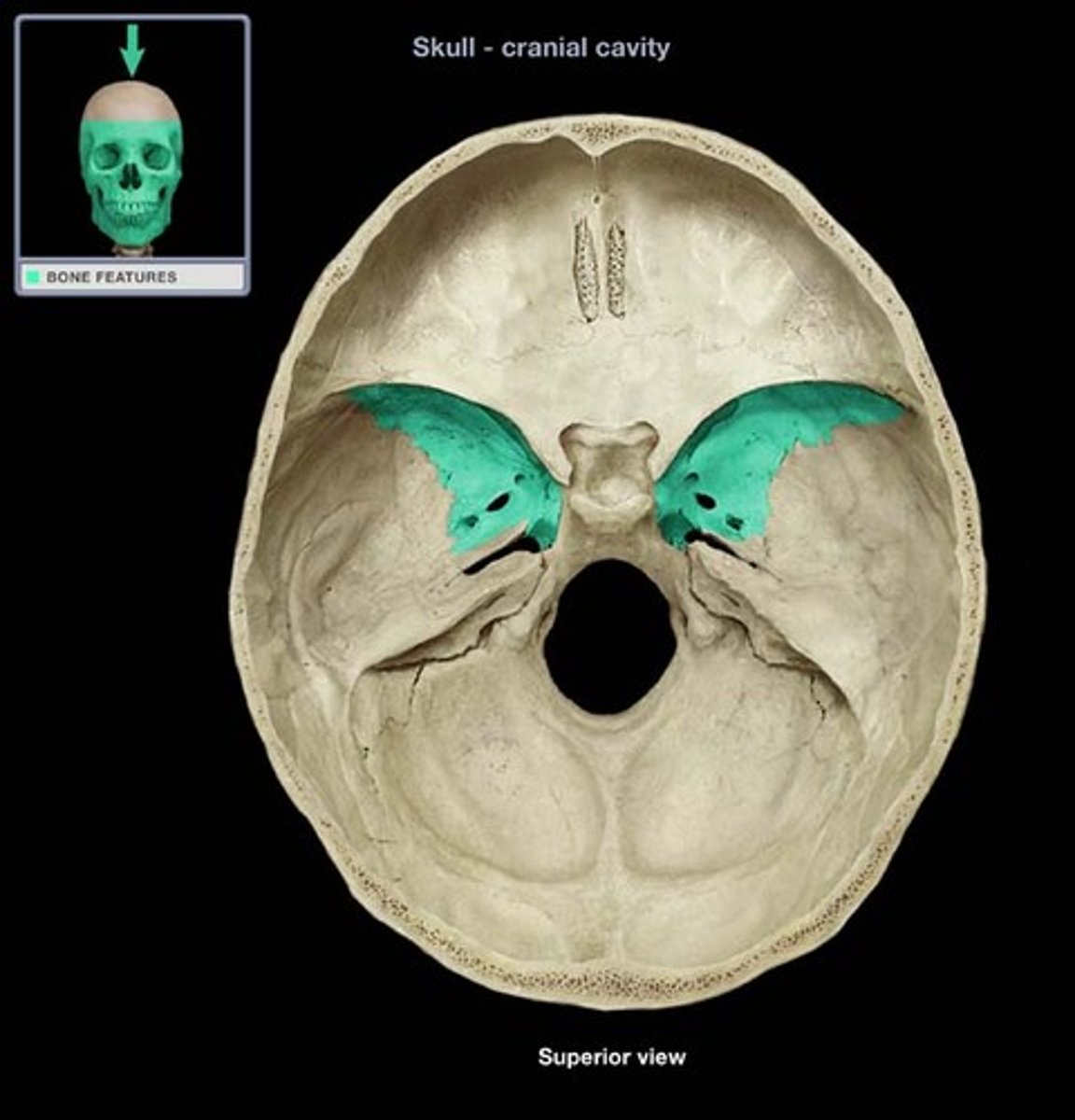
Sphenoid bone (greater wing)
flag-shaped bony areas, forms the middle cranial fossa, interior walls of the skull
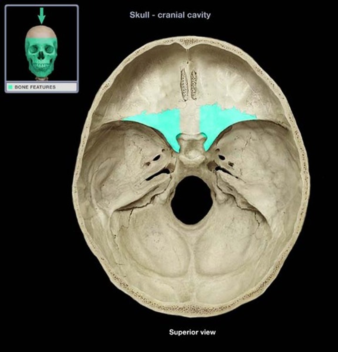
Sphenoid bone (lesser wing)
hornlike, form the floor of the anterior cranial fossa, and medial walls of the orbits
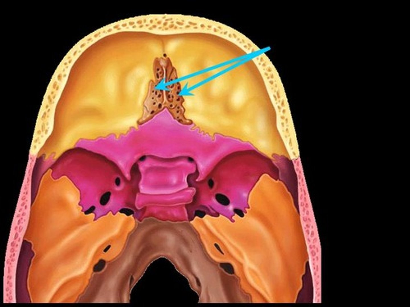
Crista galli
Triangular process to which membranes that cover the brain attach. Means "rooster's comb".
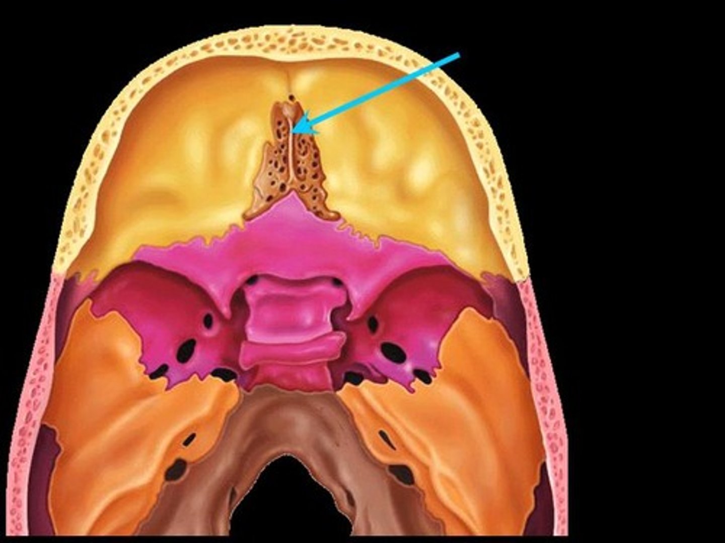
Ethmoid Bone (perpendicular plate)
behind nasal bone and in front of sphenoidal sinuses

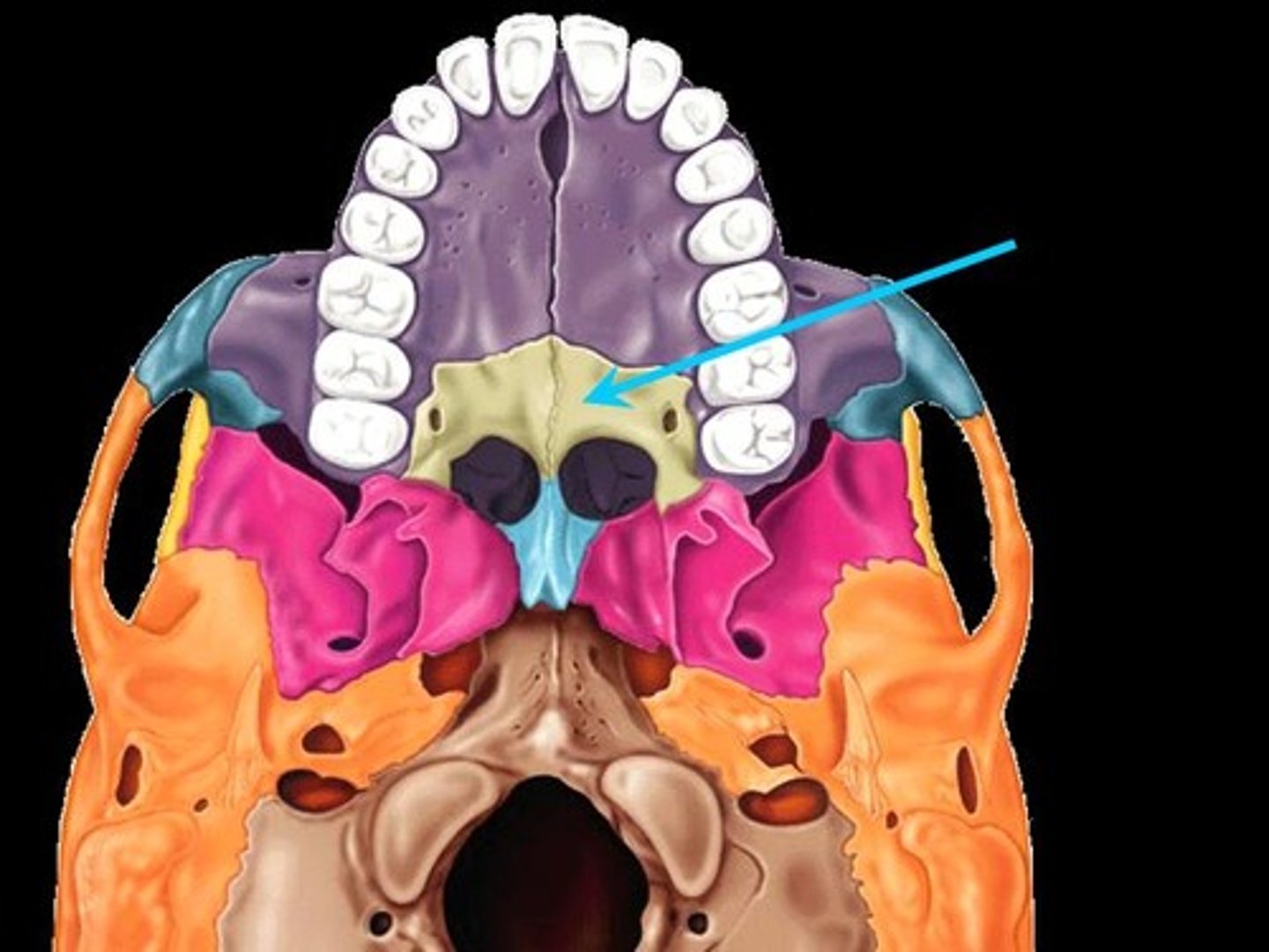
Palatine bone
either of two irregularly shaped bones that form the back of the hard palate and helps to form the nasal cavity and the floor of the orbits
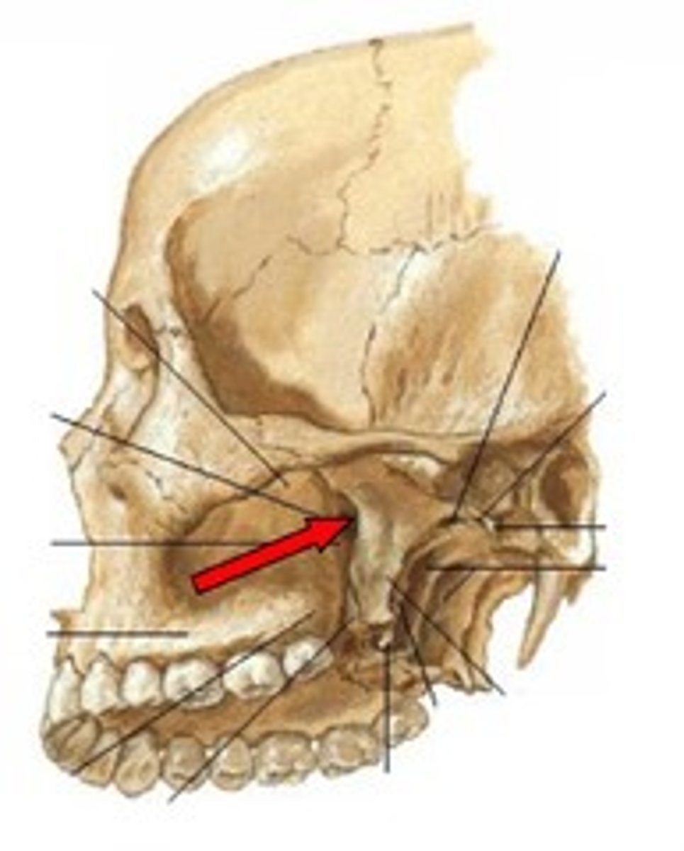
Pterygoid process of sphenoid bone
The land mark on the sphenoid bone for the attachment of many muscles of mastication...

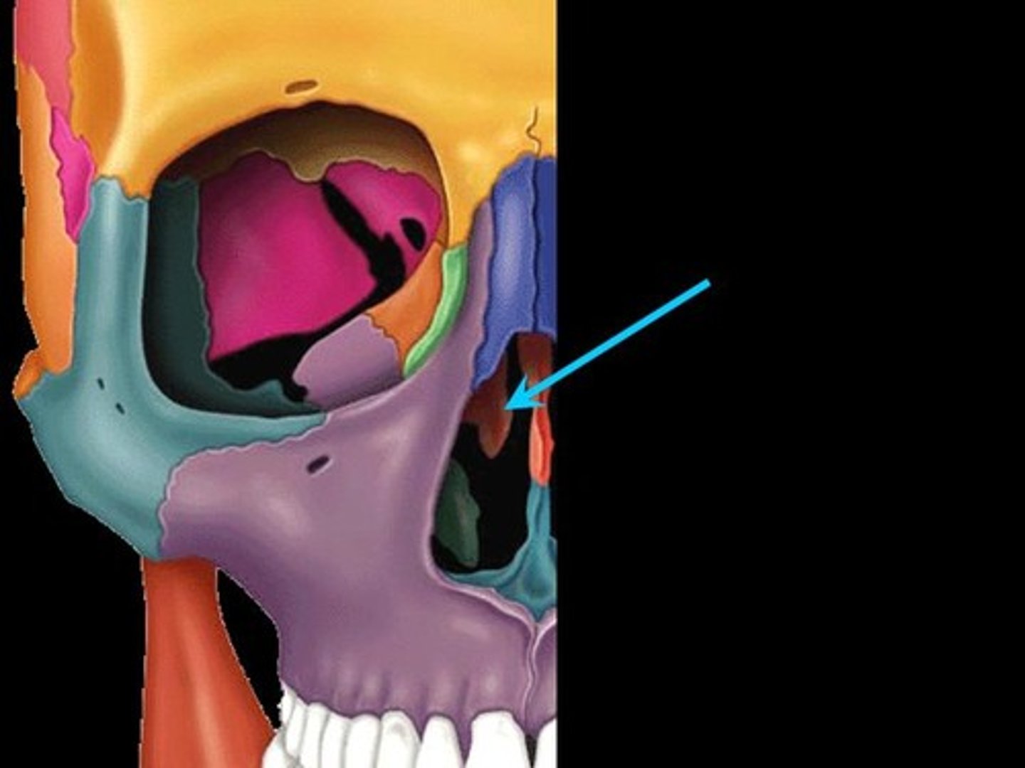
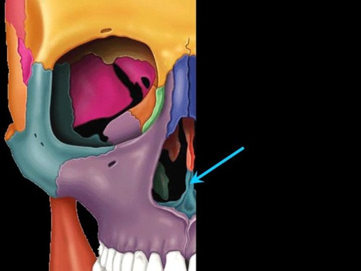
pterygomaxillary fissure
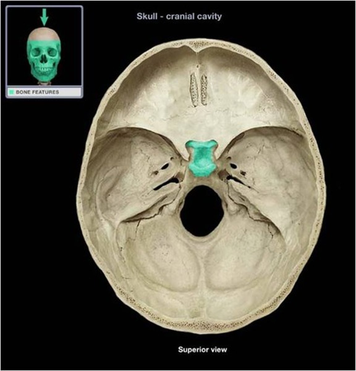
Sella turcica of sphenoid bone
houses the pituitary gland
Internal acoustic meatus
A passage for CN VIII from the inner ear to the brain.
Foramen Ovale
oval shaped
Foramen Spinosum
small foramen slightly posterior and lateral to foramen ovale
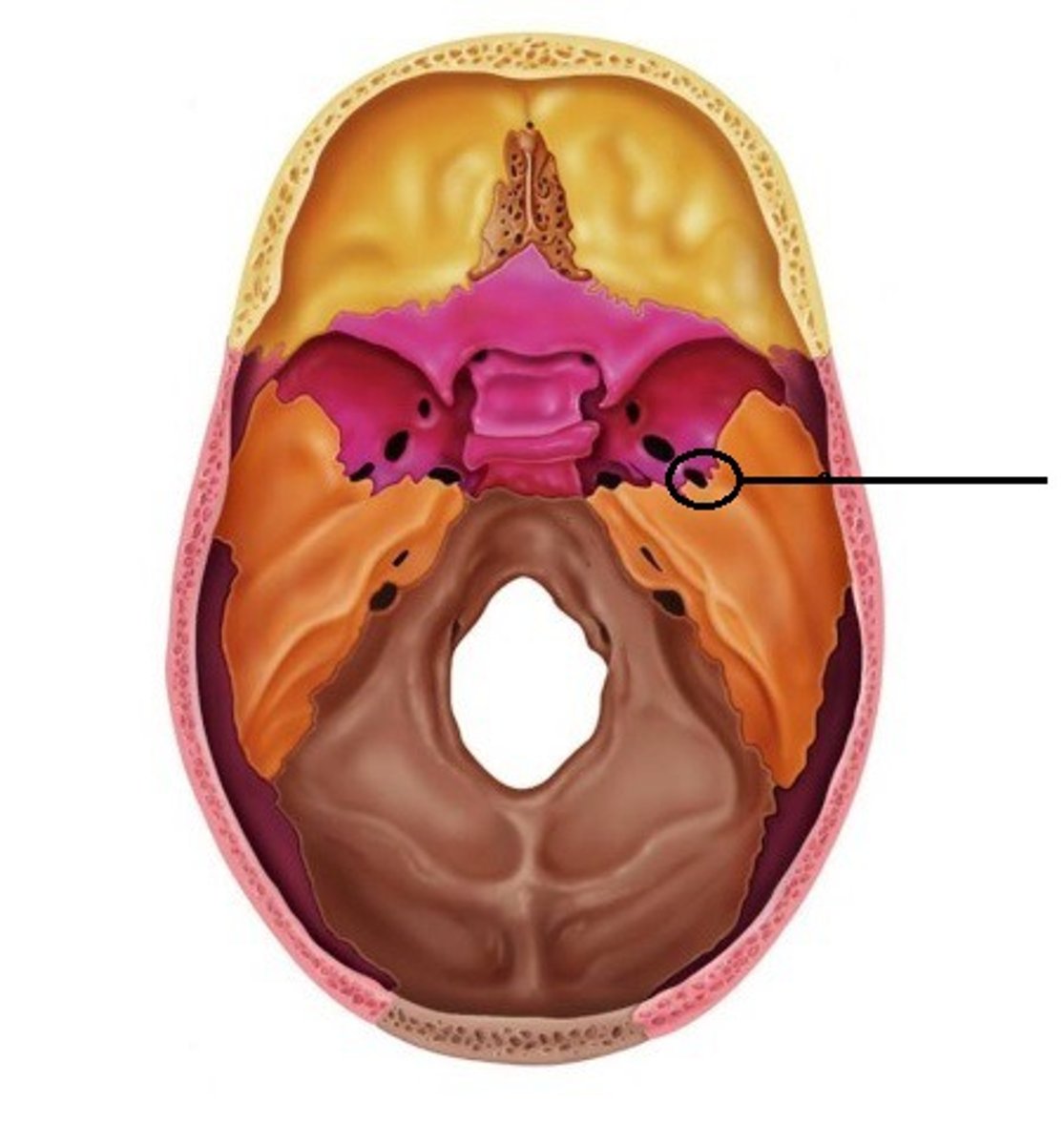
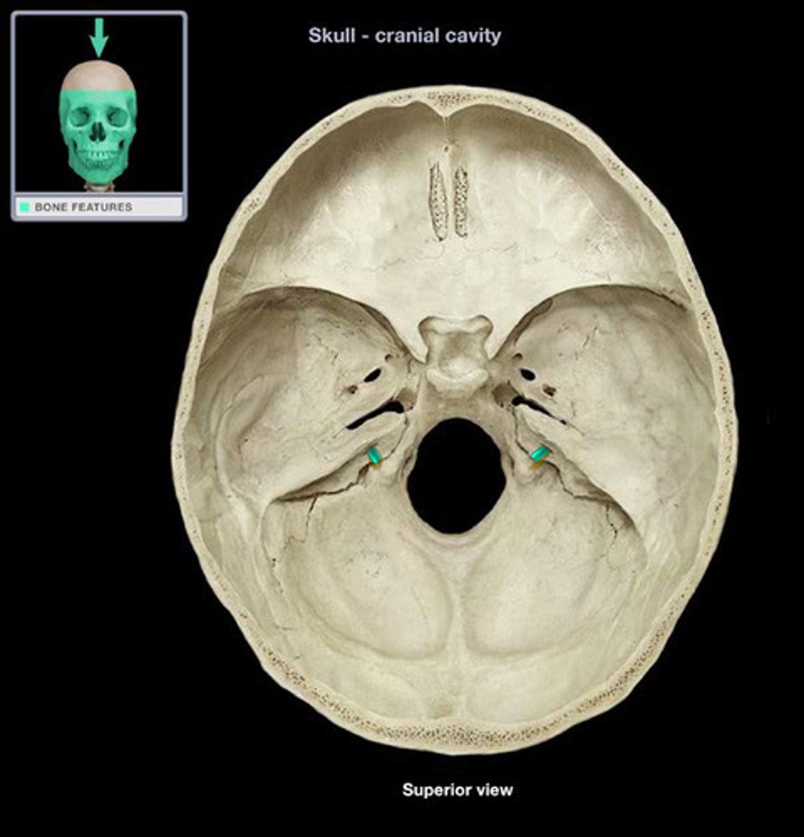
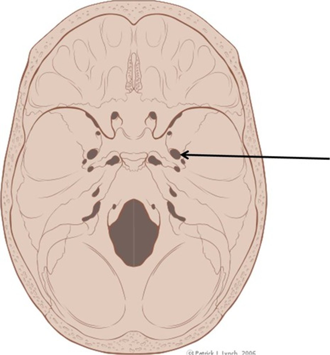
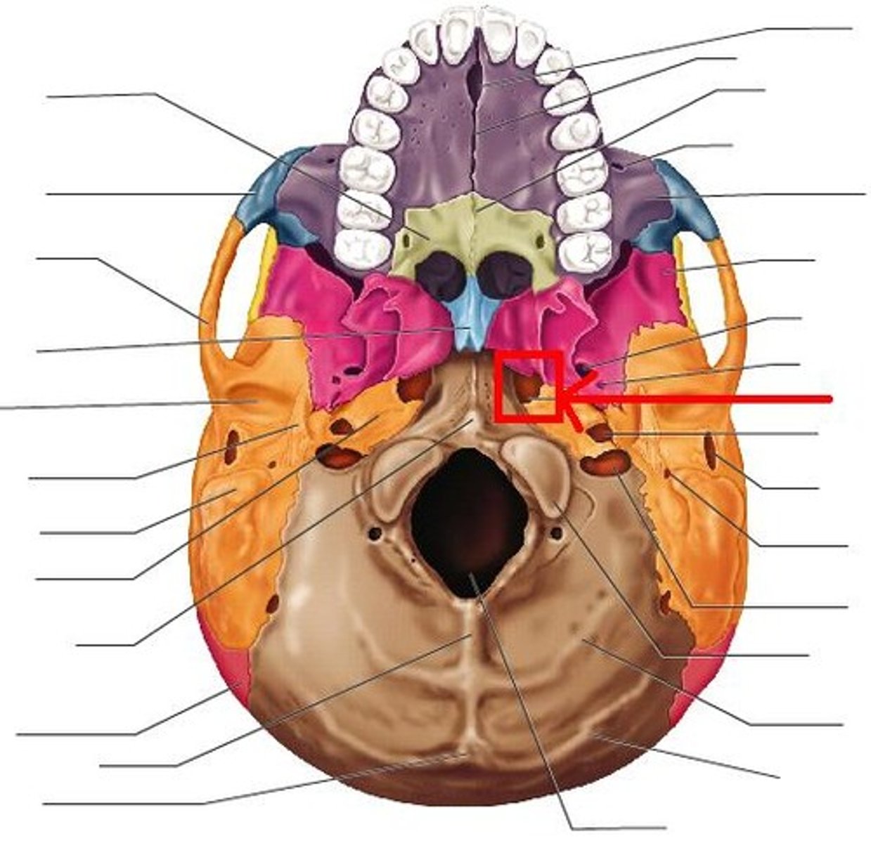
Foramen Lacerum
Jagged opening between the petrous temporal bone and the sphenoid bone
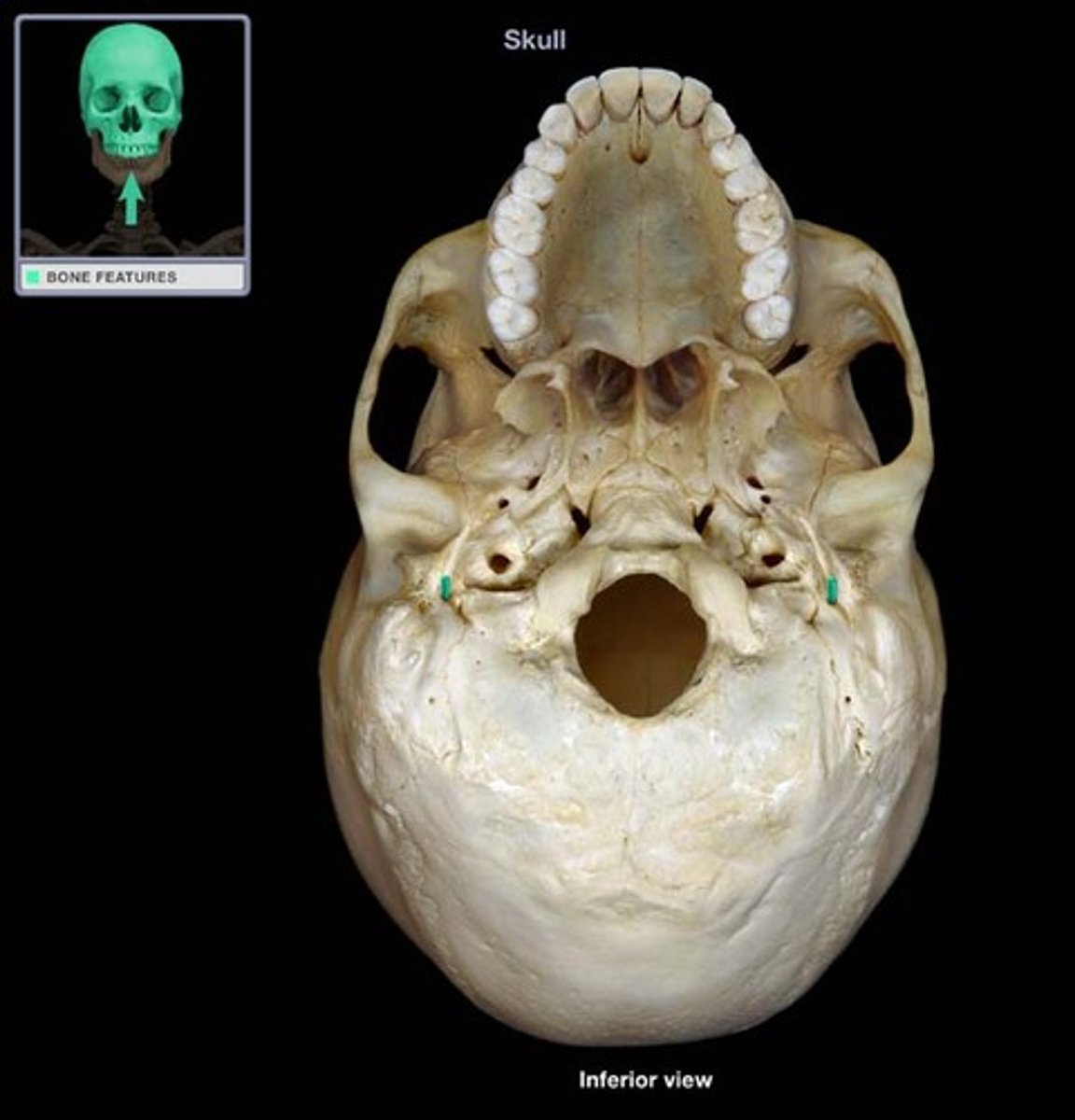
Carotid Canal
anterior to jugular foramen
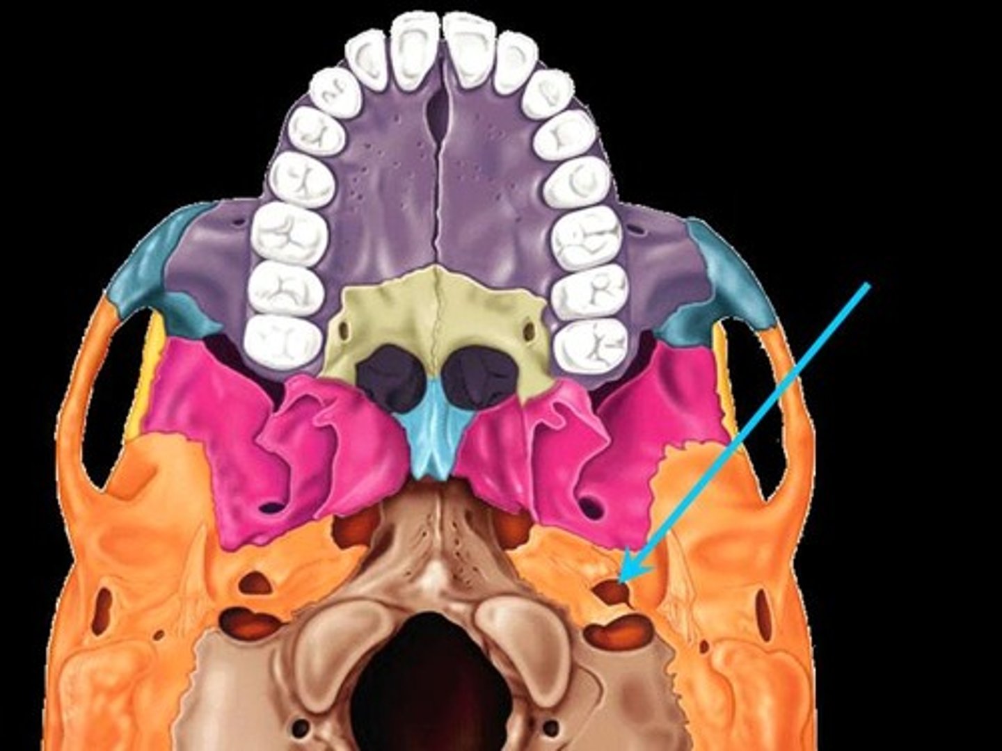
Stylomastoid Foramen
posterior to external acoustic meatus, between styloid and mastoid processes
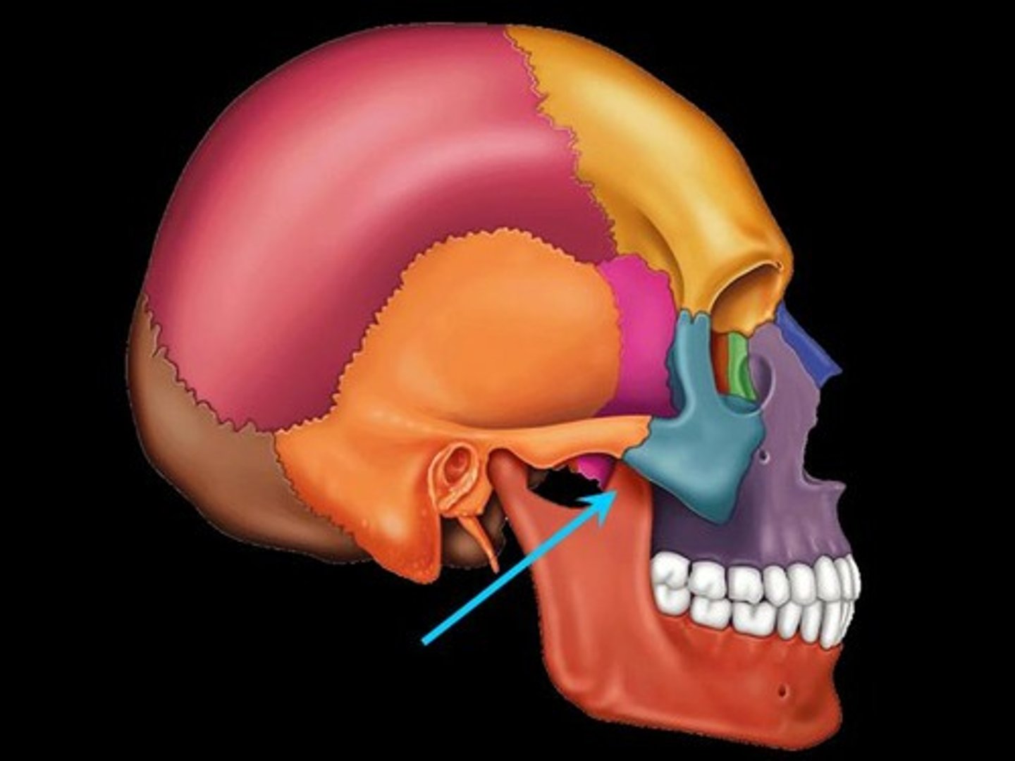
infratemporal fossa
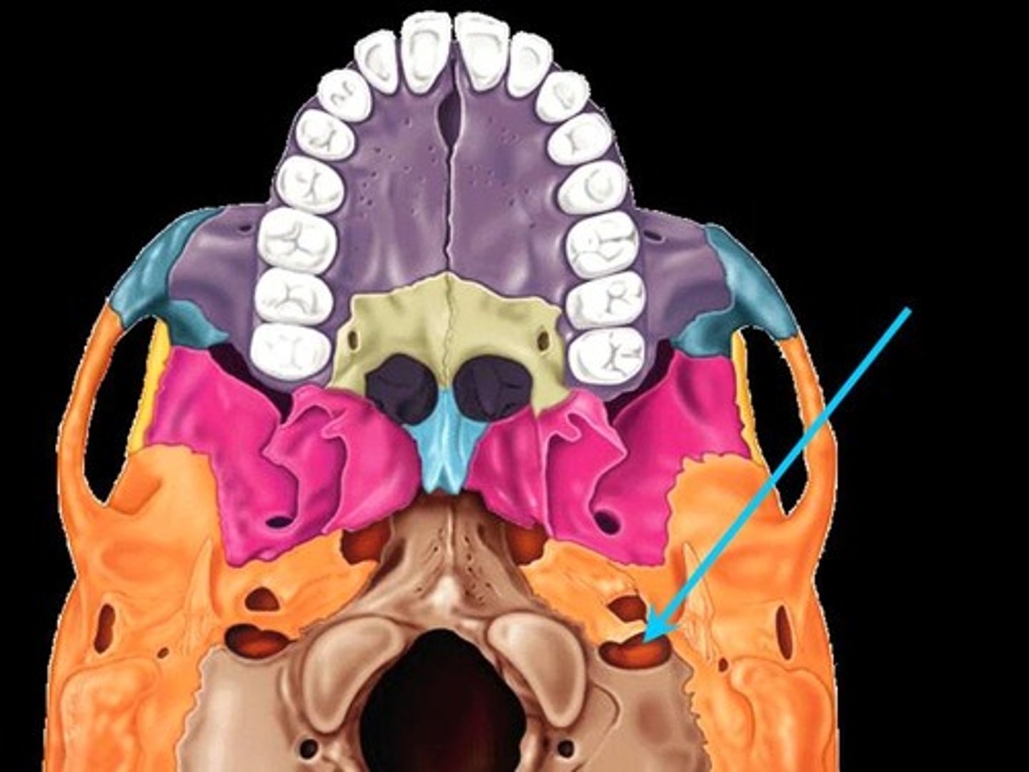
Jugular Foramen
posterior to carotid canal
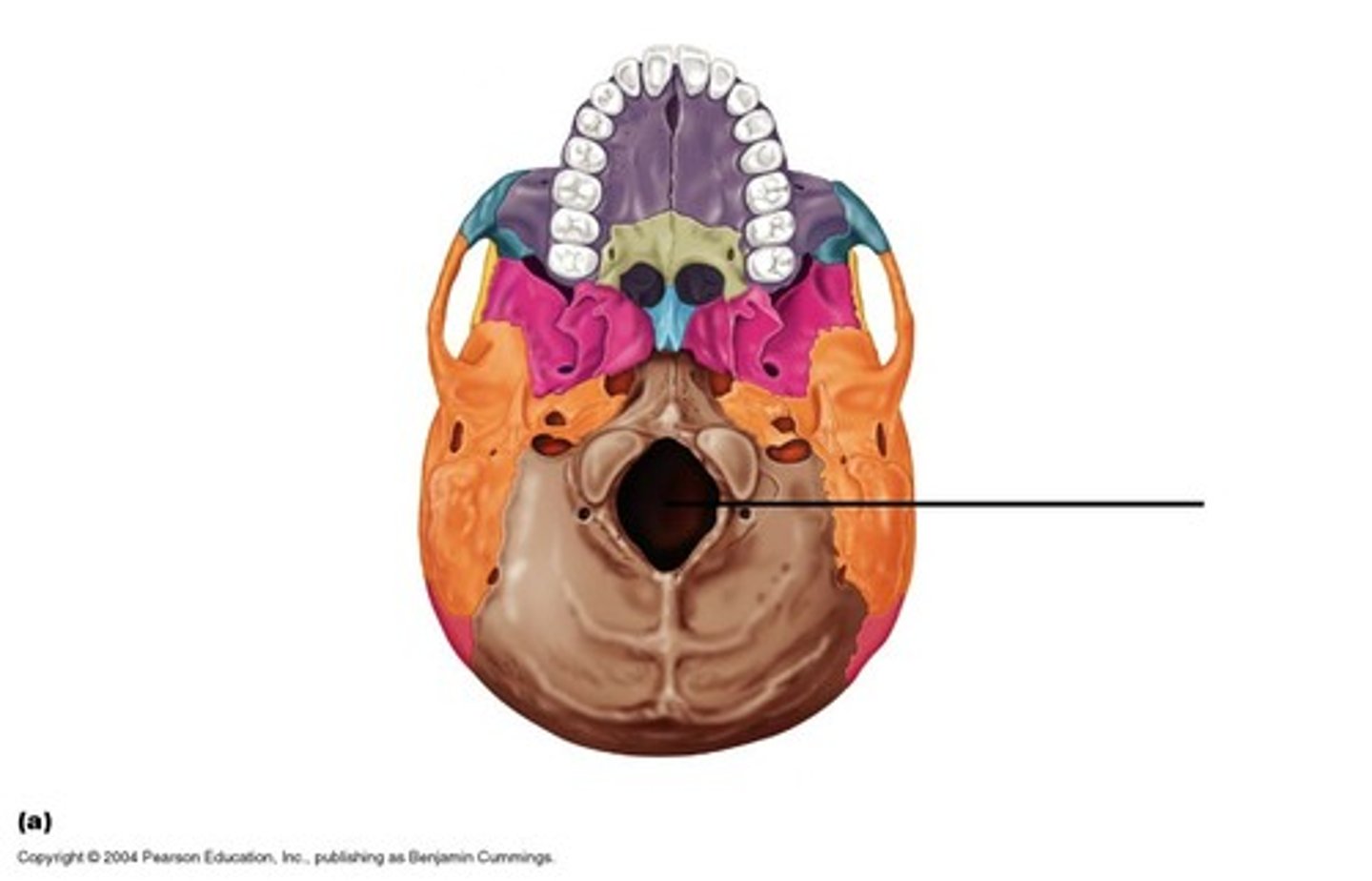
Foramen Magnum
hole at base of skull
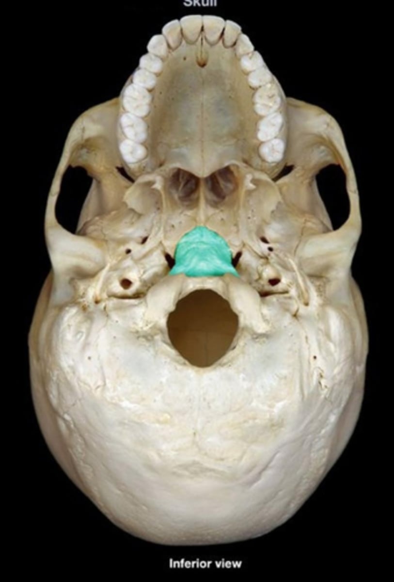
Basilar part of occipital bone
region of the occipital bone anterior to foramen magnum
Mandibular Fossa
the depression in the temporal bone into which the condyle of the mandible fits
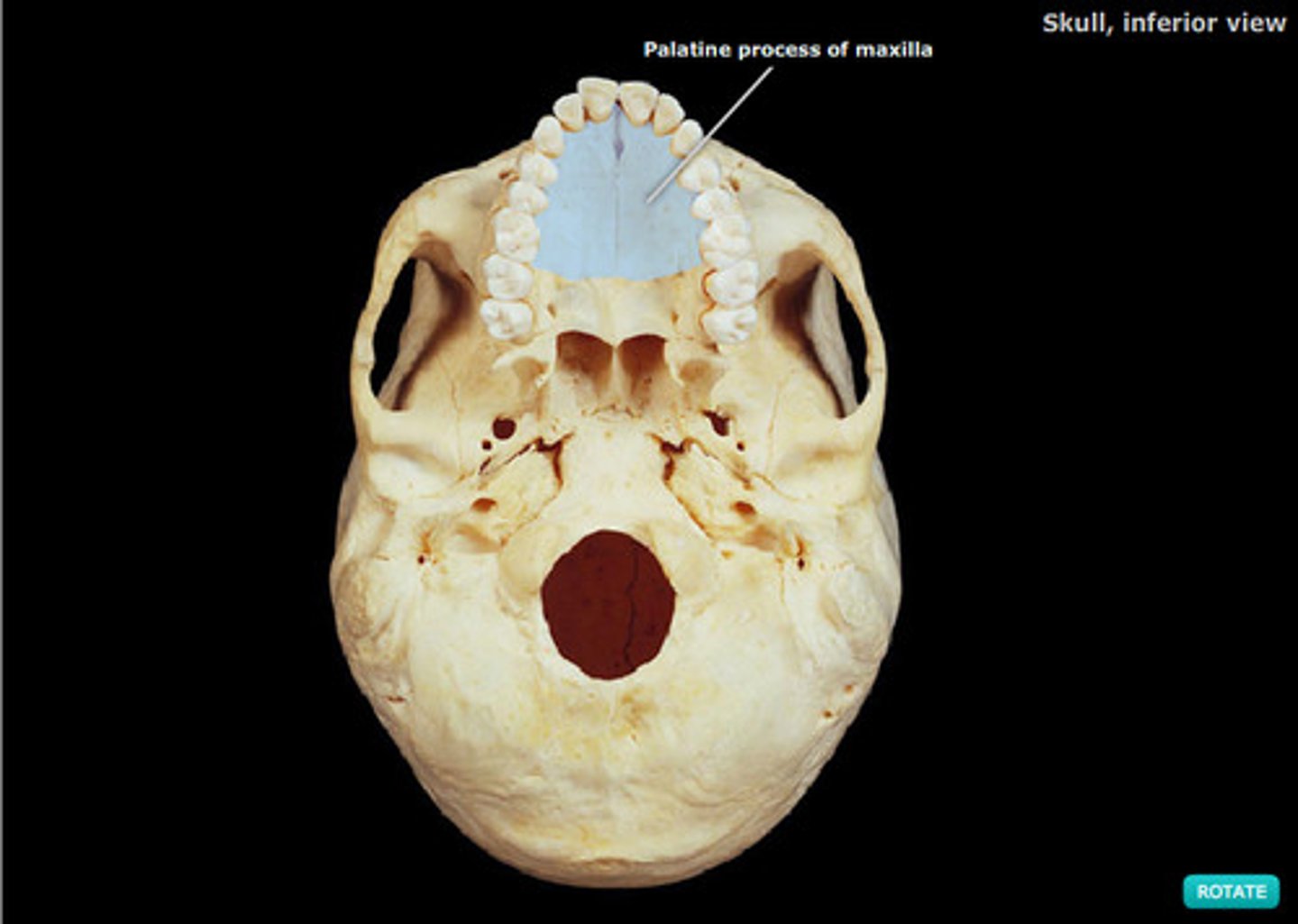
Maxilla (palatine process)
roof of the mouth
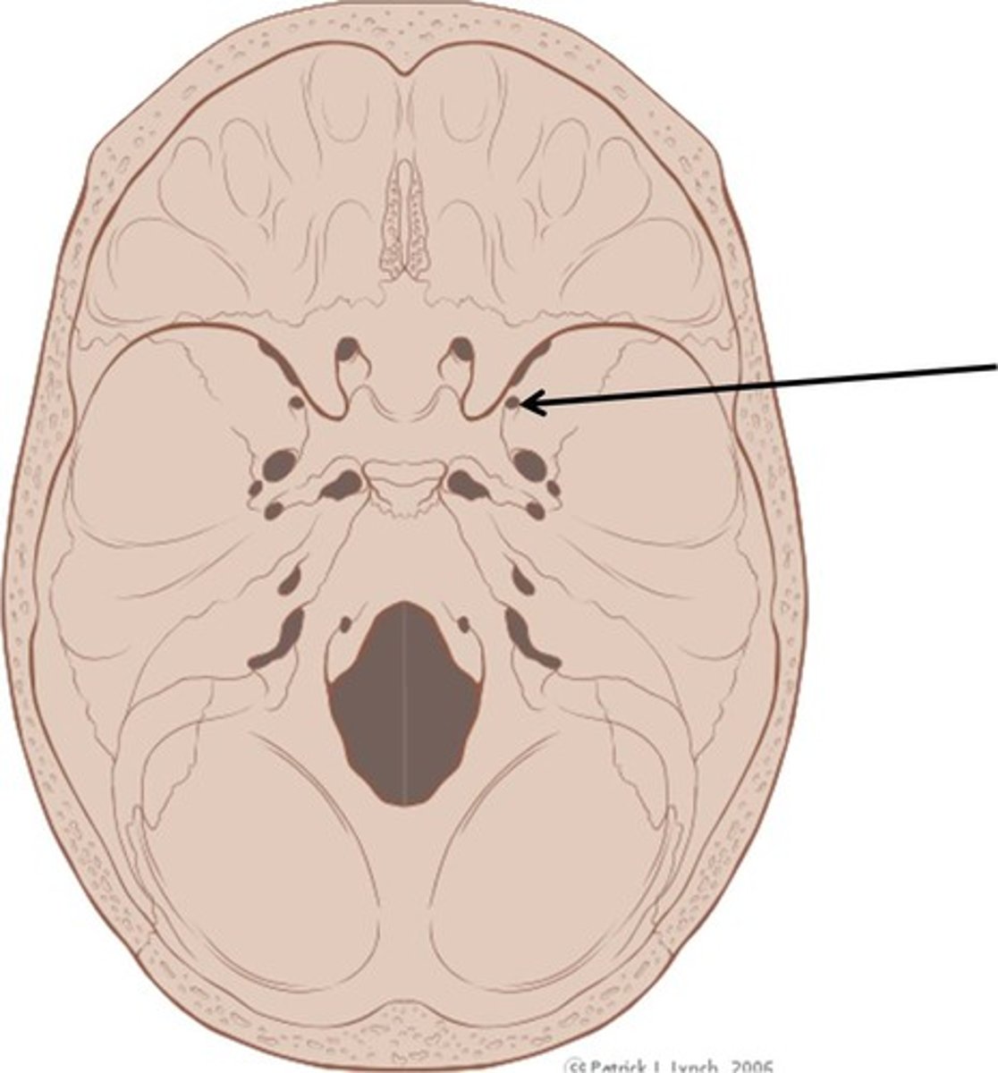
Cribriform Plate
small, flattened areas with numerous small openings, located to either side of the midline in the floor of the anterior cranial fossa; formed by the ethmoid bone
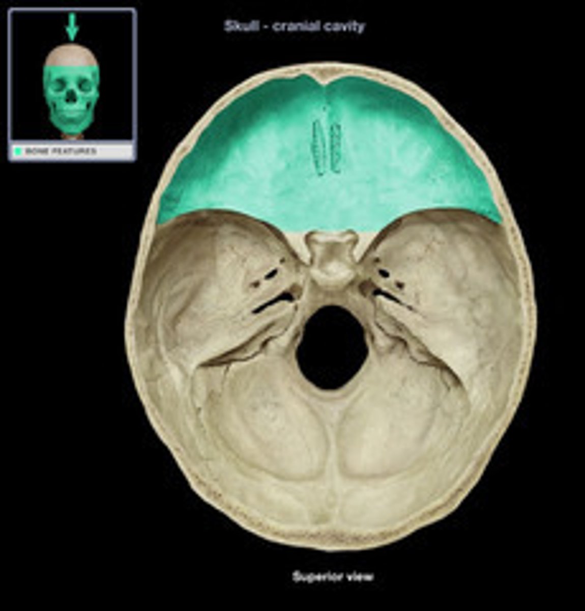
Anterior Cranial Fossa
supports the frontal lobes of the brain
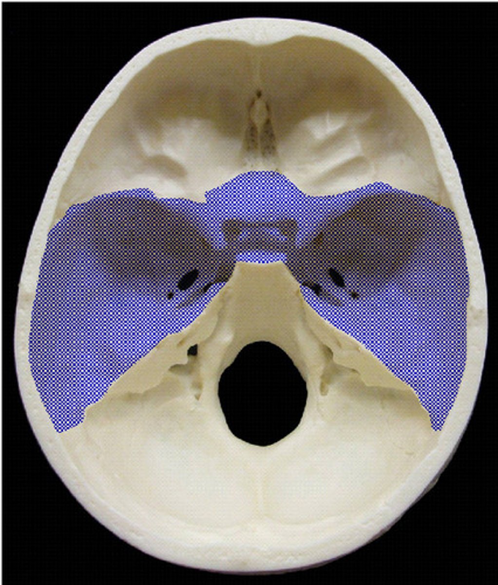
Middle Cranial Fossa
holds the temporal lobes of the brain, lateral location
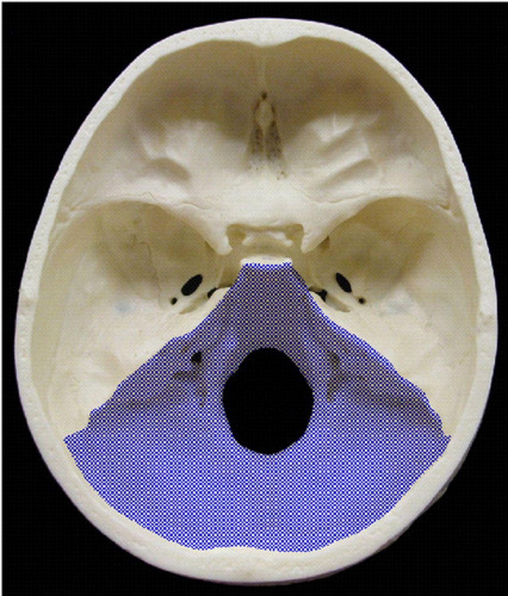
Posterior Cranial Fossa
contains the cerebellum
mandibular condyle
Hypoglossal Canal
superior and medial to condyles
superior nuchal line
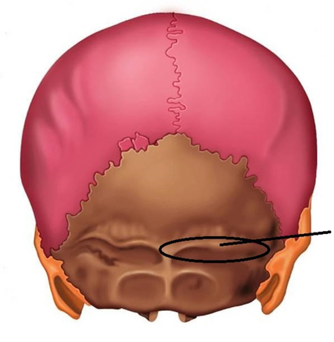
inferior nuchal line
foramen rotundum
Cribriform Foramina
holes

orbit
eye holes
anterior fontanelle (soft)
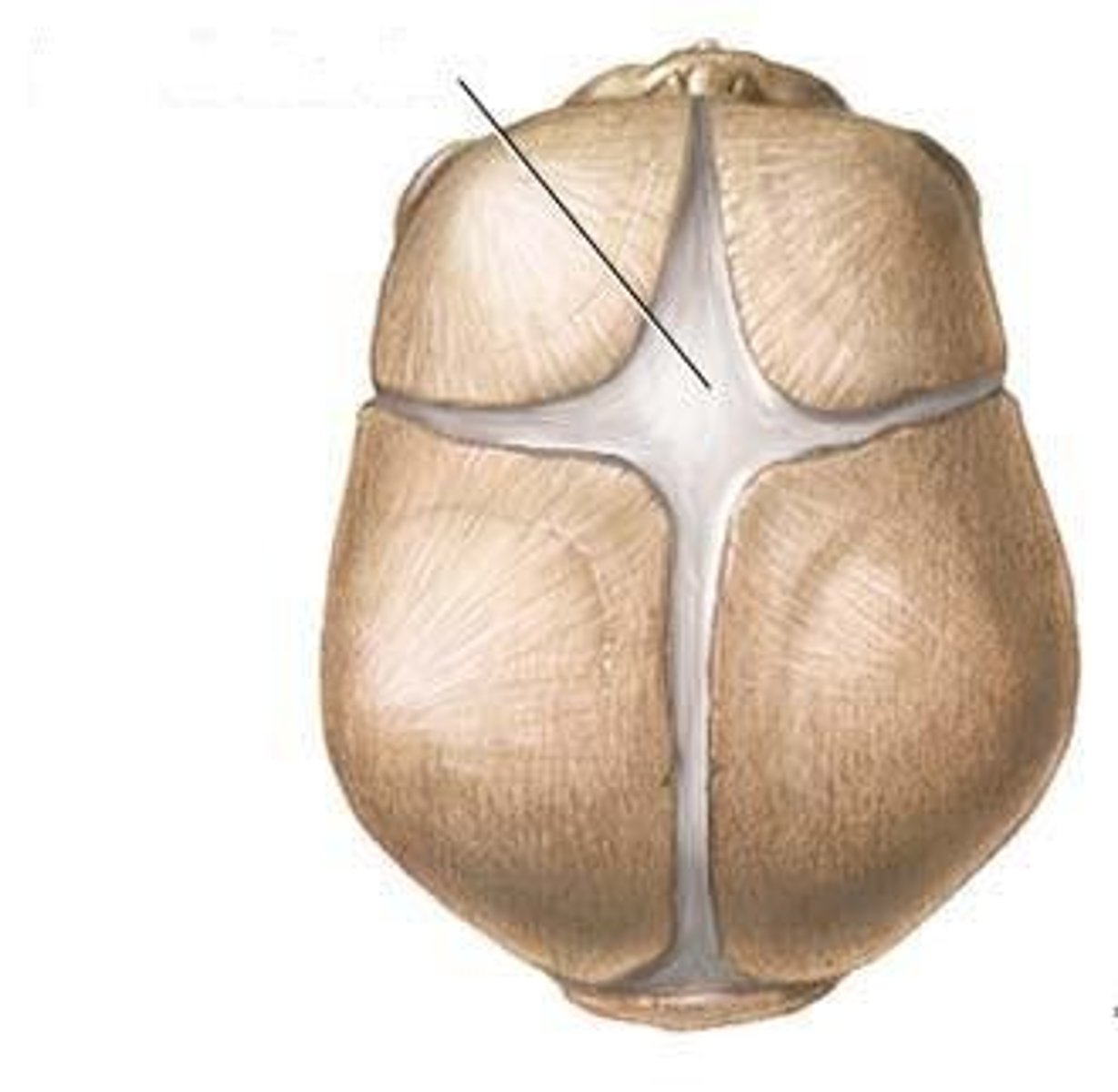
bregma suture (hardened)
vault of the skull
stylomastoid foramen
Not studied (81)
You haven't studied these terms yet.