Introduction to Anatomy and Musculoskeletal Systems
 Call Kai
 Learn
Learn Practice Test
 Spaced Repetition
 Match
Match Flashcards
 Knowt Play
1/93

There's no tags or description
Looks like no tags are added yet.
Name | Mastery | Learn | Test | Matching | Spaced | Call with Kai | Chat |
|---|
No analytics yet
Send a link to your students to track their progress
94 Terms
What are the three approaches to studying anatomy?
Regional: based on organization of the body into parts (we do this at UTMB due to cadaver stability)
Systemic: Anatomical study organized by organ systems that work together to carry out functions.
Clinical: Emphasizes aspects of structure and function of the body important to clinical practice. Uses both regional and systemic elements.
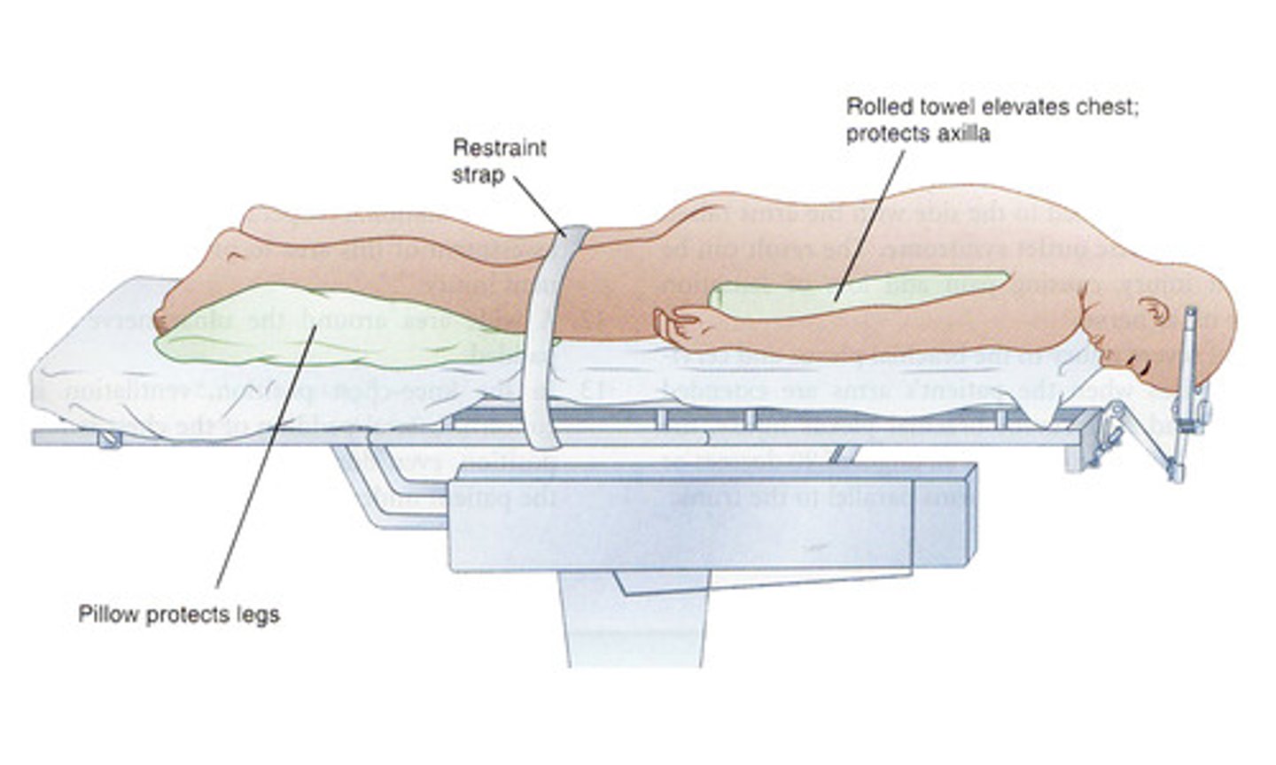
Supine
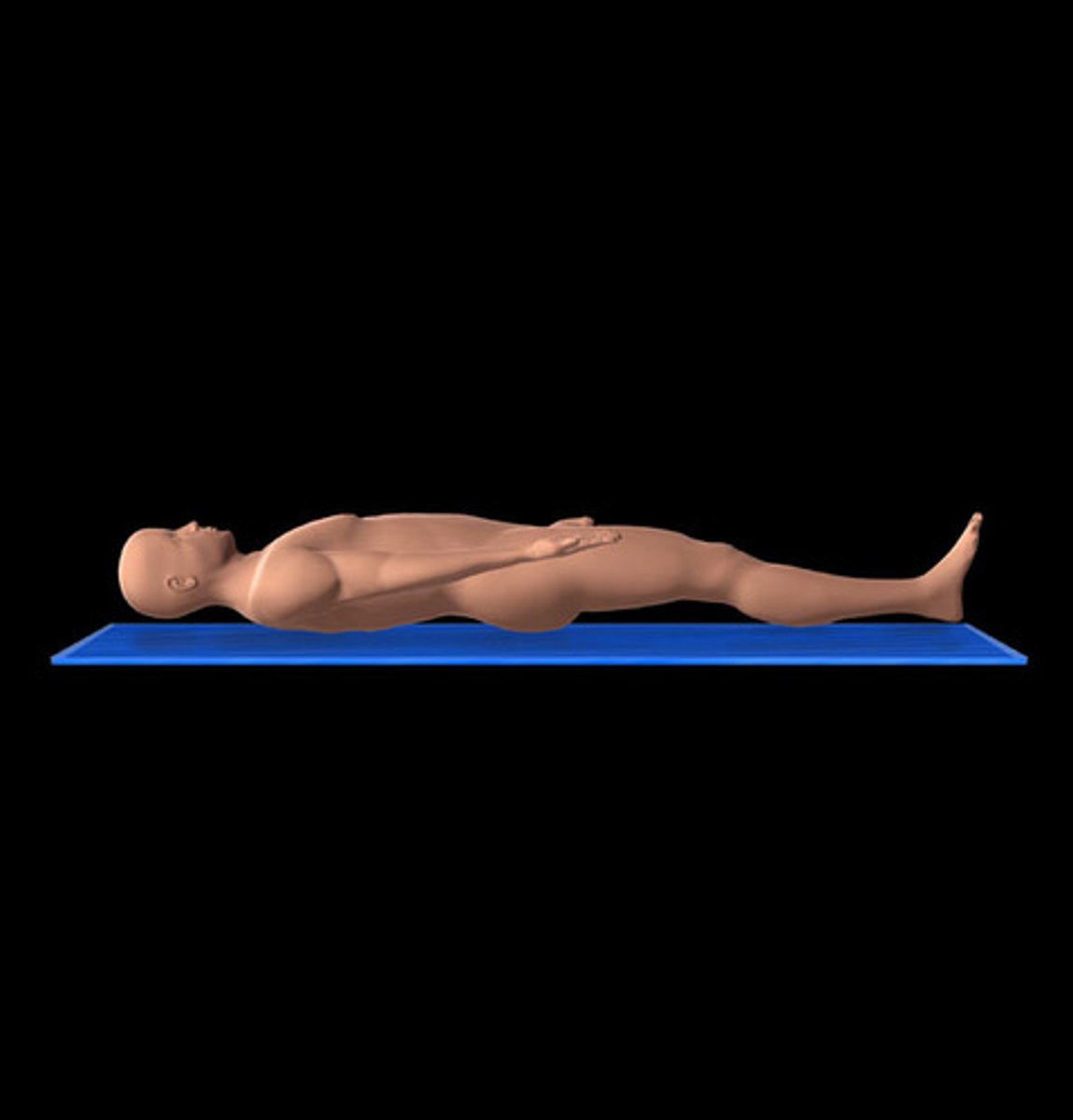
Prone
Anatomical position
Refers to the body position as if the person were standing upright with the:
Head, gaze (eyes), and toes directed anteriorly
Arms adjacent to the sides with palms facing anteriorly
Lower limbs close together with the feet parallel
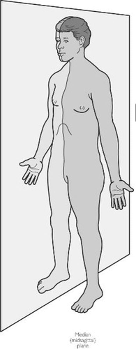
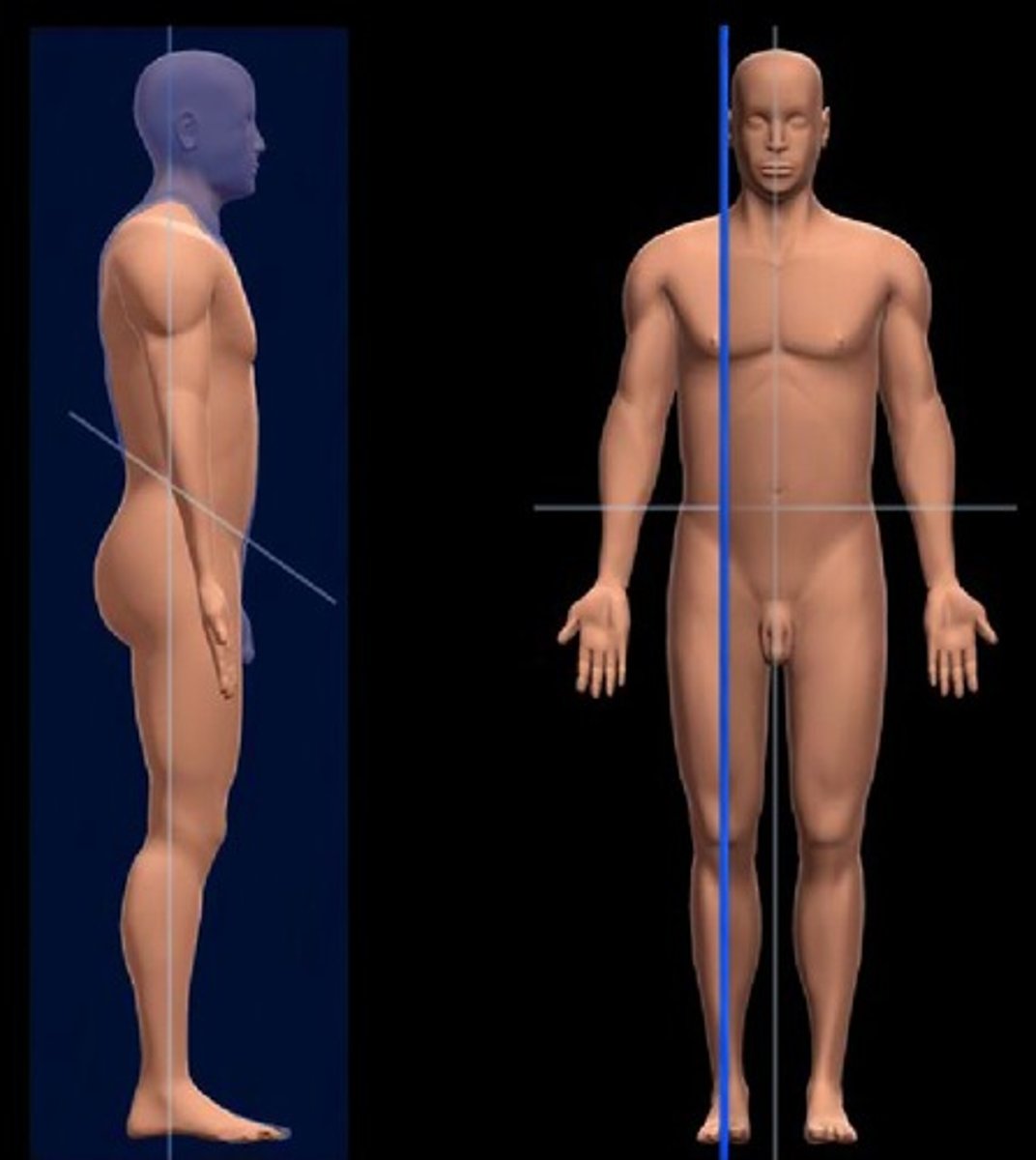
median plane
a type of sagittal plane that divides the body into equal left and right halves, may be by body part (ex. the median plane of the right hand)
sagittal plane
any verticle plane down the body that divides it into left and right portions
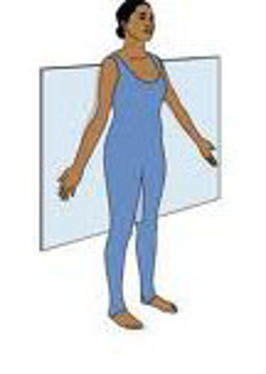
frontal plane
Divides the body into front and back portions. AKA coronal plane. May be done to certain body parts (ex. the frontal plane of the feet)
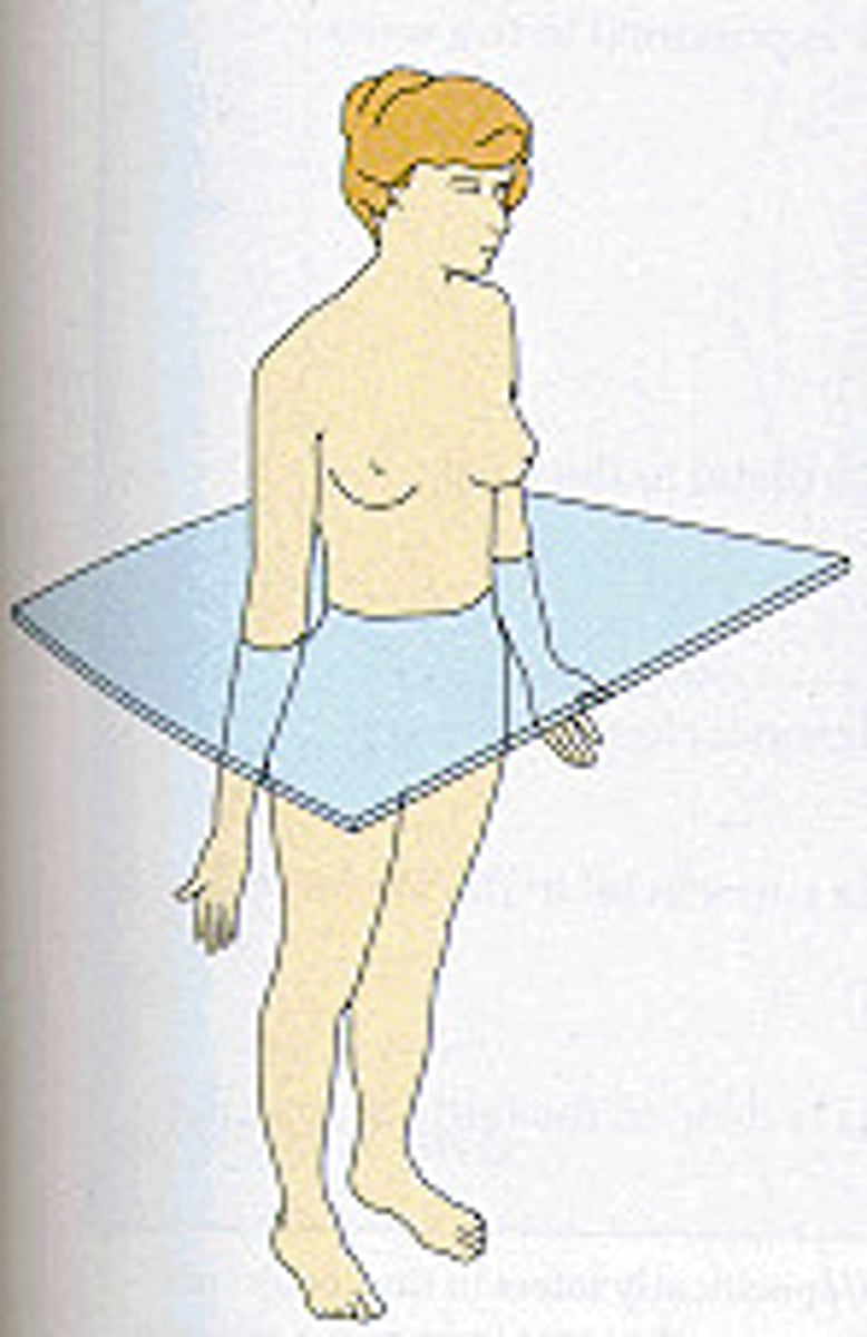
Transverse plane
horizontal division of the body into upper and lower portions
Longitudinal section
cut through the long axis
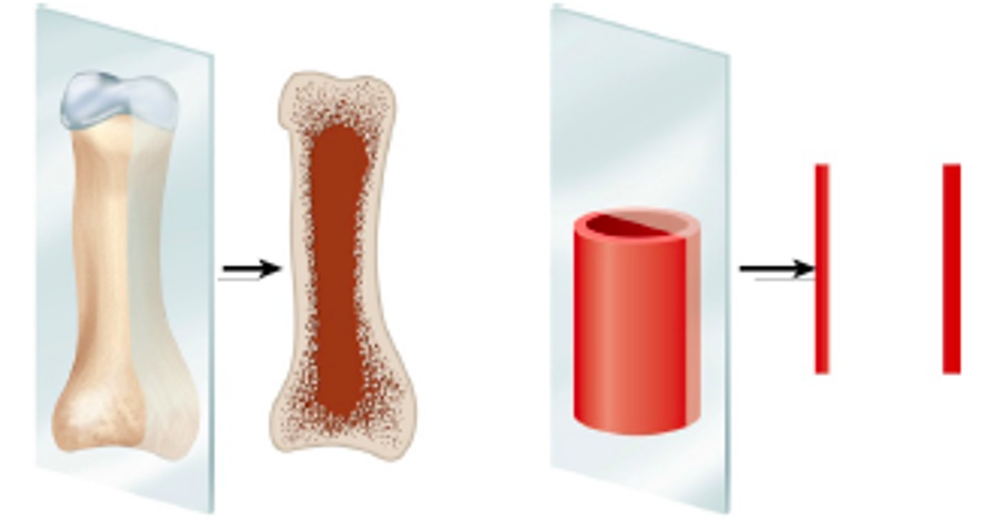
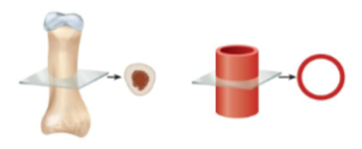
transverse section
cut made along a horizontal plane to divide into upper and lower regions
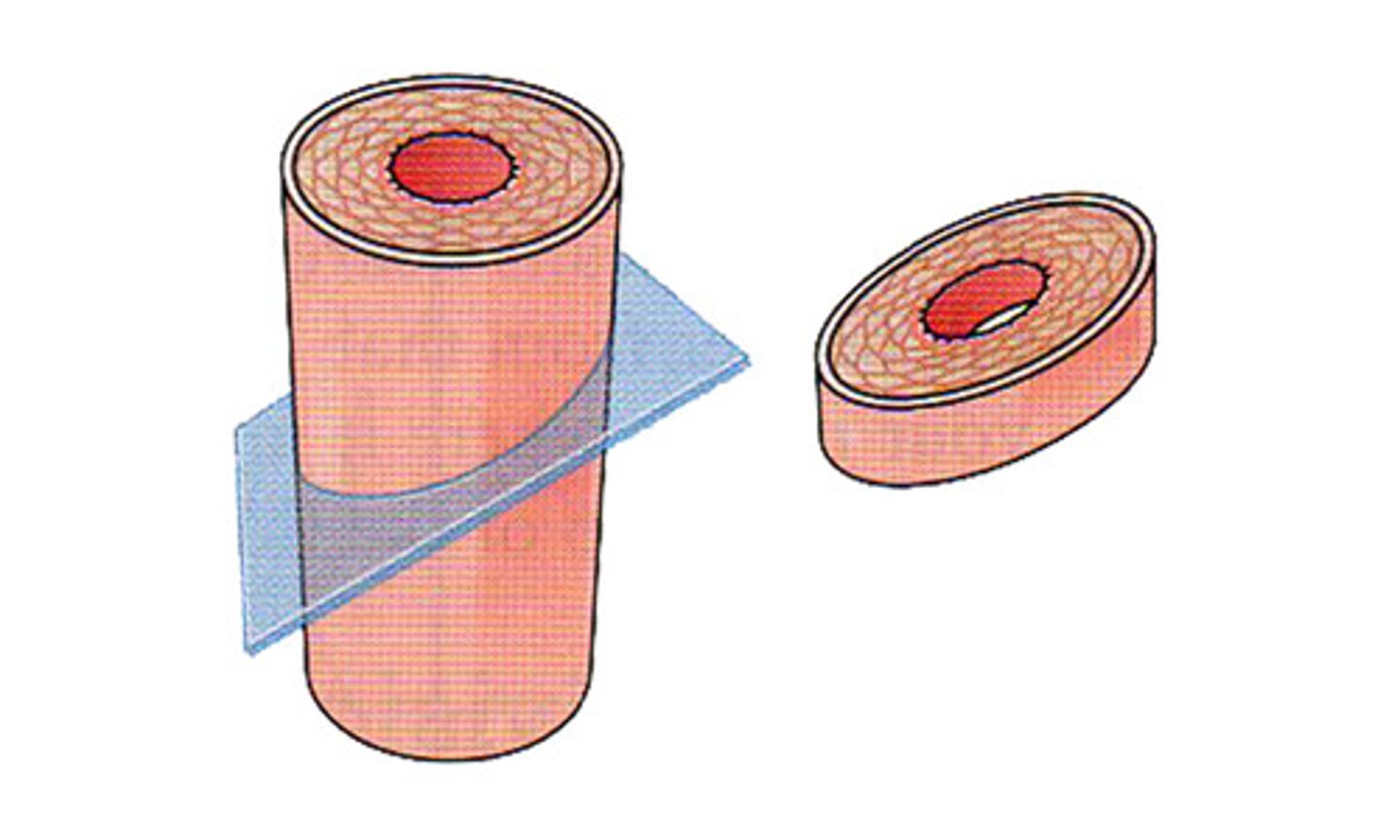
oblique section
cuts made diagonally between the horizontal and the vertical planes
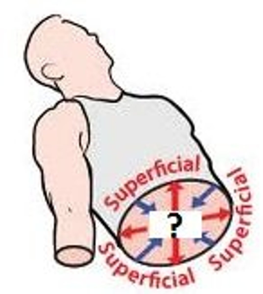
Superficial
near the surface
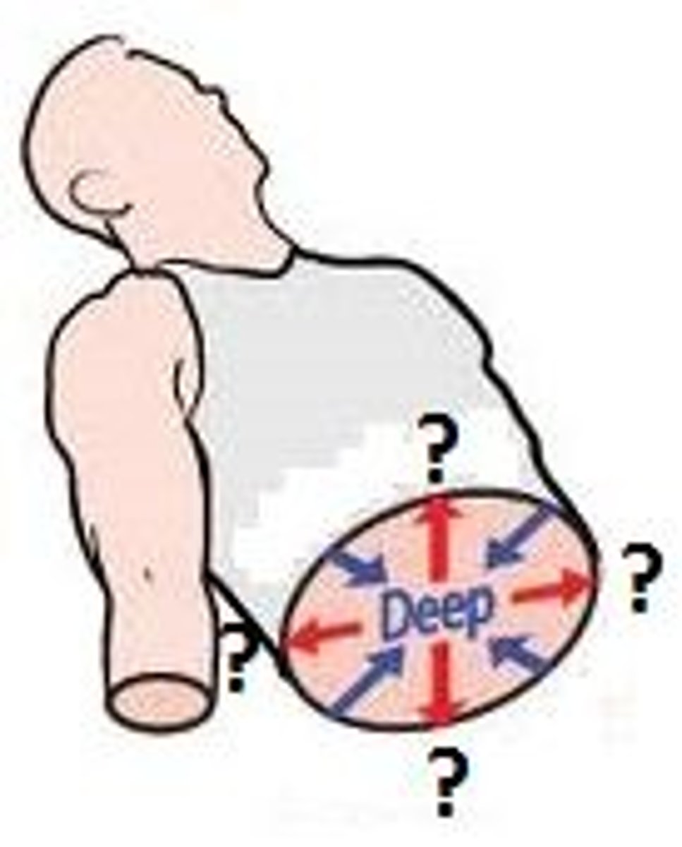
intermediate
between superficial and deep structure
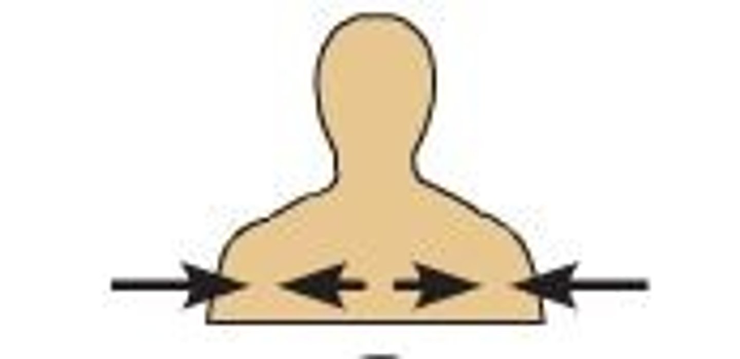
deep
farthest from the surface of the skin
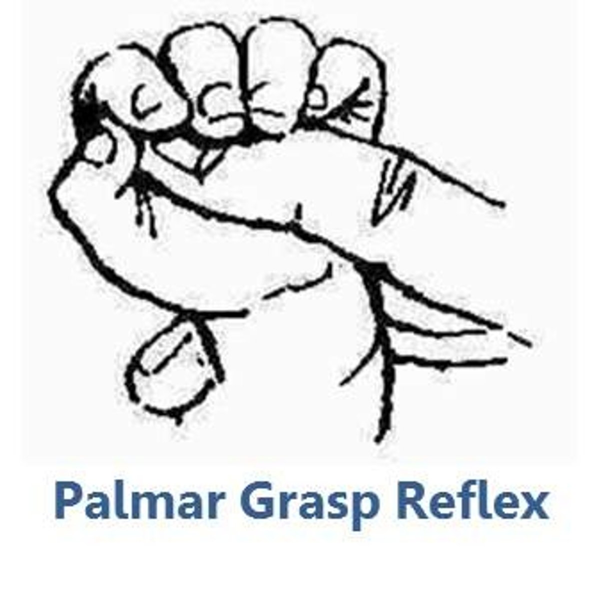
palmar
anterior hand (palm)
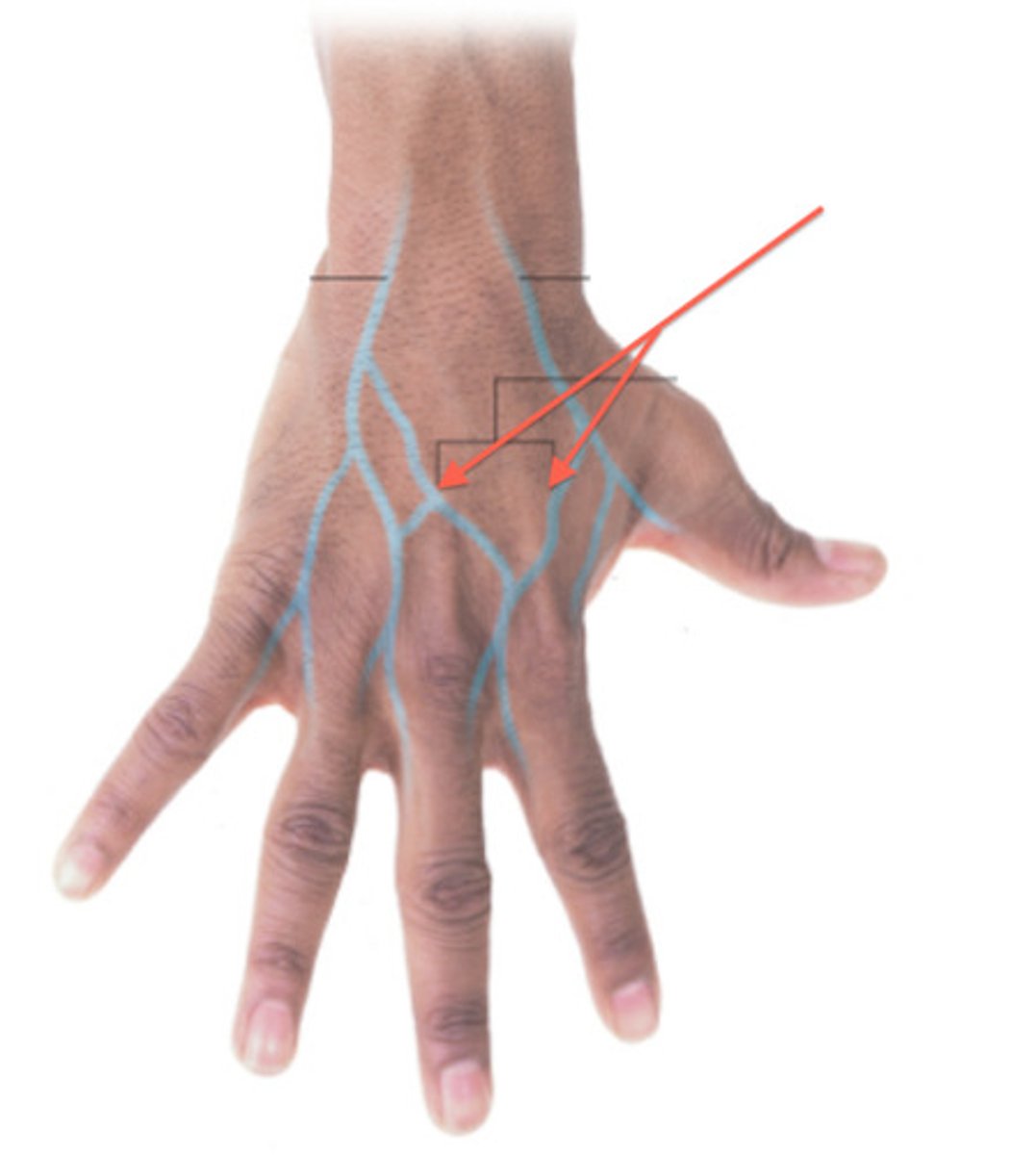
dorsal
posterior
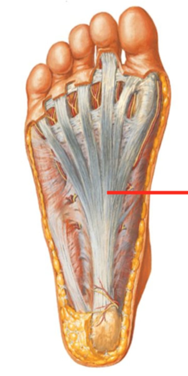
plantar
sole of foot
retrusion
moving a part of the body posteriorly (as in tucking in the chin)
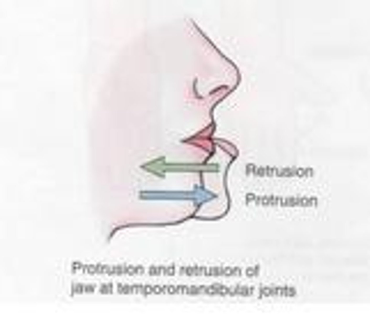
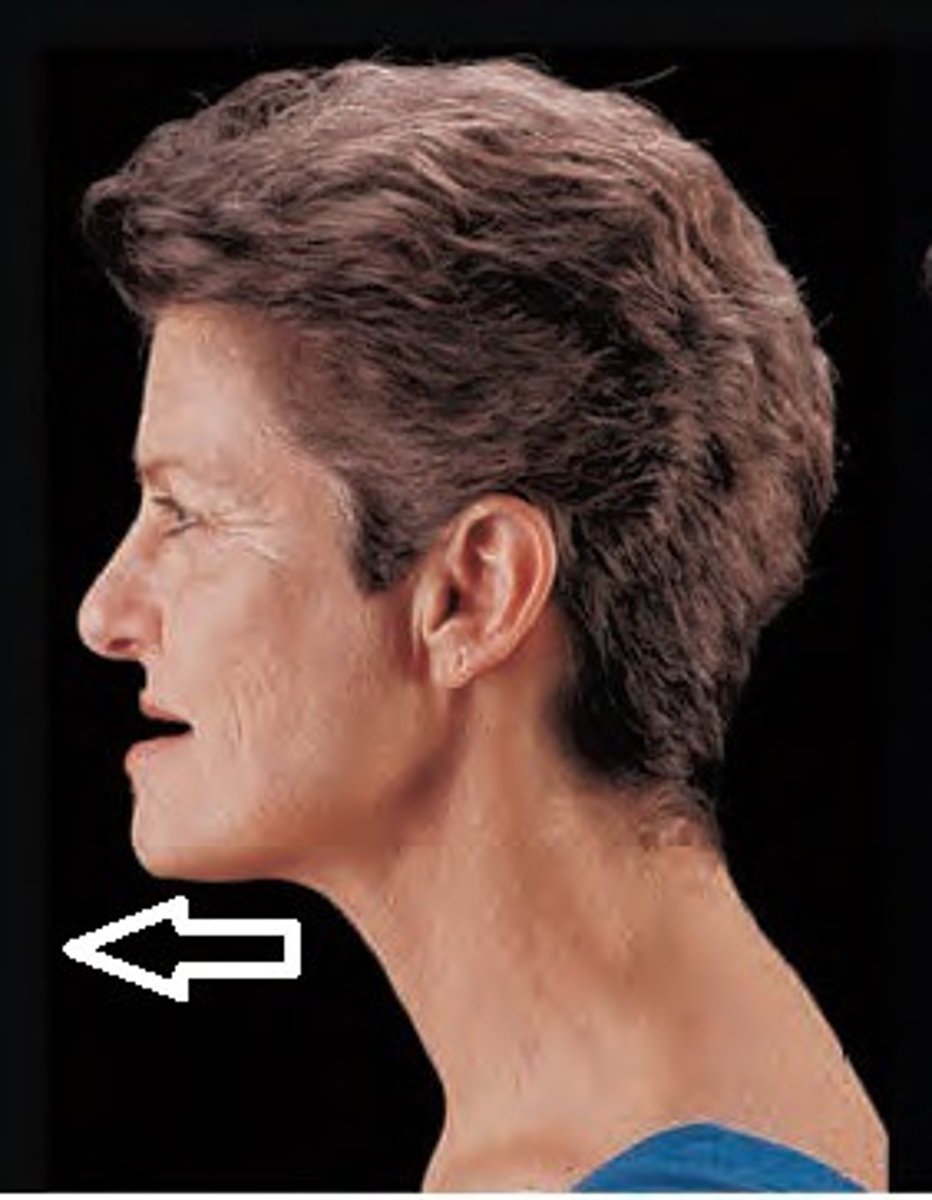
protrusion
the state or condition of being thrust forward or projecting
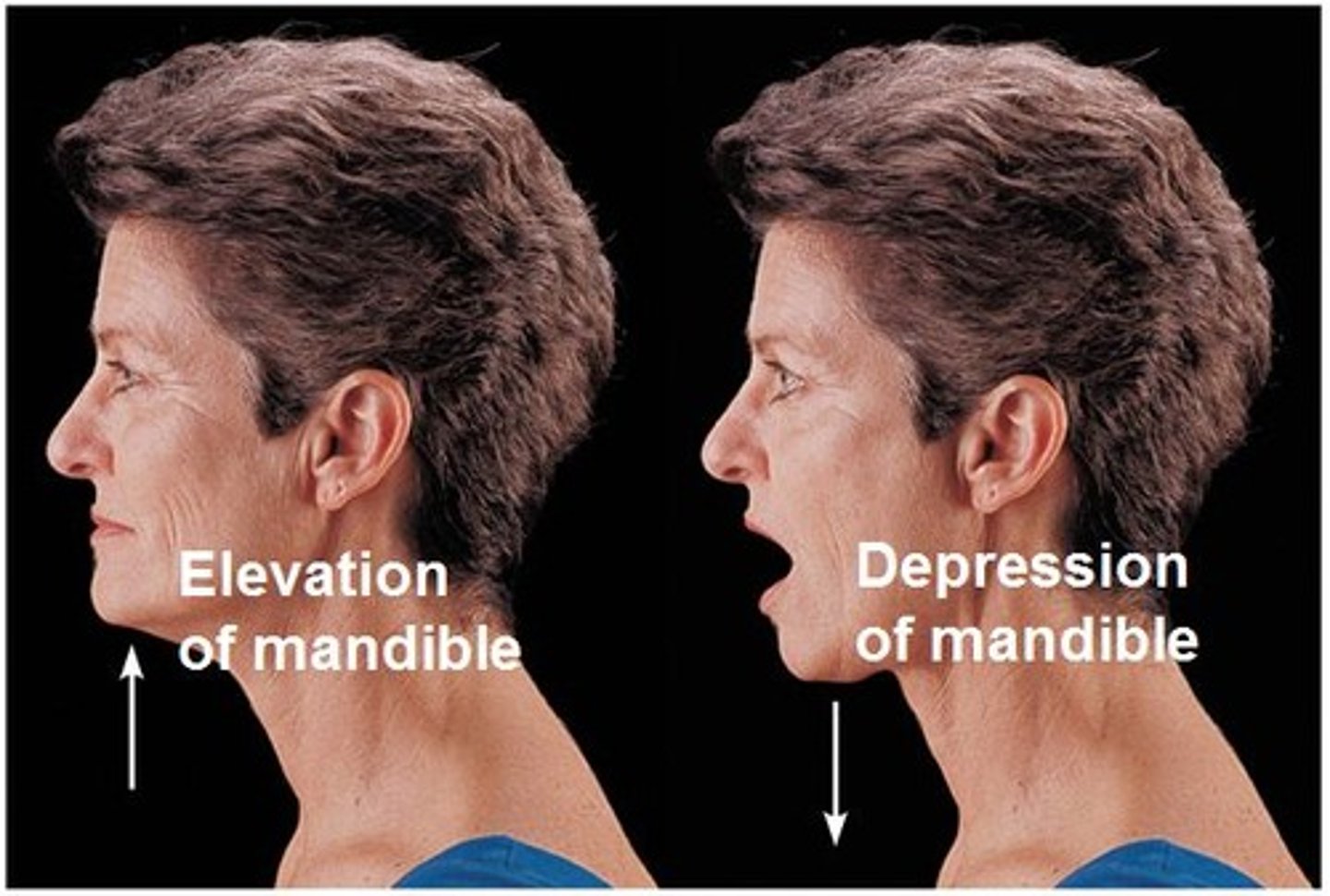
elevation and depression
protraction
moving a body part forward and parallel to the ground
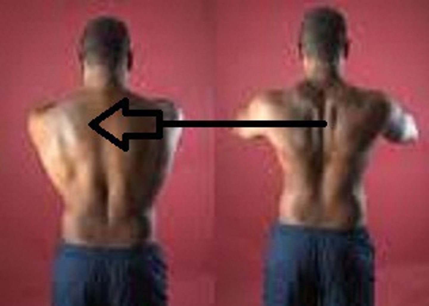
retraction
moving a part backward
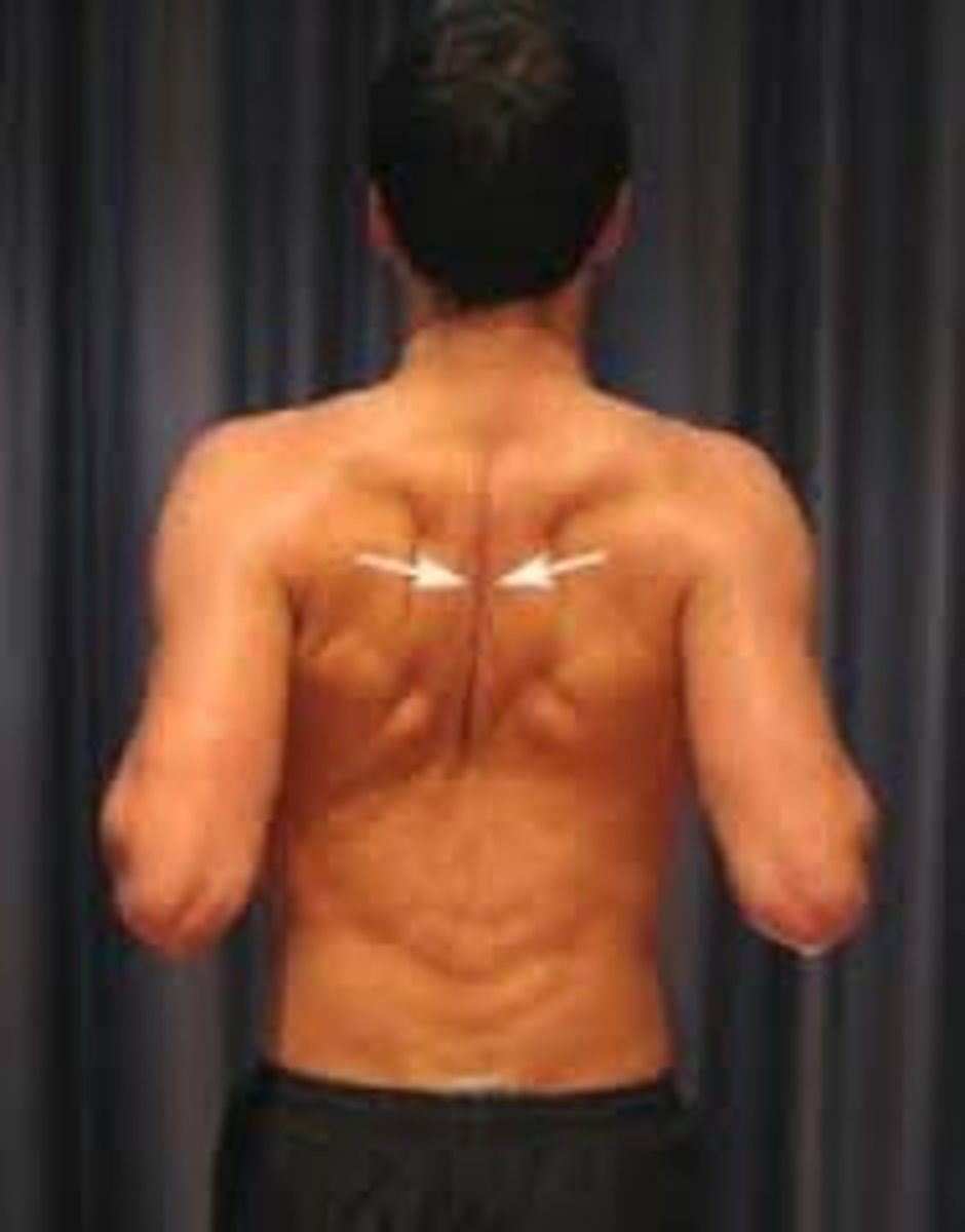
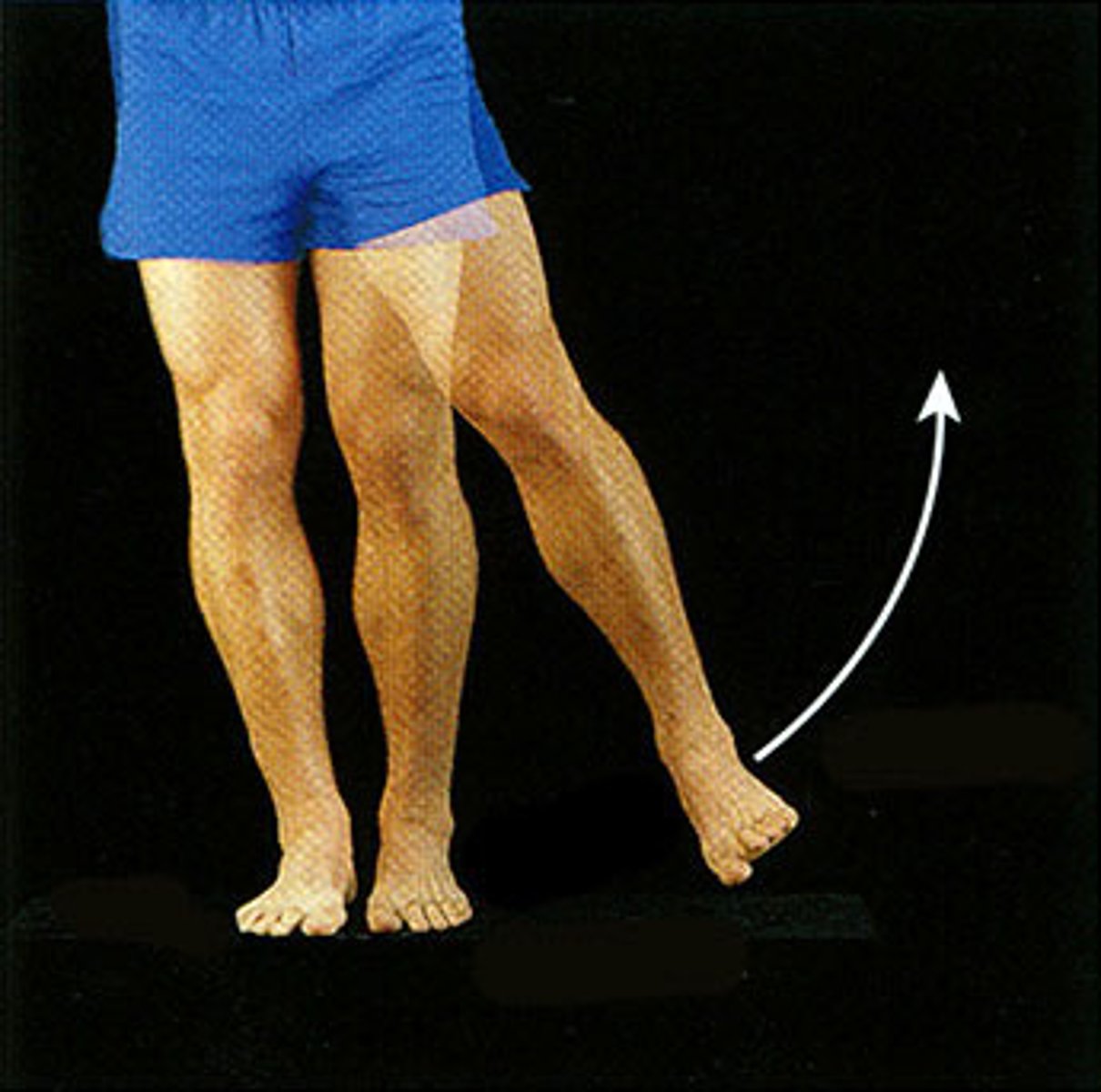
abduction
Movement away from the midline of the body
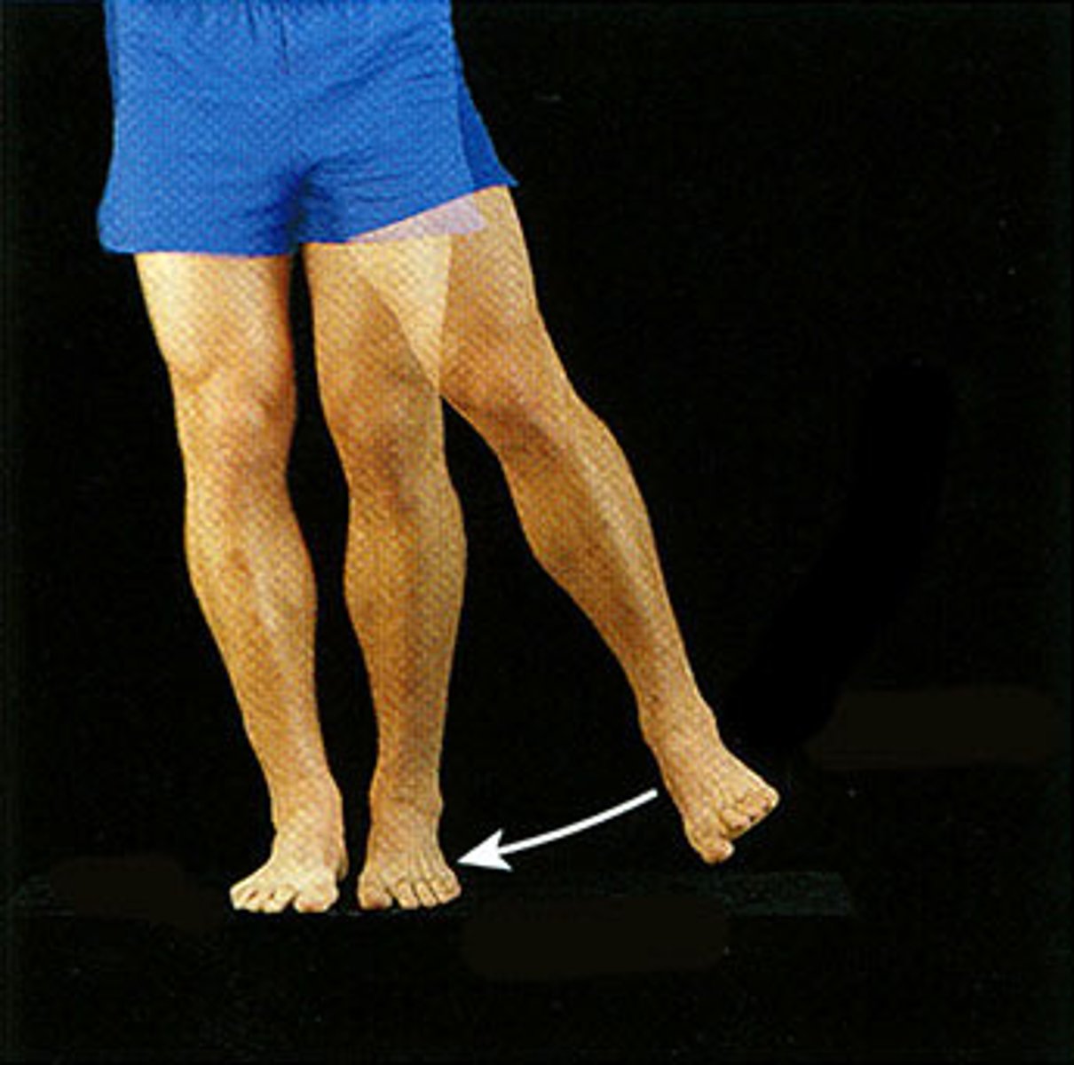
adduction
Movement toward the midline of the body
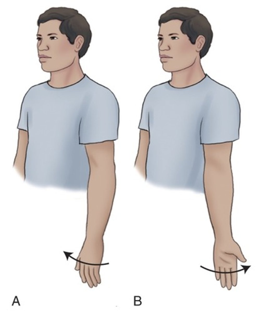
medial rotation vs lateral rotation
rotation towards the midline of the body vs away
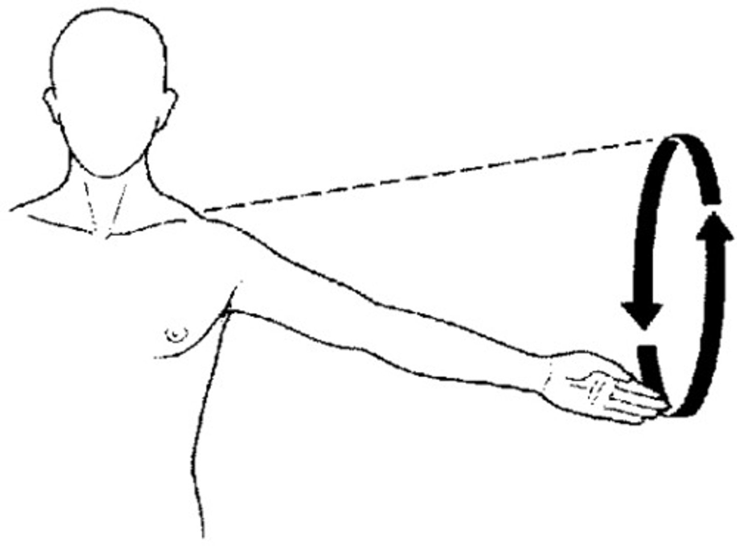
circumduction
circular movement of a limb at the far end
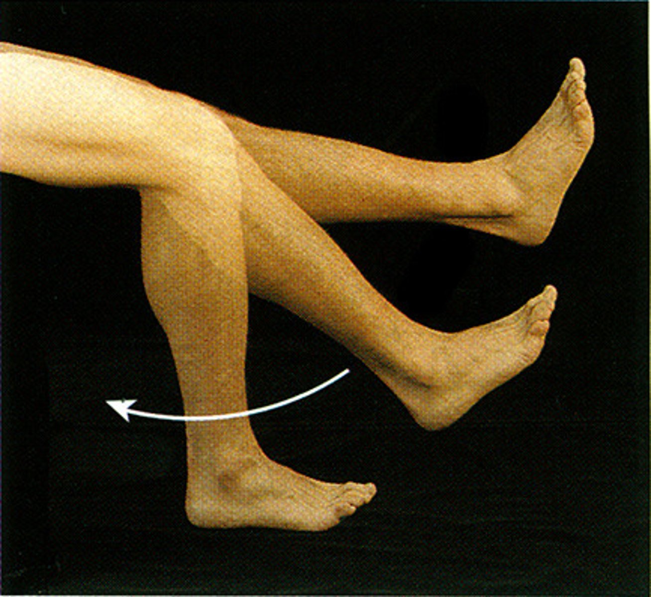
extension
increases the angle of a joint
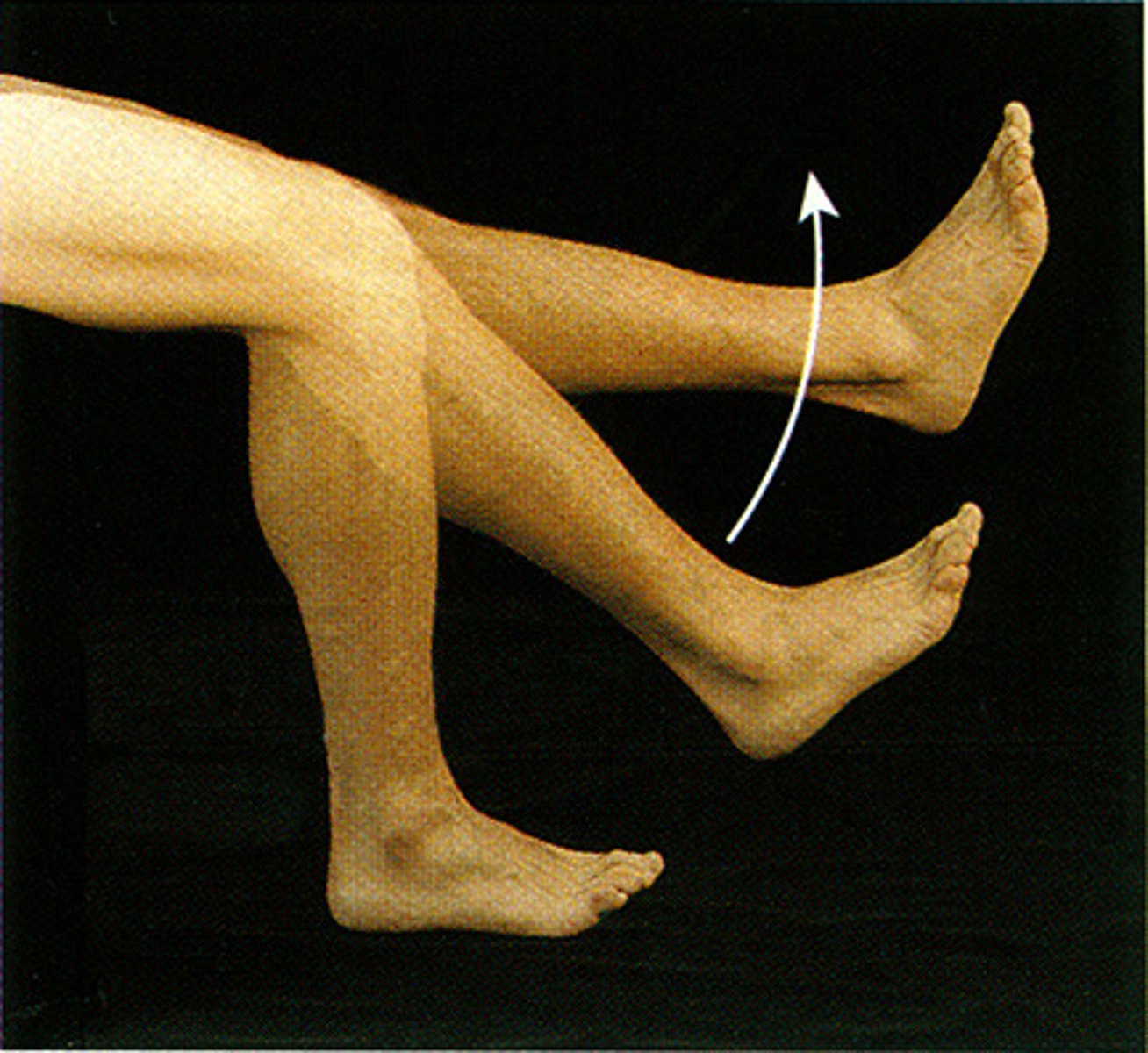
flexion
Decreases the angle of a joint
dorsiflextion
bends the foot upward at the ankle (3)
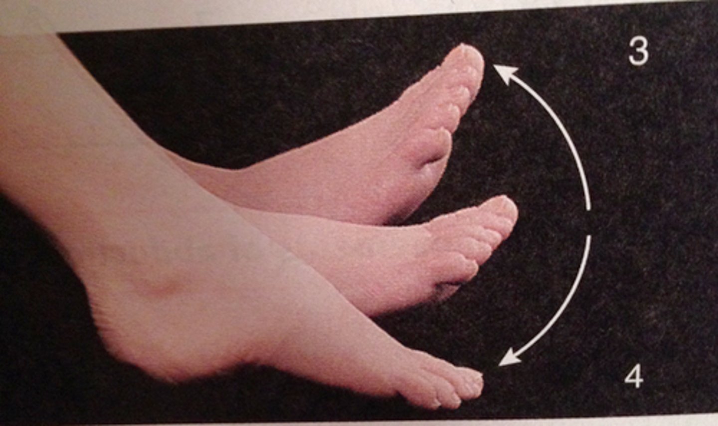
plantarflextion
Ankle movement pointing the foot downward
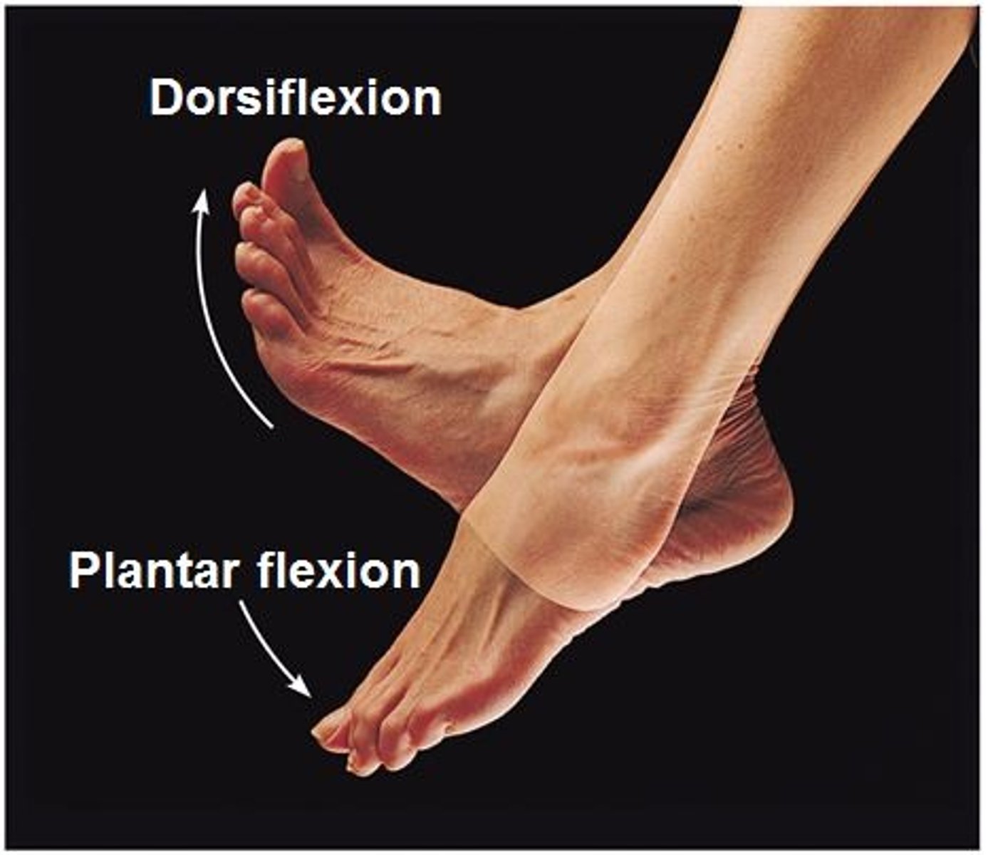
eversion
turning the sole of the foot outward
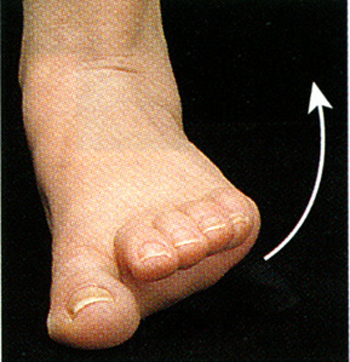
inversion
Turning the sole of the foot inward
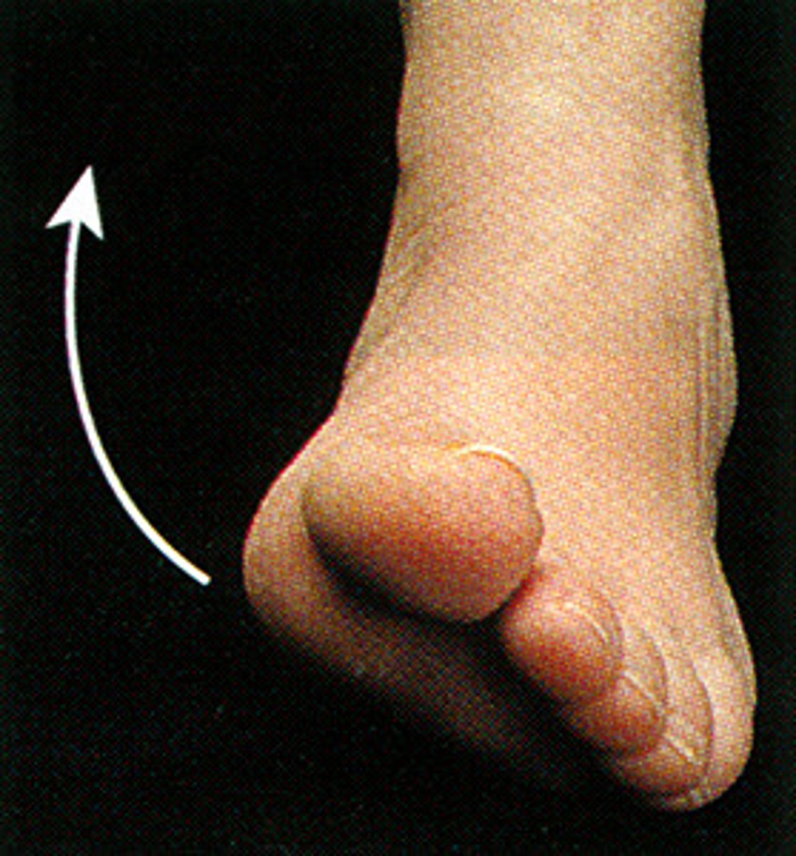
pronation
turning the palm downward
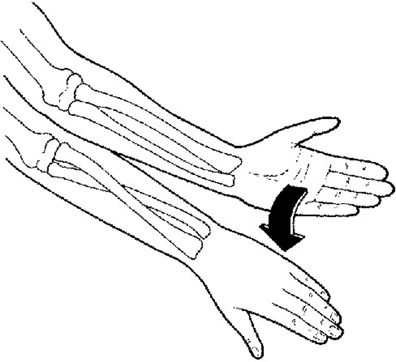
supination
movement that turns the palm up
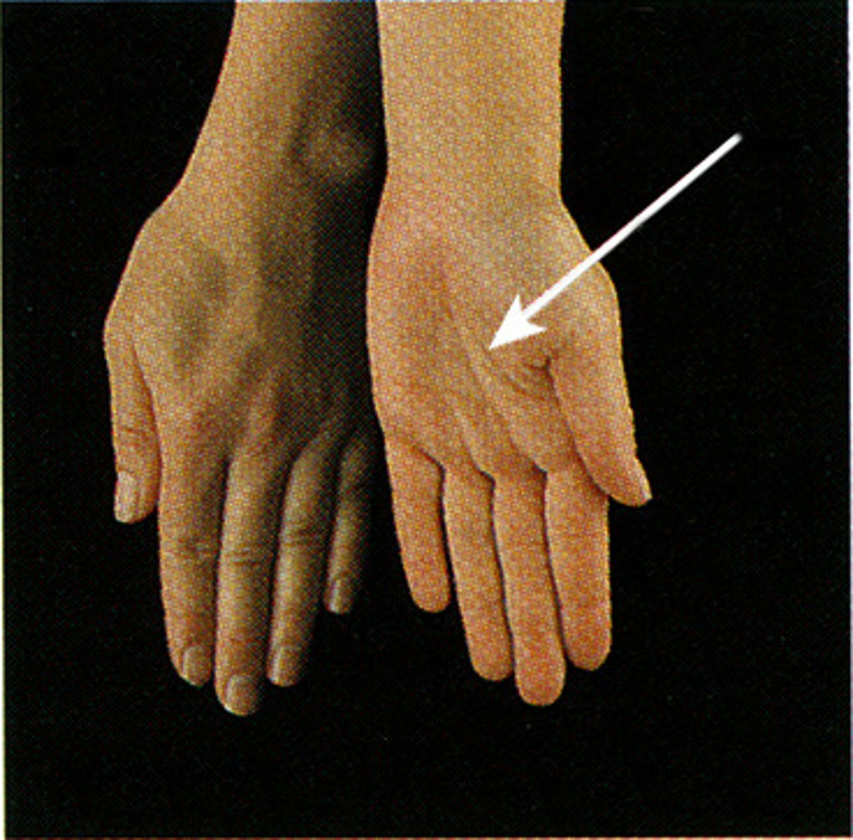
abduction of the fingers
spreading fingers apart
adduction of the fingers
brings them together again
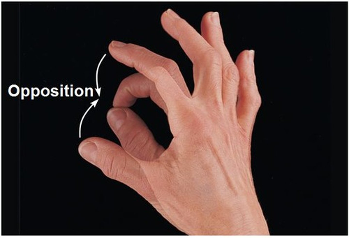
opposition
Movement of the thumb to touch the fingertips
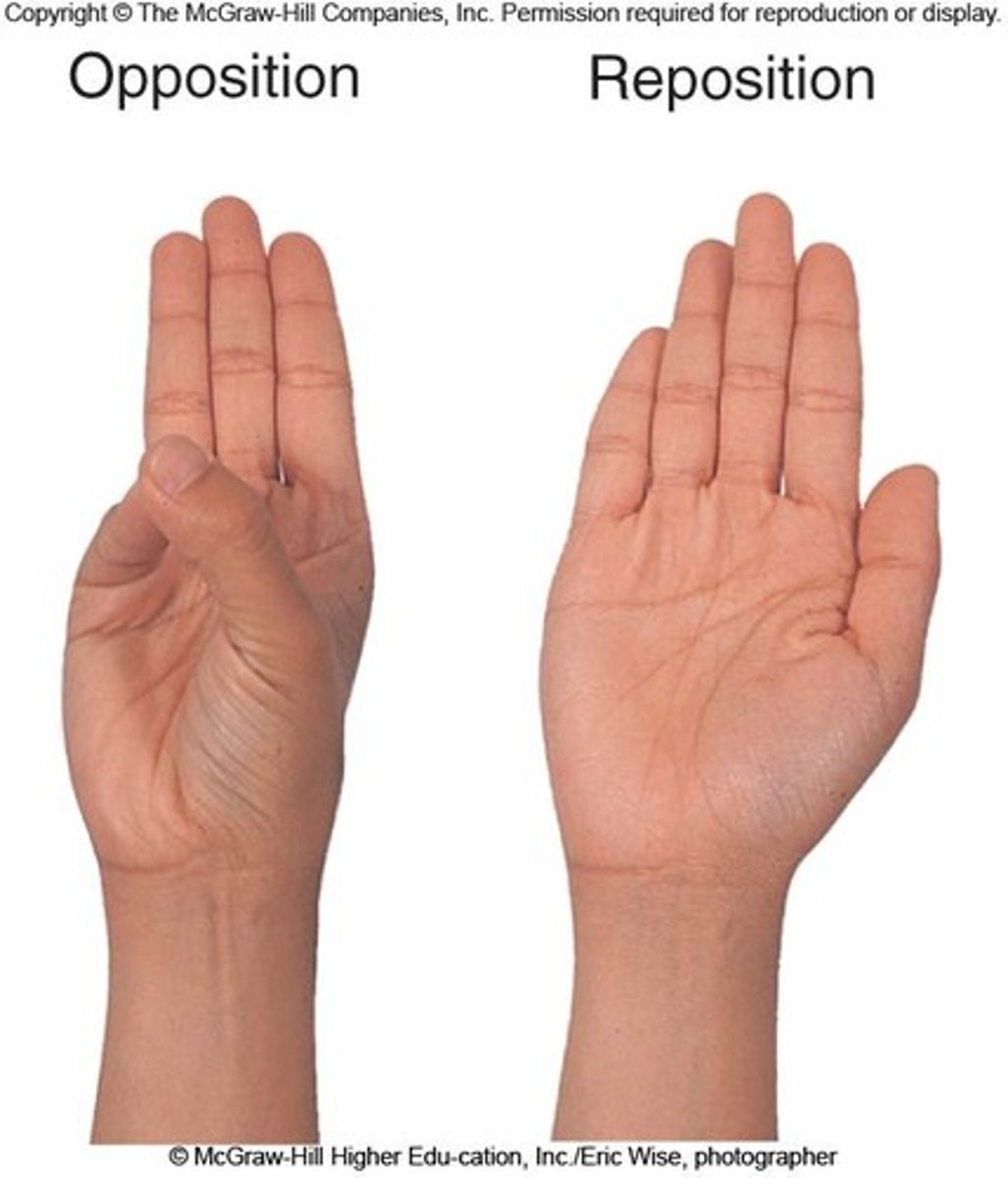
reposition
return to anatomical position
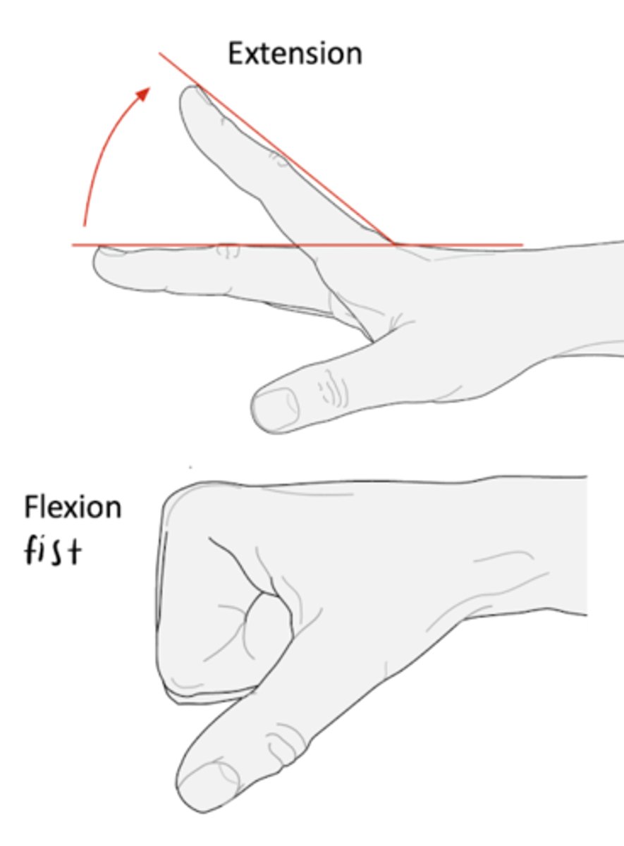
flexion and extension of digits
congenital abnormality or birth defect
A structural, functional, or metabolic abnormality present at birth. Also called congenital anomalies. These may be caused by environmental or genetic factors, or by a combination of the two. Structural abnormalities are generally referred to as congenital malformations, and metabolic abnormalities as inborn errors of metabolism.
Jobs of the skin
largest organ in the body
protects it from protection, containments, extreme heat, conveys sensation, synthesis and storage of vitamin D
What are the layers of the skin across the majority of the body?
Epidermis (basal layer is the regenerating layer), dermis, subcutaneous tissue, deep fascia, skeletal muscle
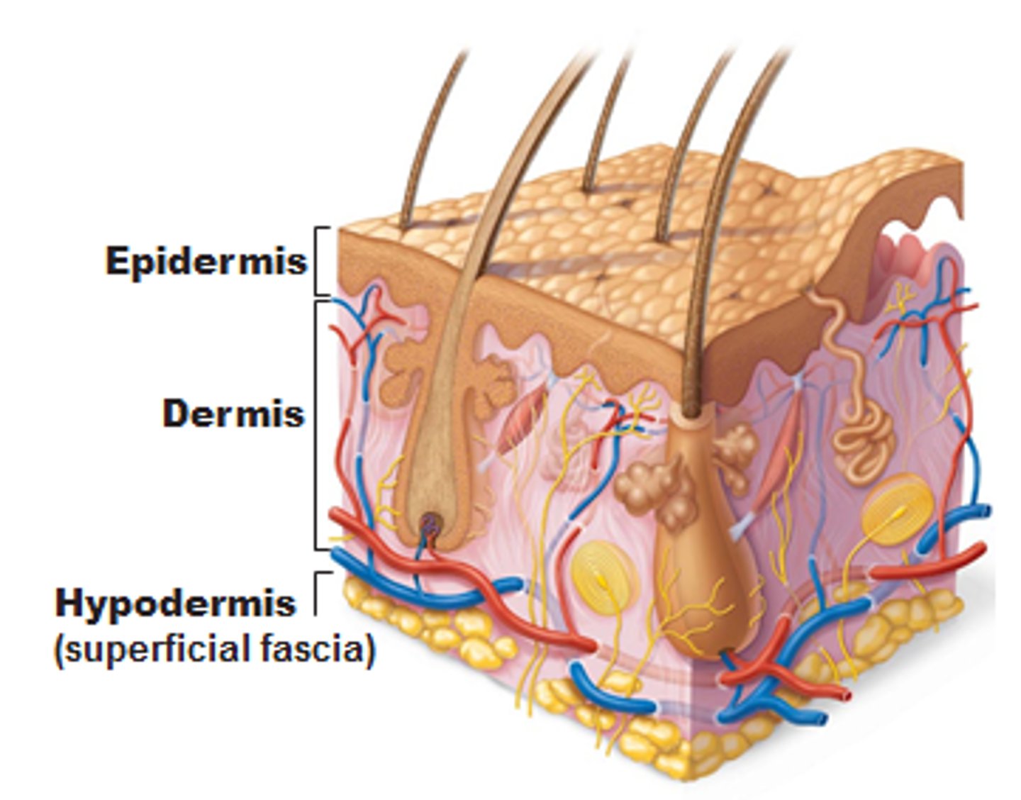
What does the basal layer of the epidermis do and where is it located?
Base of the epidermis right before the dermal-epidermal junction. Made of cuboidal germinative keratinocytes, anchors outer skin to inner skin, selective filter, controls cell proliferation/regeneration.
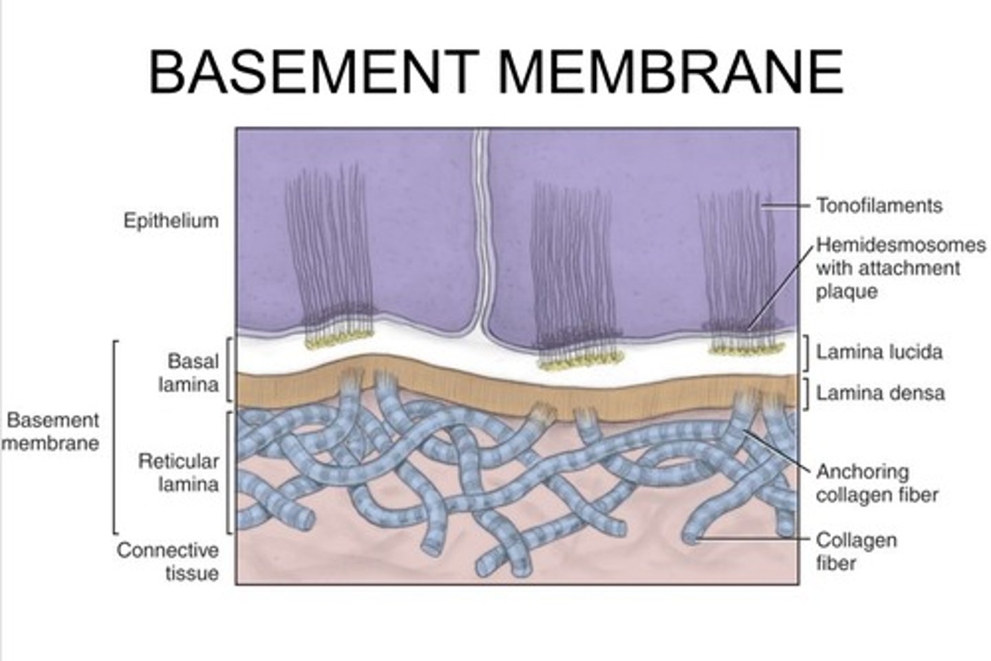
What are the layers of the basement membrane?
Basal keratinocytes connect to the lamina lucida, connects to the lamina densea, which connects the the superficial dermis.
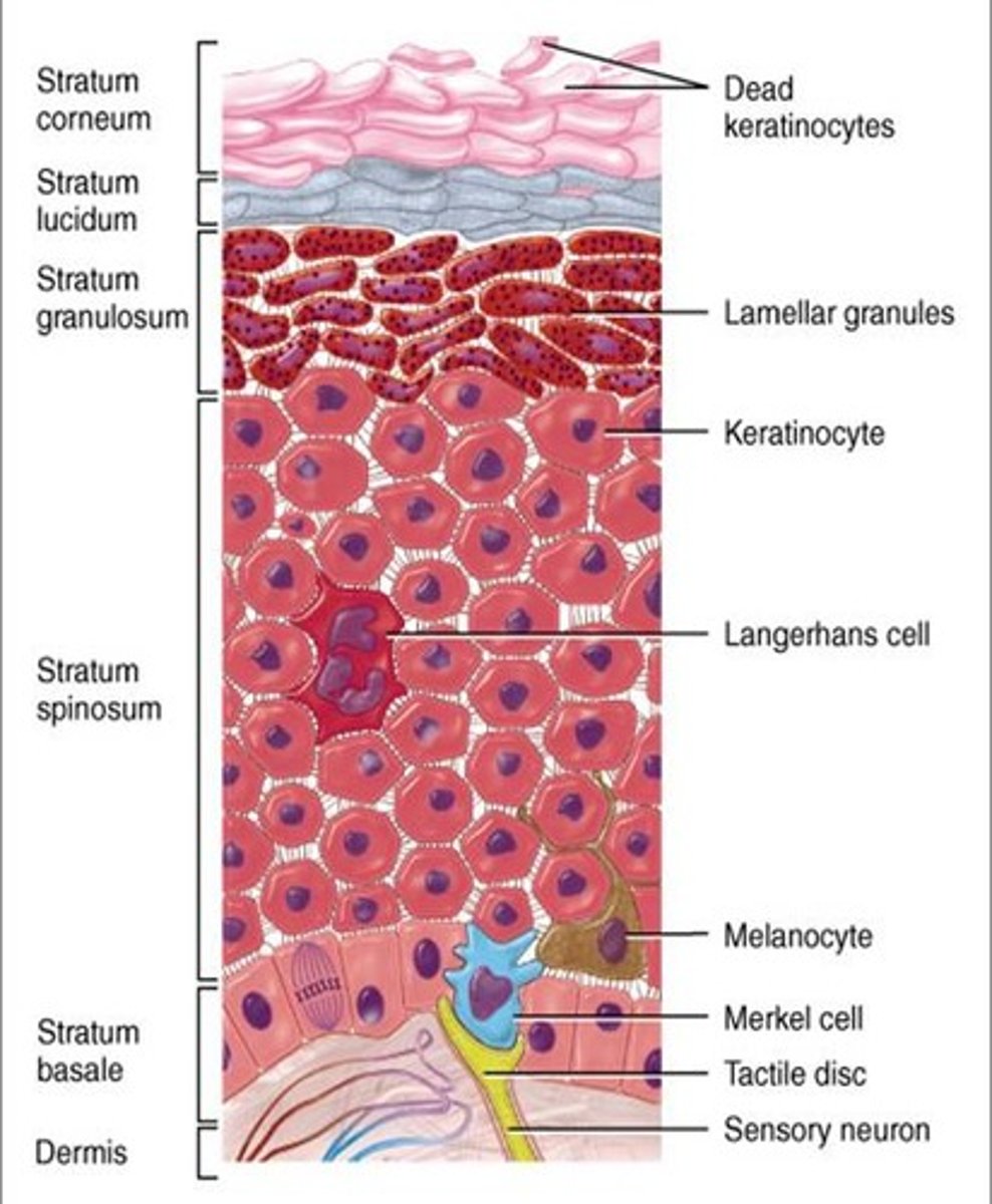
Melanocytes
epidermal melanin producing unit, disperses melanin pigment packed into melaosomes. Typically located right before the basement membrane of the epidermis.
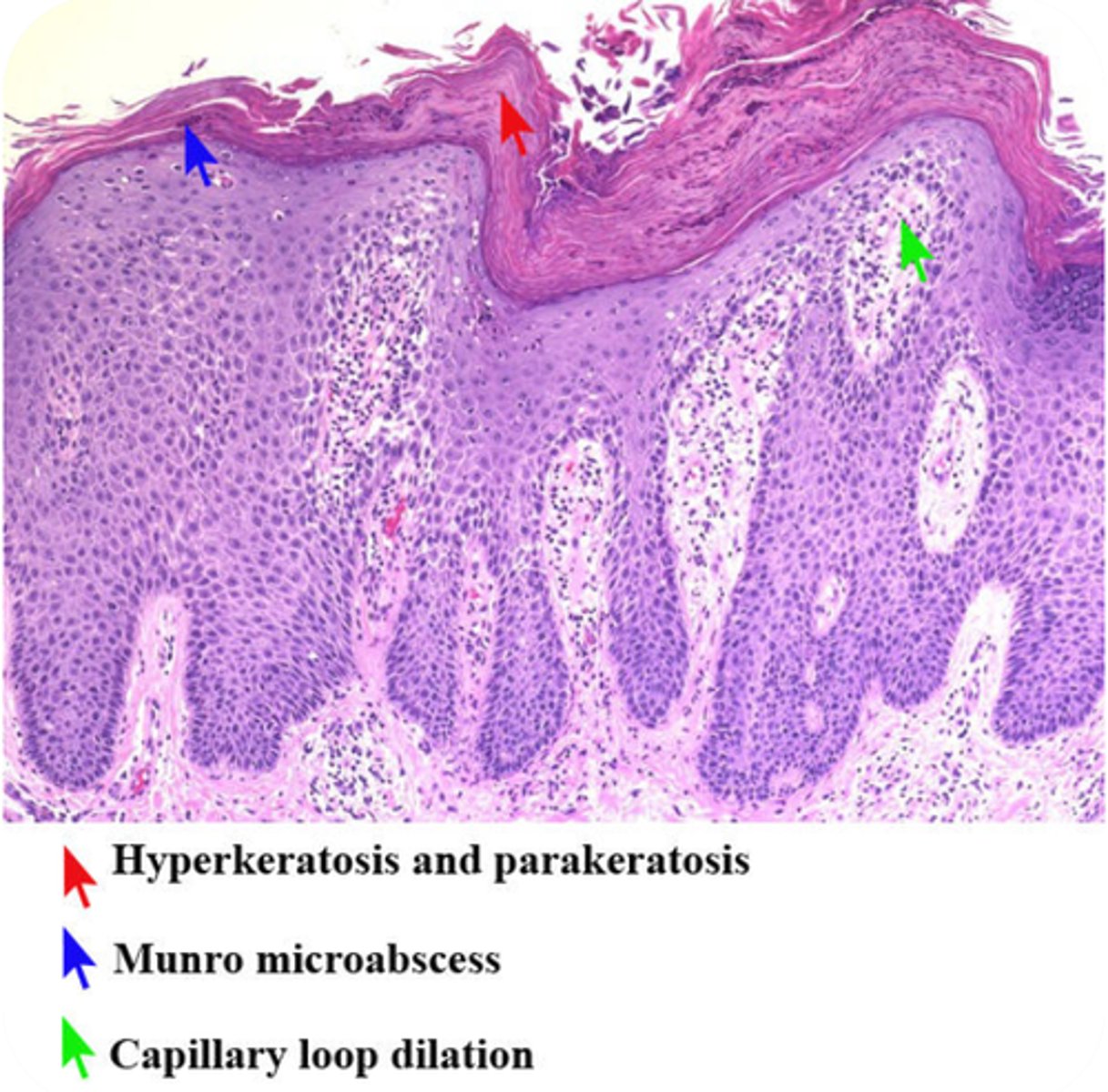
Psoriasis
Inflammatory skin disease in which the epidermis is thickened as a result of elongation of rete ridges due to excessive epidermopoiesis (epidermal proliferation)
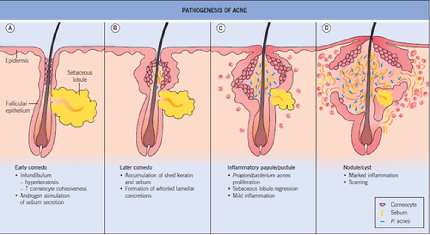
Acne
Pores surrounding hair are clogged by excess sebum and dead skin resulting in perifollicular inflammation and eventual rupture of the follicular wall.
What causes skin burns?
thermal trauma
ultraviolet
ionizing radiation
chemical agents
What are the levels of burns we talked about?
Superficial: effects only the epidermis. Red, painful, dry, no blisters (e.g., sunburn). Heals in 3-6 days without scarring.
partial thickness: Encompasses the epidermis and part of the dermis. Very painful, swollen, and forms blisters. Heals in 7-21 days.
deep: Destroys all skin layers and can reach fatty tissue. Appears white, charred, or leathery and requires surgical treatment.
How do we estimate the body surface area affected by a burn?
The rule of 9s, the body is divided into area that are approximately 9% or multiples of 9%. Allows us to estimate fluid loss, infection risk, and other patient risk factors.
What are the three factors that increase the risk of death from burn injury?
1. age older than 60
2. parital-thickness and full-thickness burns over 40% of body surface area
3. the presence of inhalation injury
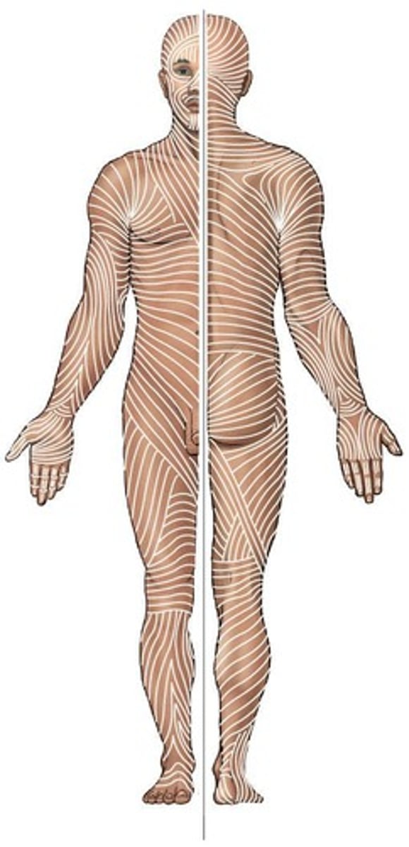
What are tension lines?
Lines of collagen within the skin across the body. Incisions made across these lines are more likely to gape and increase healing time while those made parallell have better outcomes.
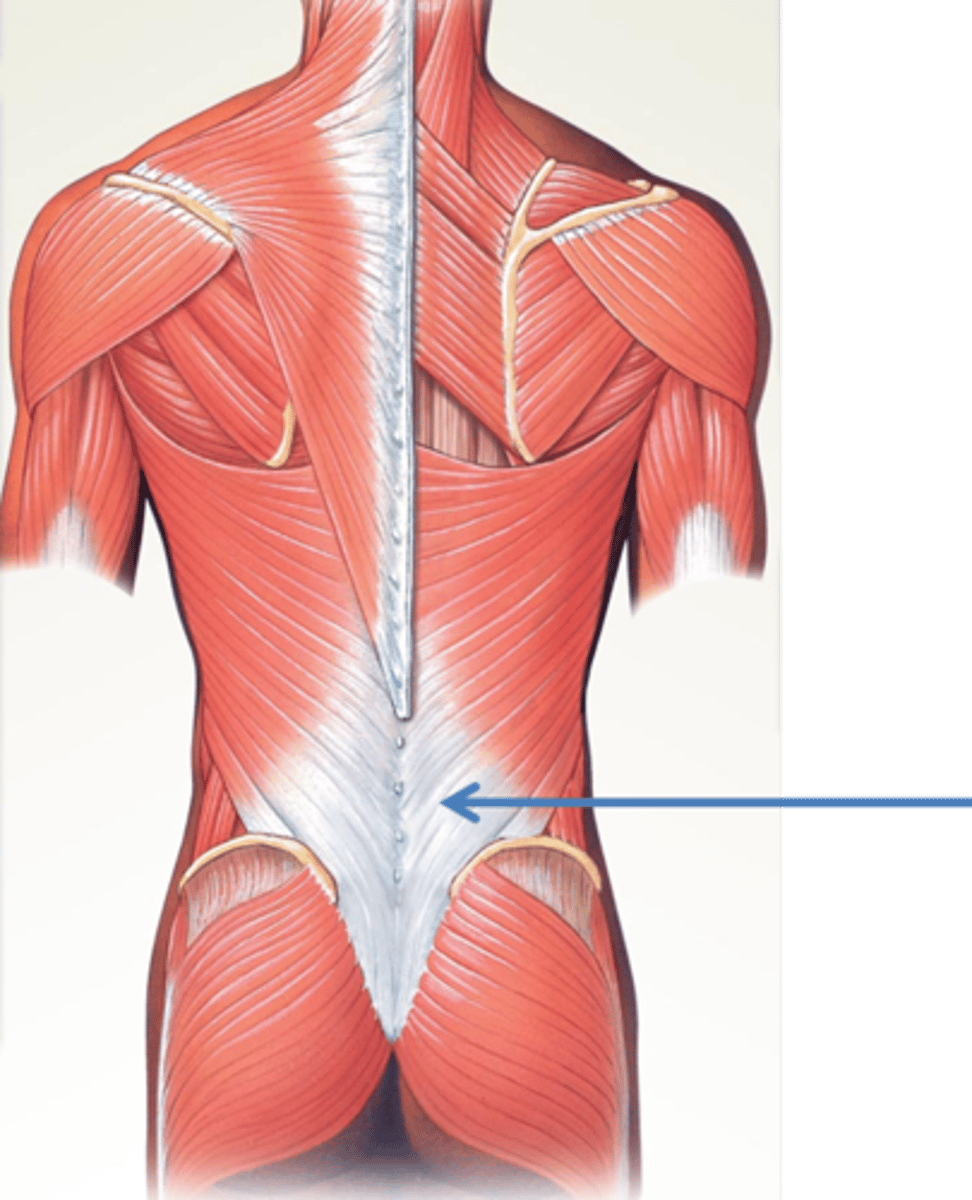
What is fascia?
Fascia constitute the wrapping, packing, and insulating materials of the deep structures of the body.
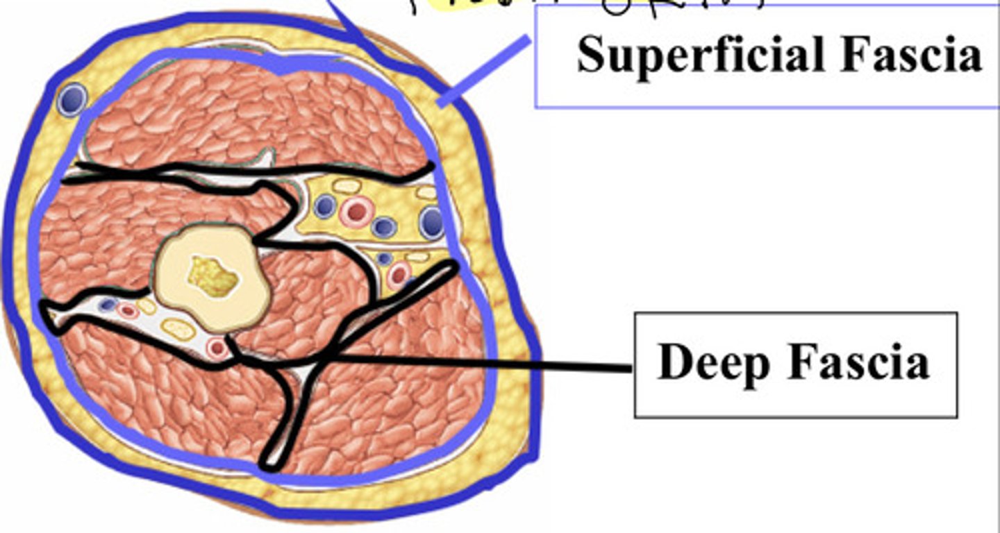
What are the two types of fascia?
Superficial: subcutaneous fascia
Deep: dense, organized connective tissue
What makes up the skeletal system? What are it's functions?
Bones, cartilage, and joints
Functions include: support, protection of vital organs, a mechanism (with muscles) for movement, storage of calcium and other salts, storage of growth factors and cytokines, a source of blood cells.
What are the classification of bones?
Long bone (ex. humerus)
short bone (ex. carpals)
flat bone (ex. parietal)
irregular bone (ex. vertebra)
sesamoid bone (ex. patella)
What are the two types of bones?
compact bone and spongy (trabecular) bone
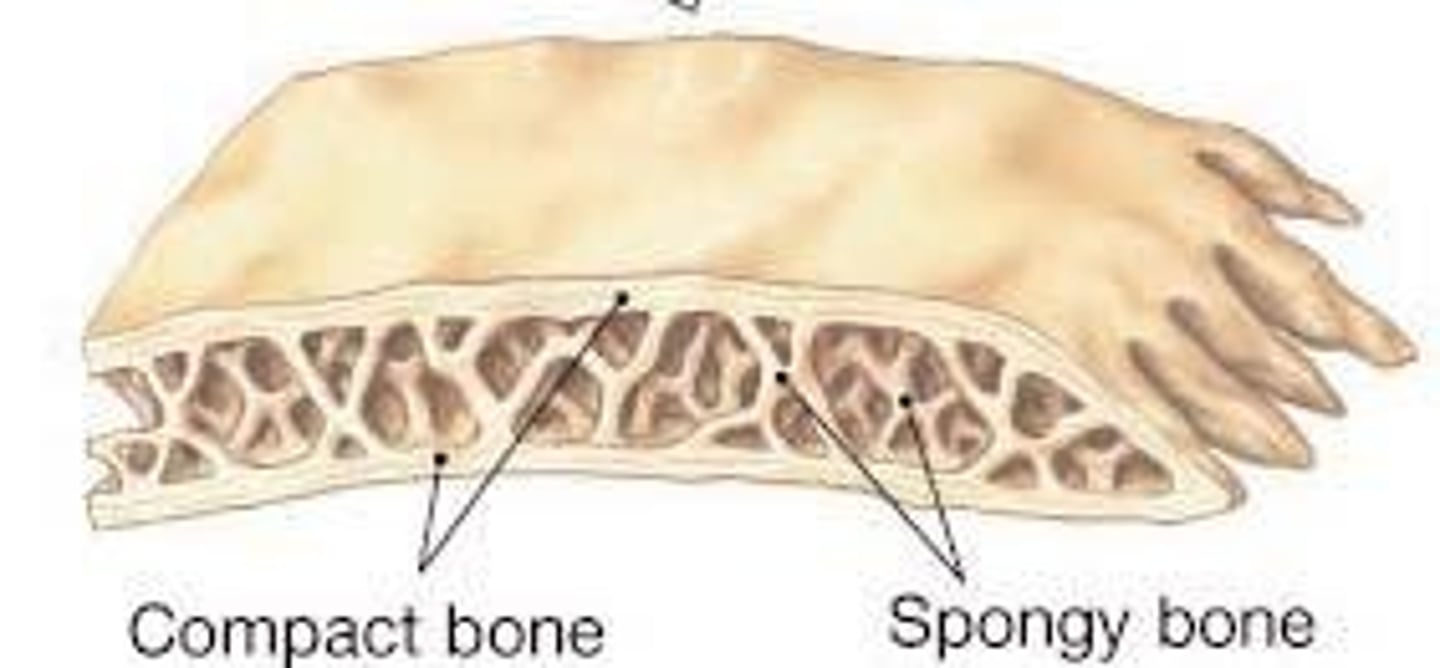
What regions are long bones divided into?
epiphysis
epiphysial plate
metaphysis
diaphysis
epiphysis
the ends of long bones, which develop from secondary ossification centers
epiphysial plate
the site of growth in length; it contains cartilage in actively growing bones
metaphysis
the site where the bone's shaft joins the epiphysis and epiphysial plate
diaphysis
the shaft of a long bone, which represents the primary ossification center and the site where growth in width occurs
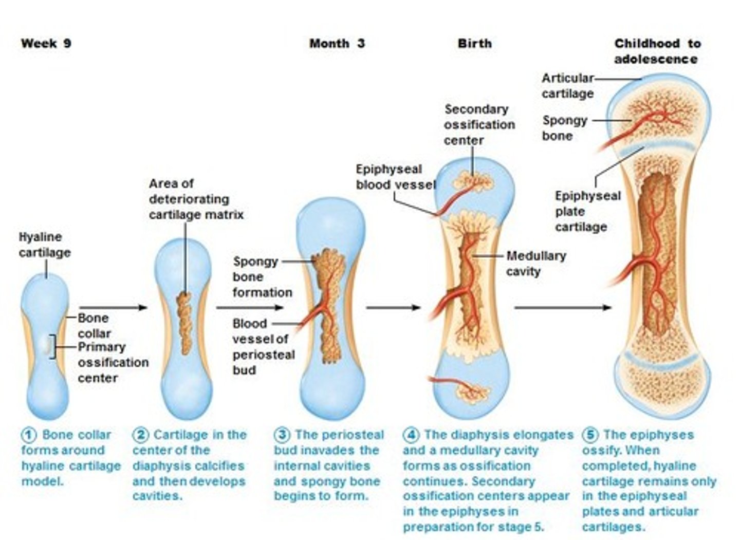
How does a long bone grow and develop?
process called endochondral ossification
1. fetal hyaline cartilage model develops
2. cartilage calcifies and periosteal bone collars form at diaphysis
3. primary ossifcation centers form at diaphysis
4. secondary ossification centers form at epiphysis
5. bone replaces cartilage, except the aricular cartilage and plates
6. ephiphyseal plates ossify = at puberty
How to we identify age from x rays?
The presence of epiphyseal plates vs. lack of
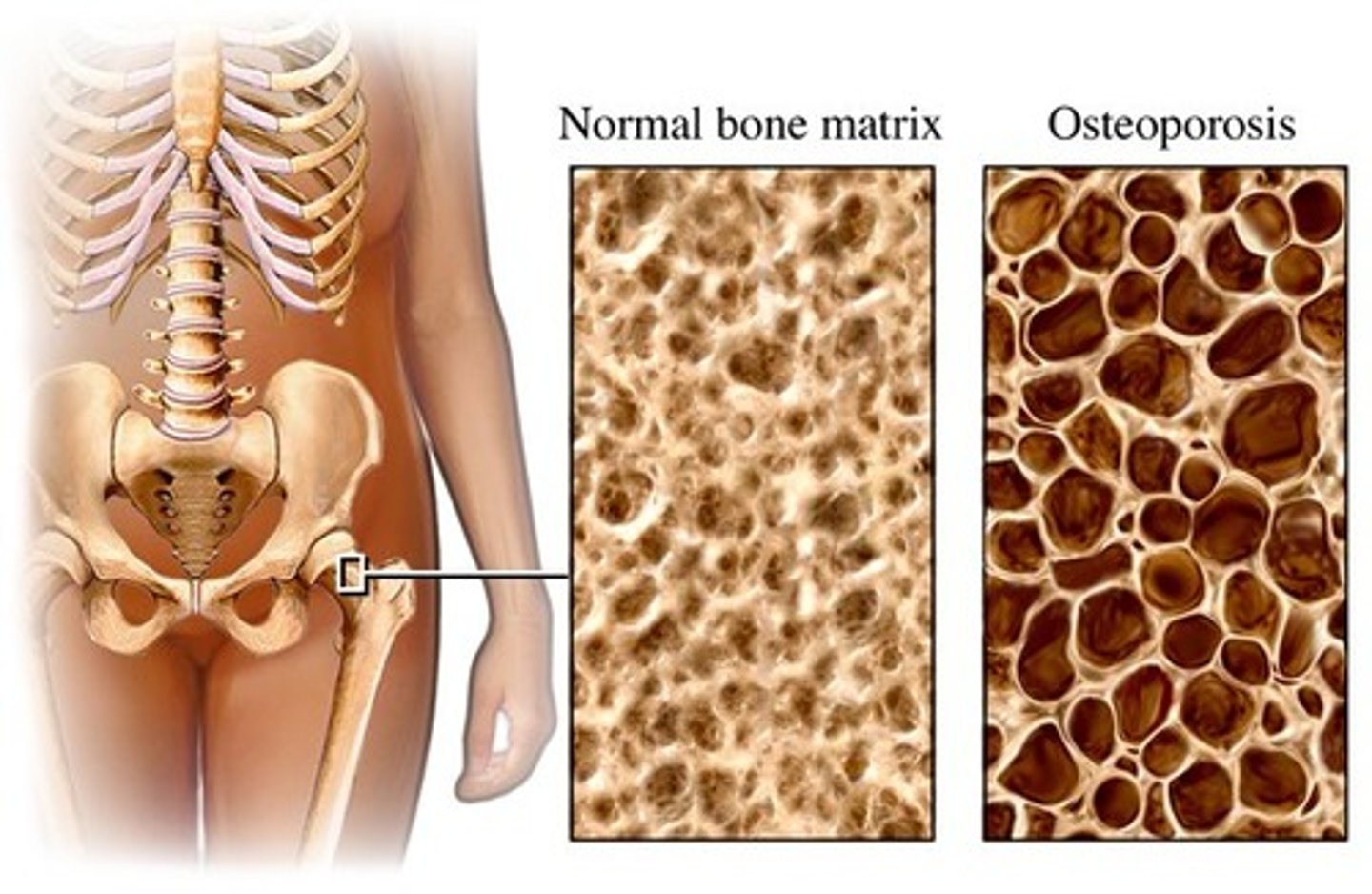
Osteoporosis
During the aging process (60+), the organic and inorganic components of bone decrease = reductio in bone quality and atrophy of skeletal tissue, bones become brittle and easily fracture. Assessed via bone scanning.
Describe the neonatal cranium.
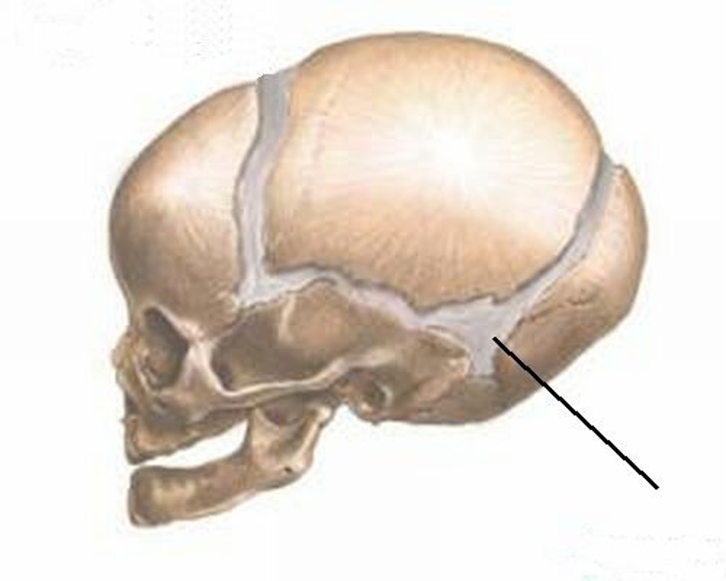
Bones of the skull separated by fibrous joints called sutures (Coronal suture: between the frontal and parietal bones. Sagittal suture: between the two parietal bones. Lambdoid suture: between the parietal bones and occipital bone. Metopic/frontal suture: between the two halves of the frontal bone.) These sutures meet at fontalles (soft membrane covered spaces)
Sutures narrow by 6 months and interlock within 2nd year, ossify by 2nd decade
What are the three types of joints?
Synovial
fibrous
cartilaginous
synovial joints definition and types
united by a joint (articular) capsule (composed out of an outer fibrous layer lined by a serous synovial membrane) spanning and enclosing the joint
plane, hinge, saddle, condyloid, ball and socket, pivot
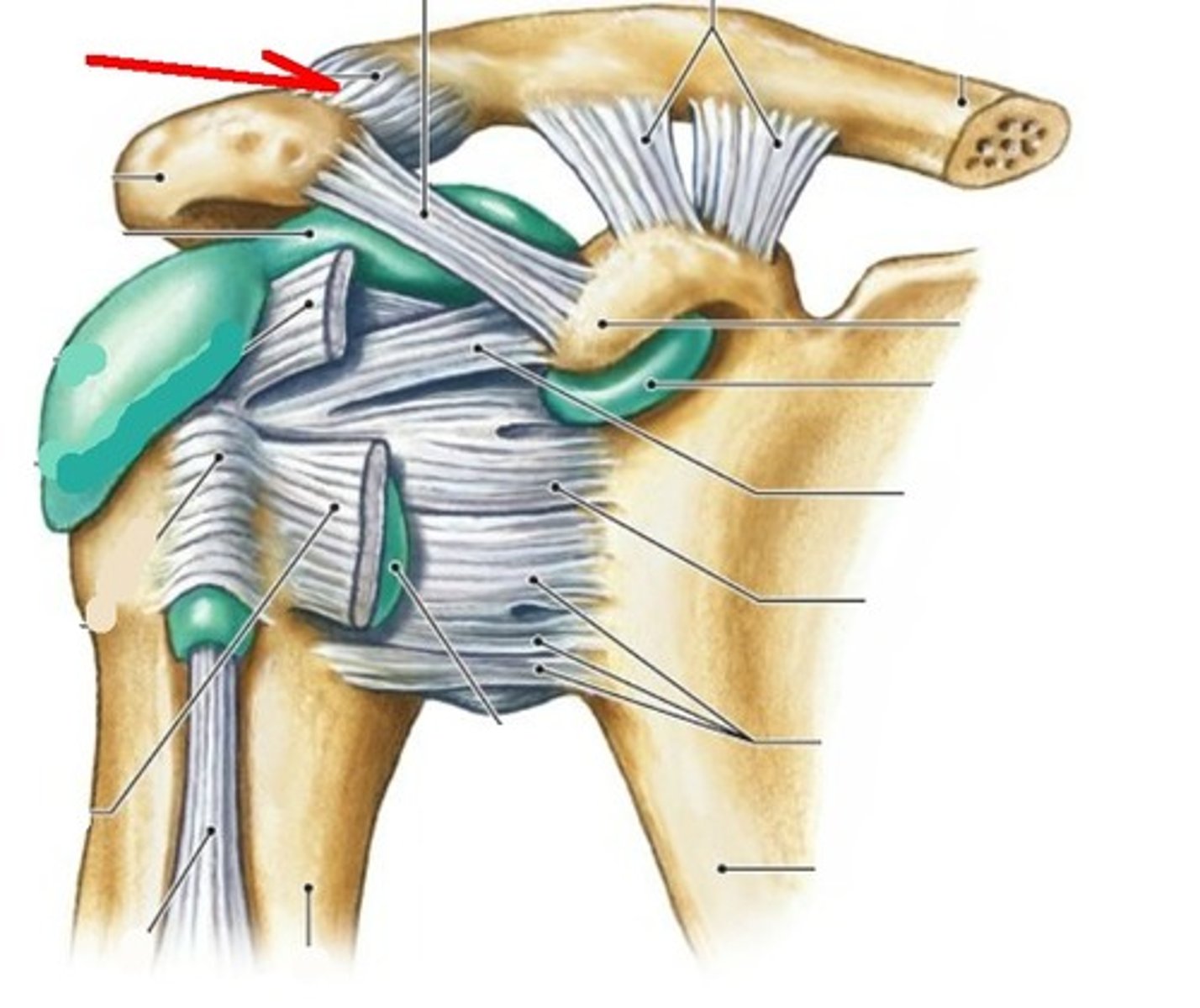
synovial: plane joints
joints that only allow simple gliding movements
ex. acromioclavicular joint
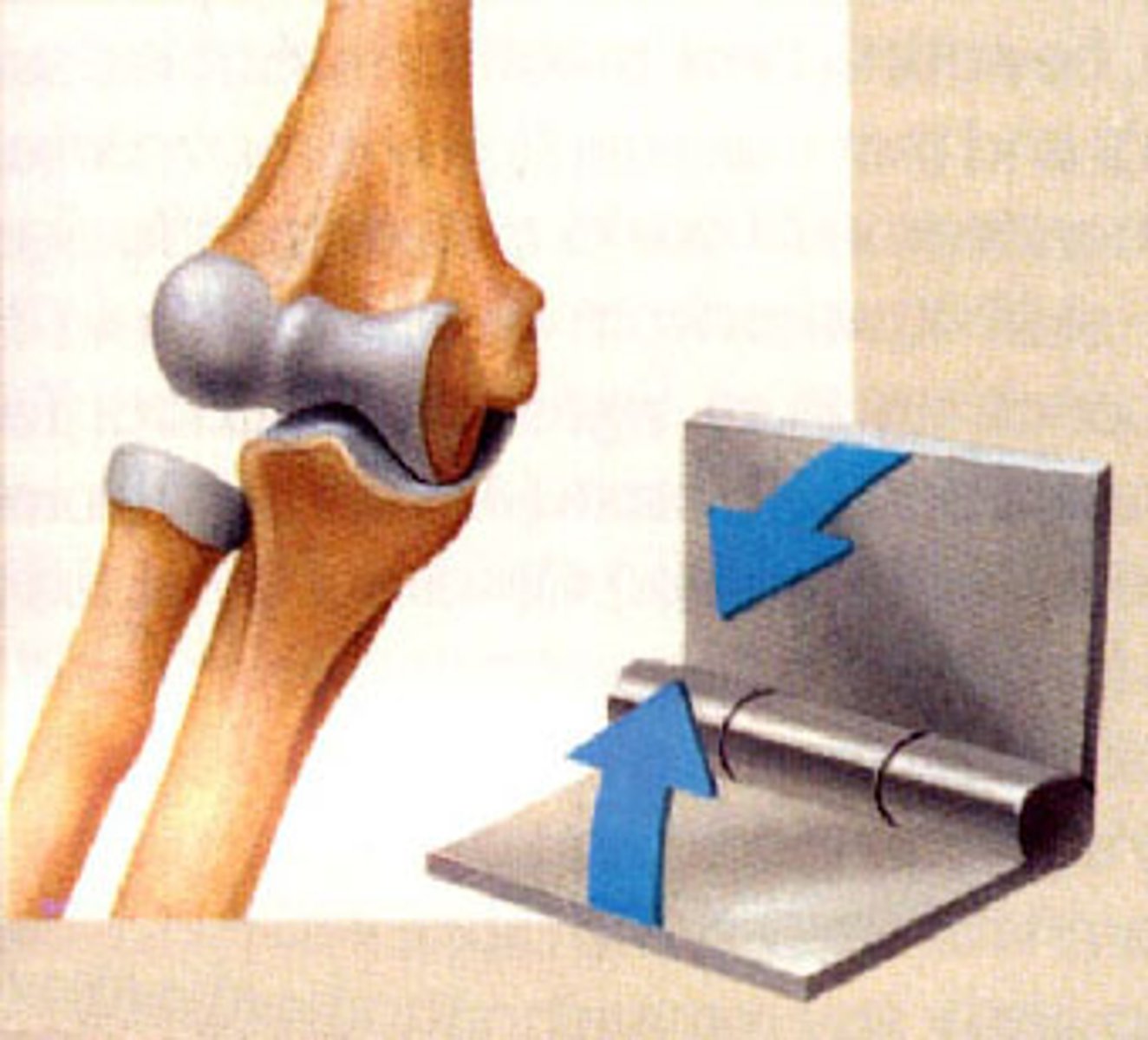
synovial: hinge
uniaxial joints for flexion and extension
ex. elbow joint
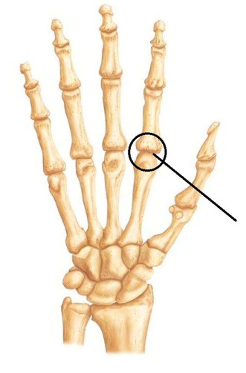
synovial: saddle
biaxial joints for flextion, extension, abduction, adduction, and cicumduction
ex. metacarpophalangeal joint
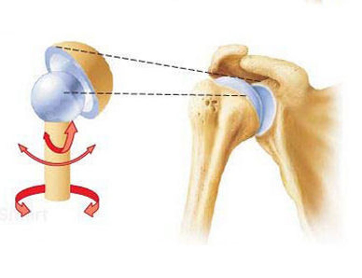
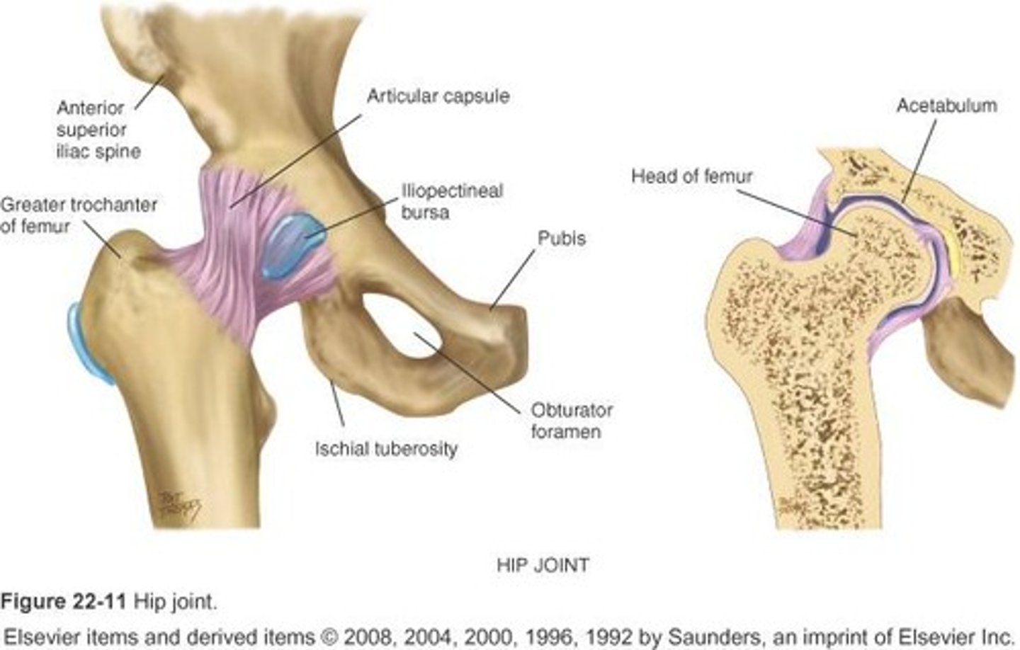
synovial: ball and socket
multiaxial joints for flextion, extension, abduction, adduction, mediolateral roation, and circumduction
ex. hip joint
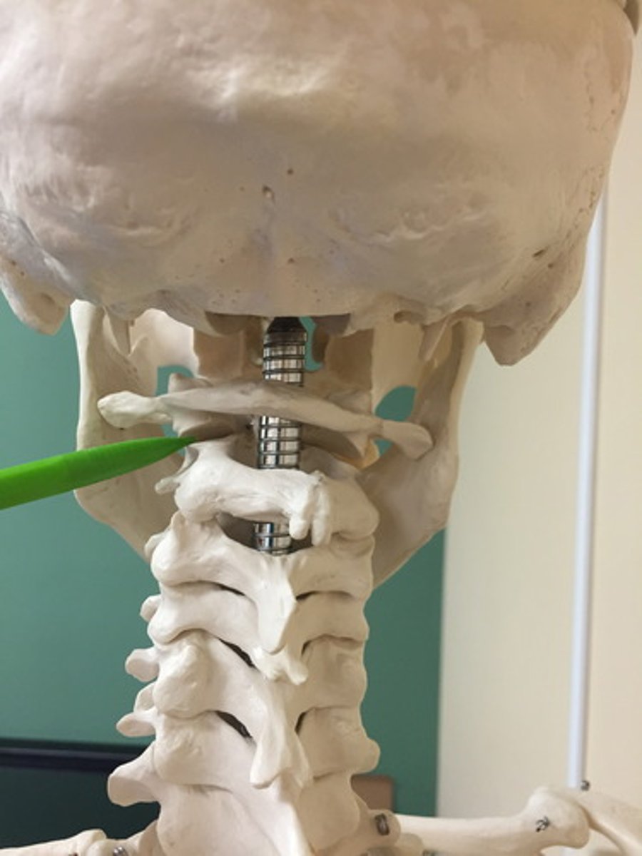
synovial: pivot
are uniaxial joints for rotation
ex. atlanto axial joint
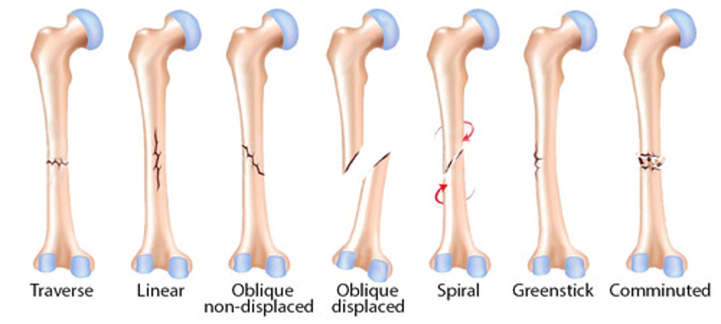
List the types of fracturs.
comminuted
spiral
transverse
greenstick
compound
oblique
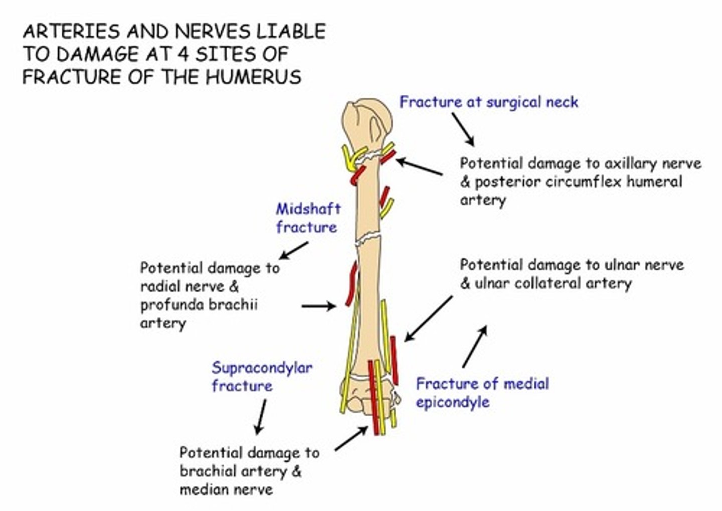
Name the complications of bone fractures seen in this image
damage to nerves
hemorrhage
excess pain
Degenerative joint disease
catch-all term for osteoarthritis, degenerative arthritis, or hypertrophic arthritis
characterized by progressive loss of articular cartilage and failure of repair
as articular cartilage is lost, joint space is narrowed, exposed bony surface = significant pain
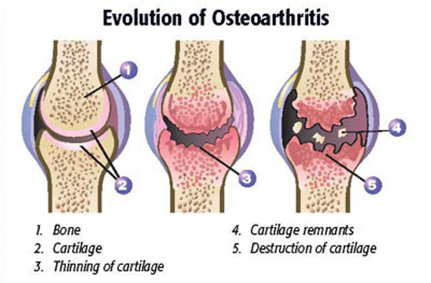
osteoarthritis
Affects any synovial joint but most often the foot, knee, hip, spine, and hand.
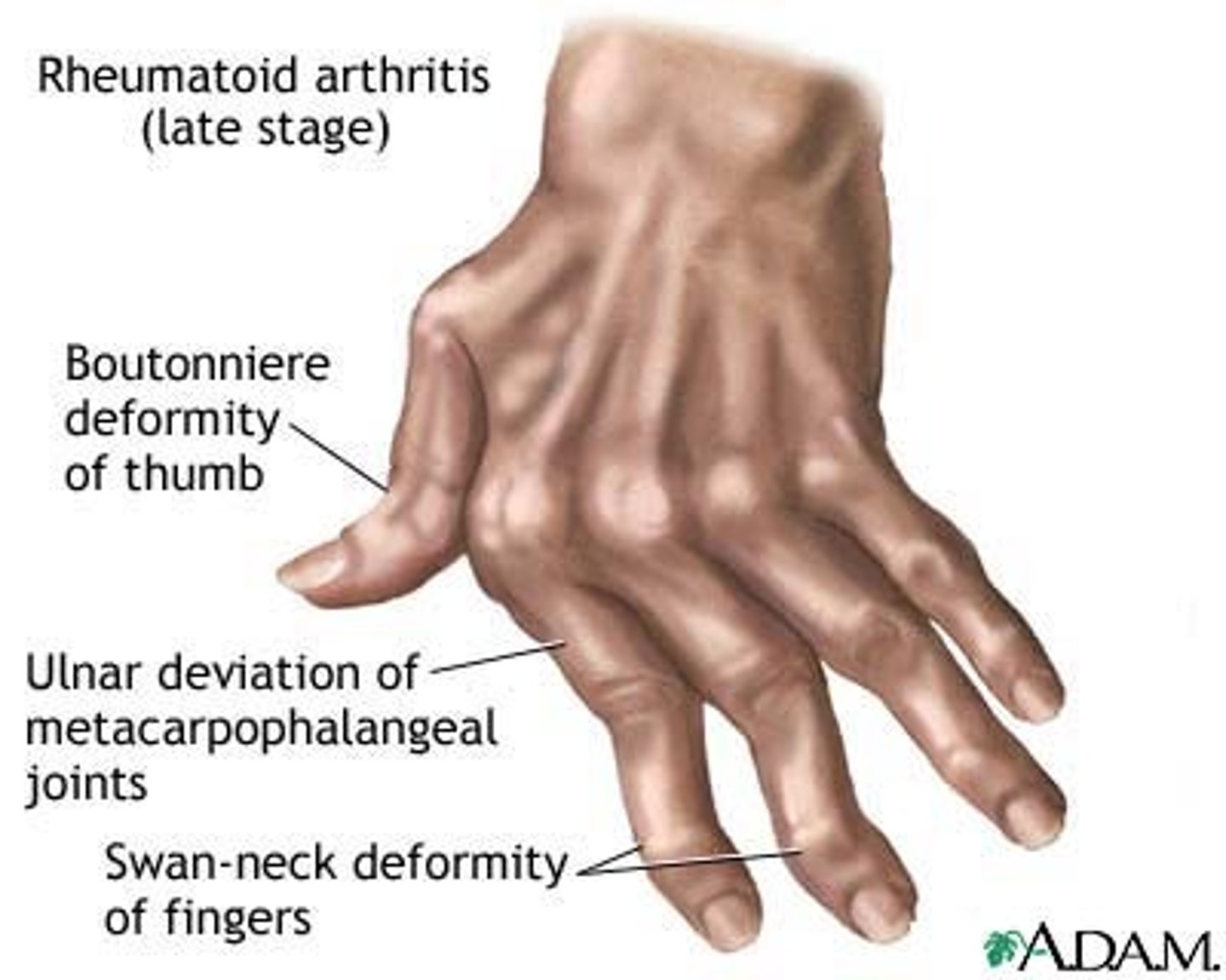
Rheumatoid arthritis
chronic autoimmune disorder that attacks the joints, producing a nonsuppurative, proliferative, and inflammatory snovitis
What are the three muscle types?
skelteal striated
cardiac striated
smooth
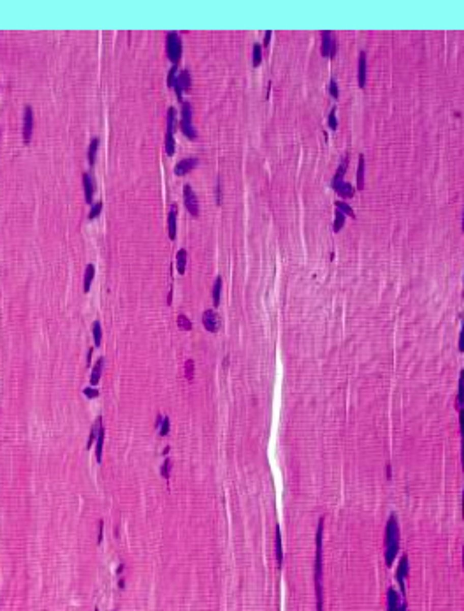
skeletal striated muscle
voluntary somatic muscle that makes up the gross skeletal muscles, moves or stabilizes bones and other structures
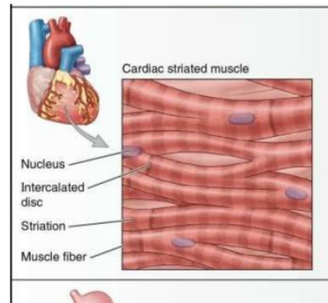
cardiac striated muscle
involuntary visceral muscle that forms most of the walls of the heart and adjacent parts of the great vessles like the aorta
smooth muscle
unstriated muscle is involuntary visceral muscle that forms part of the walls of most vessels and hollow organs, moving substances through them by sequential contractions
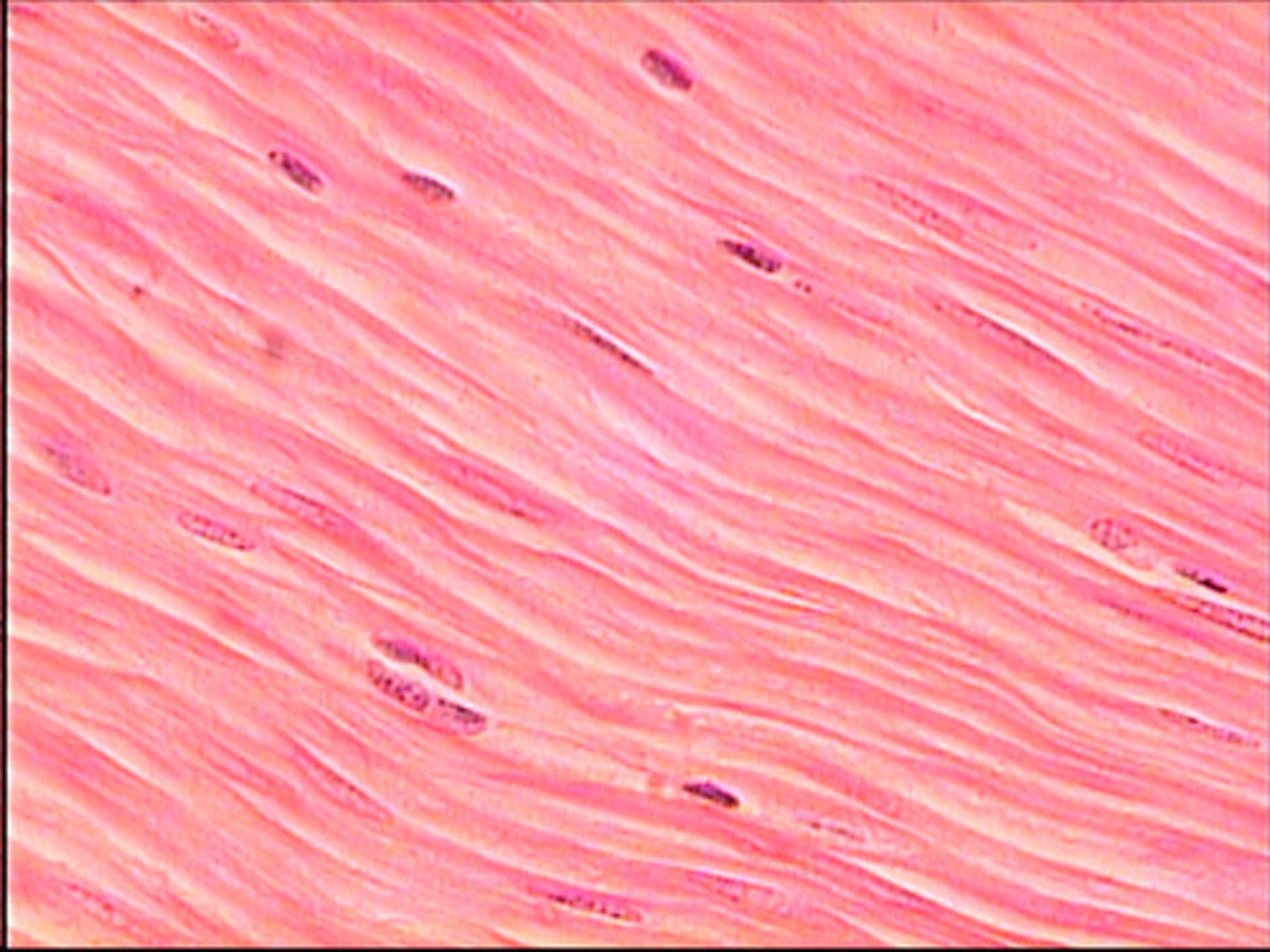
What are the rules for nomenclature of skeletal muscles?
most muscles are named on the basis of their function or the bones to which they are attached
others may be by shape, position, length, size, or number of heads or bellies
aponeuroses
Fibrous or membranous sheet connecting a muscle and the part it moves
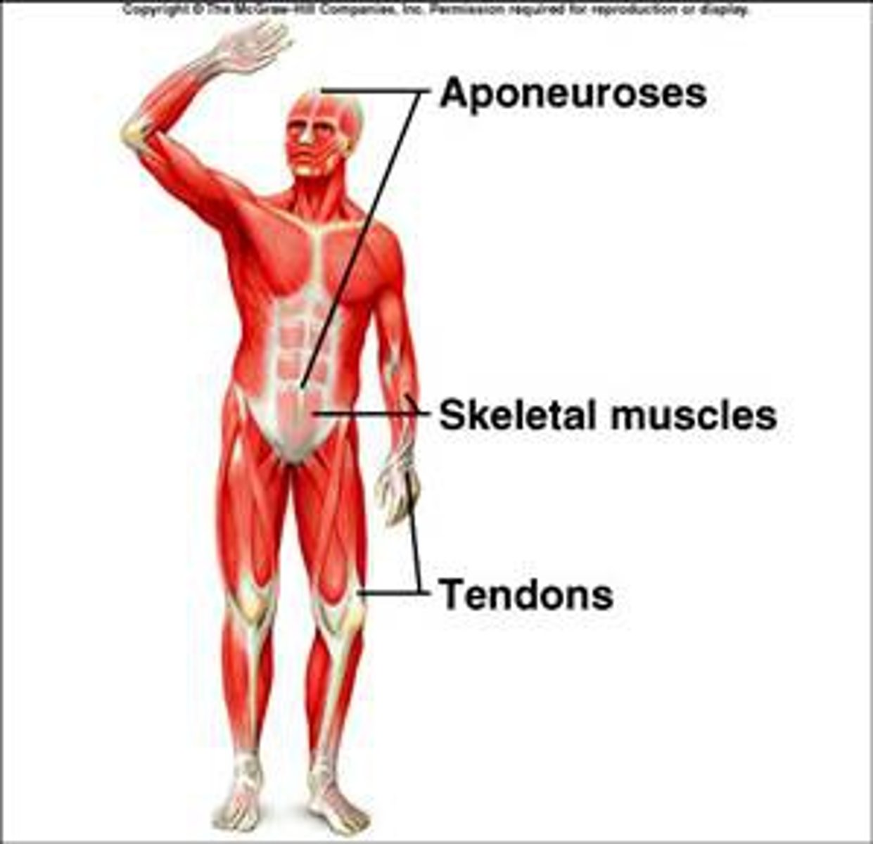
How do muscle contractions work?
**muscles pull and never push
shortens, one of attachments will be fixed while other is pulled toward it
origin is proximal and fixed, insertion is distal and moves (most of the time)
If attachment of a muscle are known, the action can me deduced.
What is the structural unit of a muscle? What is it composed of?
muscle fiber
fibers are covered by endomysium, bundled by perimysium, and the entire muscle is surrounded by epimysium
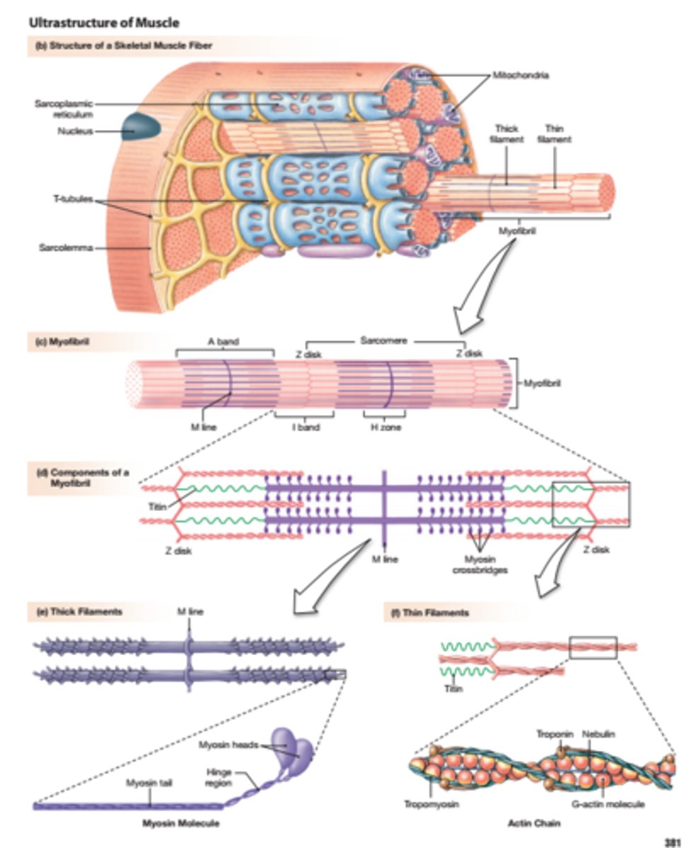
What is the functional unit of a muscle? How does it work?
motor neuron and muscle fibers = motor unit
When the motor neuron in spinal cord is stimulated, impulses case muscle fibers supplied by that neuron to contract.
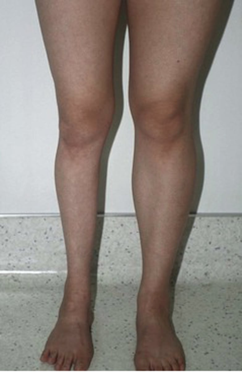
Muscle atrophy
wasting of muscles
may result from primary disorder of the muscle, from a lesion on the nerve that supplies it, or limb immbolization
cardiac muscle- what it is and how it works
striated muscle found in walls of the heart (myocardium)
Contraction of cardiac muscles activated by Purkinje fibers that form a pacemaker which is regulated by the autonomic nervous system.
Hypertrophy of the myocardium
the myocardium responds to increased demands by increasing the size of its fibers
ex. the left side of the heart is bigger than the right due to supplying blood to whole body vs. just lungs
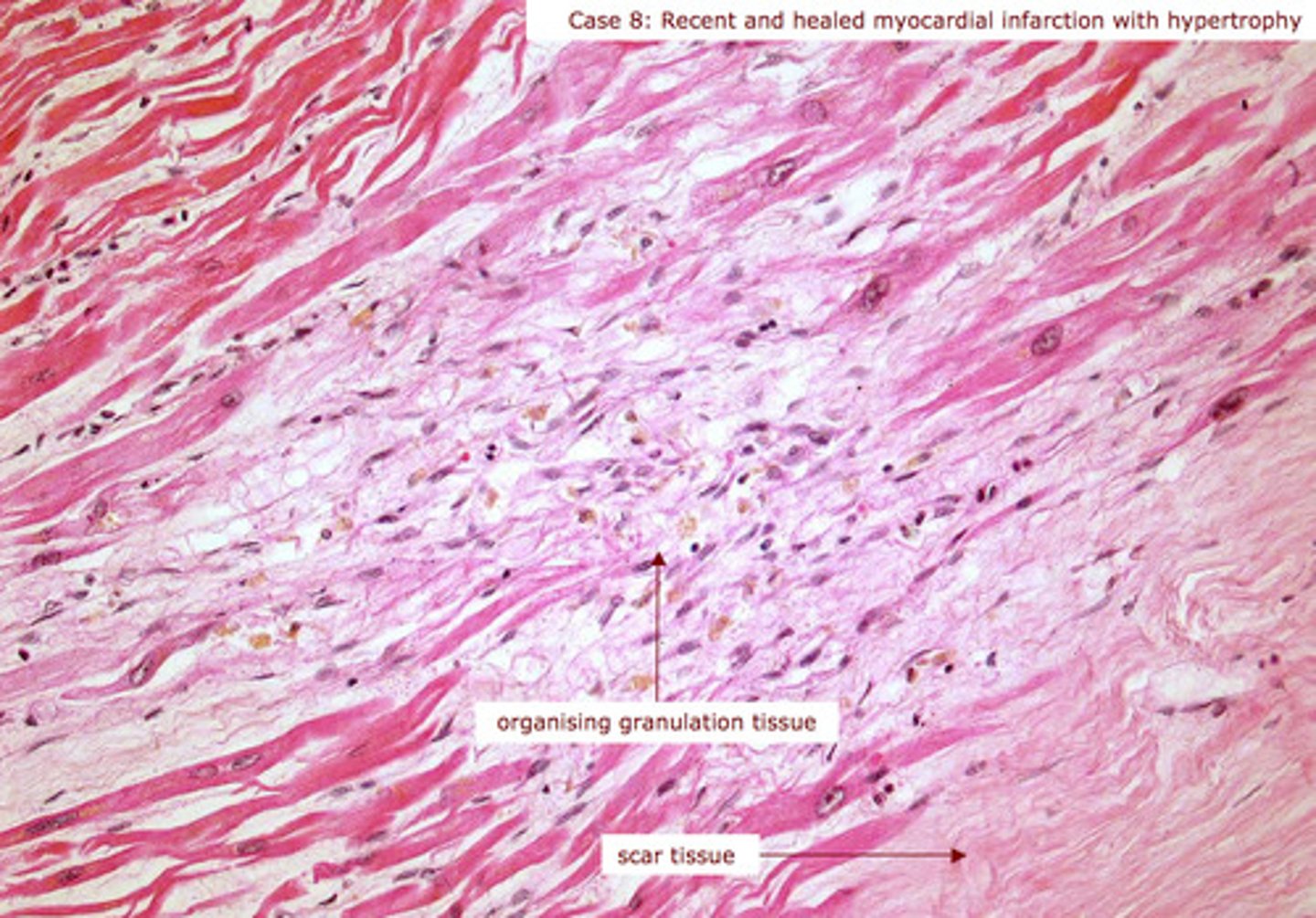
myocardial infraction
when cardiac striated muscle fibers are damaged by loss of blood supply during aheart attack, tissue becomes necrotic and the fibrous scar tissue develops to form an MI (area of myocardial necrosis)
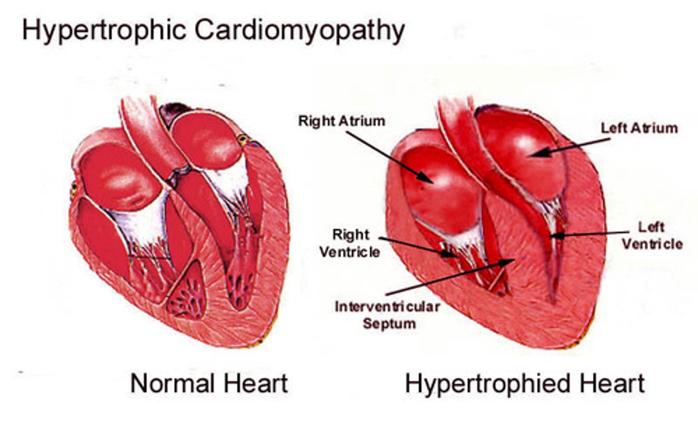
hypertrophic cardiomyopathy
A condition in which the heart muscle becomes abnormally thick and blocks blood flow
inherited genetic disease caused by mutations in the genes that make proteins in the heart muscle (sarcomeres)
smooth muscle: what is it, how does it function, etc.
no striations, occurs in most vascular tissues and walls of alimentary tract/other organs
directly innervated by the ANS and not under voluntary control.
ex. GI, uterus, urinary tract, etc.
hypertrophy and hyperplasia of smooth muscle
compensatory hypertrophy to meet increased demands
ex. smooth muscle cells in uterine wall during pregnancy increase in size and number (hyperplasia)
new smooth muscle cells may develop form pericytes along small blood vessels