7a. Urinary System Disorders
1/6
There's no tags or description
Looks like no tags are added yet.
Name | Mastery | Learn | Test | Matching | Spaced | Call with Kai |
|---|
No analytics yet
Send a link to your students to track their progress
7 Terms
Cystitis
affects the ability to urinate - infection or inflammation of the bladder
Causes -
idiopathic - unknown cause
trauma - secondary to an injury
neoplasia
stress - new people/animal, fireworks..)
Clinical Signs -
Pollakiuria - small amounts of urine
haematuria
incontinence
Dysuria / Straining
Urine Scolding
Treatment -
Assist VS with diagnostics
medication under VS direction
monitor vital signs
close observation
urinary catheterisation
assist with bladder lavage
monitor urine output
monitor behaviour
Urolithiasis
bladder stones
most common in urinary bladder - may drop into the urethra
Causes -
UTI
high dietary intake of certain minerals
disease of genetic predisposition
Clinical Signs -
Dysuria
Hematuria
Pollakuria
scalding or perineum
extended penis
distended bladder
Diagnostics -
physical examination
ultrasonography
urethral endoscopy
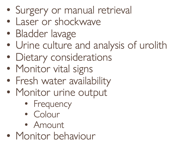
Treatment -
•Surgery or manual retrieval
•Laser or shockwave - pulse will break up stones to make them easier to pass
•Bladder lavage
•Urine culture and analysis of urolith
•Dietary considerations
•Monitor vital signs
•Fresh water availability
•Monitor urine output
•Frequency
•Colour
•Amount
•Monitor behaviour
Feline Lower Urinary Tract Disease
May be obstructive or non-obstructive
Commonly seen in:
•Overweight cats
•Young – middle-aged males
•Indoor multi-cat households where the diet is dry food
•Stressed cats
•Clinical signs
•See cystitis
Treatment – obstructive
•Urgent
•Blood tests
•Cystocentesis
•IV fluid therapy
•GA & blockage removal
•Bladder flush & catheterisation
•Medication
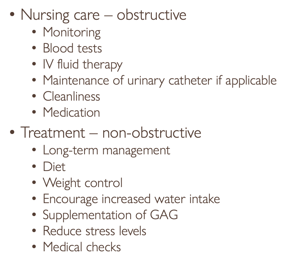
Nursing care – obstructive
•Monitoring
•Blood tests
•IV fluid therapy
•Maintenance of urinary catheter if applicable
•Cleanliness
•Medication
Treatment – non-obstructive
•Long-term management
•Diet
•Weight control
•Encourage increased water intake
•Supplementation of GAG
•Reduce stress levels
•Medical checks
Acute Renal Failure
May occur as a consequence to:
•Decreased blood flow to the kidneys (e.g hypovolaemicshock)
•Direct effect on the cells of the kidneys (e.g. toxins (antifreeze), infectious causes (leptospirosis))
•Post-renal obstruction (e.g. urethral stone)
•Chronic renal failure
Diagnostics
•Blood tests
•Urinalysis
•Radiography
•Ultrasonography
Treatment
•IV fluids
•Drug therapy
•Antiemetics
•Peritoneal dialysis
Nursing care
•Barrier nursing
•Fluid therapy
•Monitoring
•Hydration
•Bodyweight
•Vomiting
•Medication
•Diet
•Grooming and cleanliness
Chronic Renal Failure
•Very gradual onset of clinical signs as loss of renal function gets progressively worse
•Results in azotaemia (uremia)
•Accumulation in the blood of nitrogenous waste products (urea) that are usually excreted in the urine
Clinical signs -
•Symptoms when >75% of renal function has been lost
Causes include:
•Acute renal failure
•Congenital/hereditary disease (e.g. polycystic kidney disease)
•Glomerulonephritis
•Ischaemic damage
•Hypercalcaemia
•Idiopathic
Diagnostics -
•Biochemistry
•Haematology
•Urinalysis
•Radiography
•Blood pressure monitoring
Treatment -
•Treat underlying cause
•IV therapy & electrolyte supplementation
•Antiemetics
•Dietary management
•Vitamin B supplementation & Erythropoietin by injection
Nursing Care -
•Monitoring
•IV therapy and medication
•Ad lib fresh water and dietary adjustments
•Taking patient out regularly
Nephritis
inflammation of the kidneys
severe kidney infection
high body temperature
symptoms sometimes unnoticeable
loose the ability to filter toxins
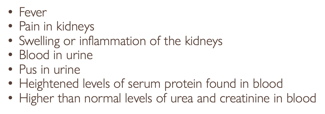
Clinical Signs -
•Fever
•Pain in kidneys
•Swelling or inflammation of the kidneys
•Blood in urine
•Pus in urine
•Heightened levels of serum protein found in blood
•Higher than normal levels of urea and creatinine in blood
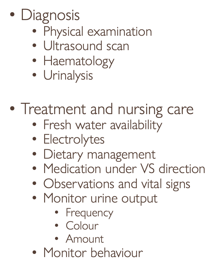
Diagnosis -
•Physical examination
•Ultrasound scan
•Haematology
•Urinalysis
Treatment and nursing care -
•Fresh water availability
•Electrolytes
•Dietary management
•Medication under VS direction
•Observations and vital signs
•Monitor urine output
•Frequency
•Colour
•Amount
•Monitor behaviour
Ruptured Bladder
Possible Causes -
trauma of the bladder wall
tear in the urachus
urinary obstructions
tear in dorsal part of bladder
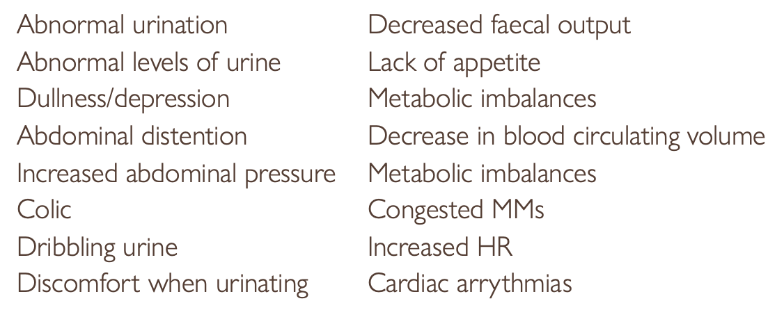
Clinical Signs -
Abnormal urination
Decreased faecal output
Abnormal levels of urine
Lack of appetite
Increased HR
Discomfort when urinating
Cardiac arrythmias
Diagnosis -
Physical examination
history of symptoms
haematology
ultrasound
cystoscopy
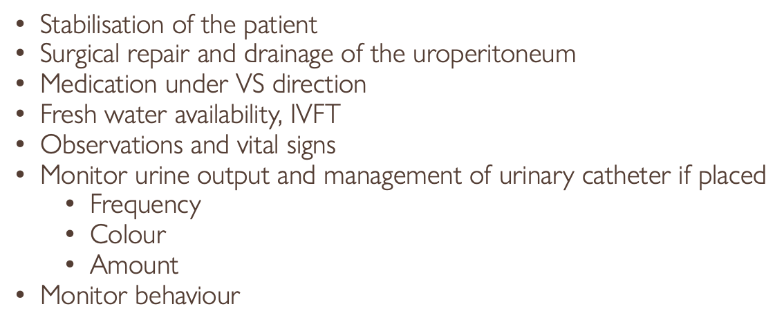
Treatment -
•Stabilisation of the patient
•Surgical repair and drainage of the uroperitoneum
•Medication under VS direction
•Fresh water availability, IVFT
•Observations and vital signs
•Monitor urine output and management of urinary catheter if placed
•Frequency
•Colour
•Amount
•Monitor behaviour