PARASITOLOGY - PROTOZOA
 Call Kai
 Learn
Learn Practice Test
 Spaced Repetition
 Match
Match Flashcards
 Knowt Play
1/84

There's no tags or description
Looks like no tags are added yet.
Name | Mastery | Learn | Test | Matching | Spaced | Call with Kai | Chat |
|---|
No analytics yet
Send a link to your students to track their progress
85 Terms
What’s Amoebiasis
All clinical conditions produced by Entamoeba histolytica
What differentiates Entamoeba histolytica from other non-virulent Entamoeba species?
Entamoeba histolytica is hematophagous
E. histolytica in the population
90% Asymptomatic carriage or subclinical presentation (No dysentery, however if subclinical infection, they can suddenly develop extraintestinal amoebiasis
10% Invasive
Describe Entamoeba histolytica’s Trophozoite and cyst forms
Trophozoite: Amoeboid shape, motile by Pseudopodia, and hematophagous
Cyst: Spherical, non-motile, and infective.
→ It has 1-4 nuclei
→ Chromatoid bodies: Semi-crystalline arrays of ribosomes - reserve for excystation)
→ Central karyosome: Inactive chromatin, each associated with its corresponding nucleus
→ Glycogen masses
Top 3 deadliest protozoa worldwide
Plasmodium spp (malaria)
Leishmania spp
Entamoeba histolytica
What is requireme
What makes the diagnosis of amebiasis difficult
Overlap with IBD
Acute amoebic dysentery clinical presentation
Frequent mucoid stools
Rectal blood release without diarrhea (Especially children)
Dysentery and Constipation alteration
Abdominal pain with tenderness
Tenesmus
Possibly: Fulminant + Necrotizing colitis
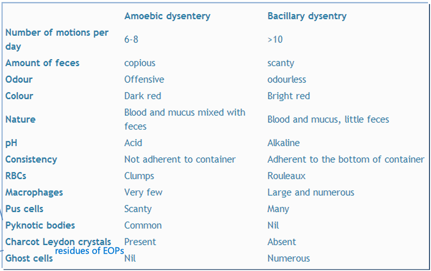
What are some features that help differentiate Amoebic vs Bacillary dysentery
In Amoebic:
Less motions/day
Lots of feces vs few
Darker color
Blood + Mucus + feces (Little feces in bacillary)
Acid pH (Alkaline bacillary)
Stools do not adhere to container (Do in bacillary)
Clumped RBCs in amoebic
Pyknotic bodies PRESENT
Charcot Leydon crystals PRESENT (Residues of EOPs)
ABSENT Ghost cells
Laboratory diagnosis of Intestinal Amoebiasis
Stools + Saline: Motile trophozoites with ingested RBCs
Stools + Iodine: Should stain Yellow: Nucleus + central karyosome + brown glycogen mass
Biopsy + H&E
Stools + Trichrome stain
How can we determine E. histolytica asymptomatic carriage
If cysts can be found in feces in least 3 stool specimens
Non-histological lab diagnosis of E. histolytica and their main purpose
Sigmoidoscopy + biopsy
Serology and PCR: To differentiate between E. histolytica and E. dispar (In saymptomatic carriage screening cuz they look identical but the latter is completely non-pathogeneic)
E. histolytica tissue bipsy apearance
Flask-shaped ulcer appearance
On screening, ulcerations look like buttons
Amoeboid dysentry sequalae
Ulcerative post-dysenteric colitis:
Edema of the mucosa + Other irregularities
Spasms
Loss of haustration → Smooth colon appearance of X-Ray (Important sign of ulcerative colitis in general)
Amoebiasis intestinal complications
Perforation (Rare)
Peritonitis (40% Mortality)
Amoeboma
Blood invasion and extraintestinal infections
Describe Amoeboma
Due to chronic infection by E. histolytica
→ Tumor-like inflammatory msses in the colon (neoplasms) that can cause intussusception and obstruction
Hepatic Amebiasis Pathogenesis
Trophozoites reach liver via the hepatic portal vein and multiply locally
They cause inflammation and endothelial damage which can cause vessel obstruction
Endothelial damage can lead to thrombosis
Liver cell necrosis due to the pathogen itself and worsened by endothelial damage and iscchemia
Hepatic amebiasis diagnosis
Imagining
Blood test: WBC/ESR/CRP/SGPT/Alkaline phosphatase
Liver Aspirate microscopy
Anchovy sauce-like pus: Mix of sloughed liver tissue and blood, and contains NO TRPHOZOITES
Which regions of a liver abcess have the most trophozoites?
Peripheral zones
Complications of hepatic amoebiasis
Rupture of abscess into:
→ Right pleural cavity
→ Right lung
→ Peritoneal cavity
→ Below diaphragm
(Can be fatal)
Metastasis to lungs, skin, spleen, brain
Luminal Amebisis treatment
Paromomycin (Humatin)
Diodoquin
Furamide
Cyst passers
Invasive amebiasis treatment
Metronidazole (Flagyl) or Tinidazole + A lumen-acting drug
*Note: Metronidazole is actually activated by one of E. histolytica’s fermentative enzymesEmetine or DehydroEmetine for severe disease (Fulminant & Necrotizing colitis)
Amebiasis prevention
Hygene, safe water drinking, avoid raw vegetables and fruits
Note: If kept moist, cysts can survive even if refregirated for up to a month
Note: Cysts are fairly resistant to drinking water chlorination
Giardia lambia habitat in body
Duodenum and upper jejunum
Giardia lambia Trophozoite morphology
Tennis-racket-shape, dorsally convex, and ventrally concave with sucking disc.
2 Oval nuclei with karyosome
2 axostyle traversing the body longitudinally
2 coma-shaped parabasal that lie perpendicular to the axostyle
8 flagella (4 pairs), that grant motility
Giardia cyst morphology
Oval, the axostyles are diagonal, parabasal bodies still pependicular, 4 nuclei, flagellar reminants in the cysts (Retrated flagella)
G. lambia pathogenesis
The cysts are ingested
The cysts cross the stomach easily because they are acid-resistant
Once in the small intestine, the alkaline-sensitive cysts excyst and release alkaline-tolerant trophozoites
The G. lambia trophozoites bind to the mucosa with their sucking discs
Overtime, this causes abnormal villous architecture
Mucoid diarrhea and Stearrhoea due to fat absorption impairment
Encystation in Large intestine
Name and explain the symptoms caused by Giardiasis
Duodenitis: Caused by mechanical irritation by the pathogen
Mild or Severe Diarrhea: The pathogen’s attachment and exotoxin production triggers cytokine production and inflammation which increases gut permeability, mobility, and electrolyte release (Increased water content)
Mucoid Diarrhea: Inflammation and cytokine release triggers hypersecretion of goblet cells
Villous Atrophy and Blunting of Brush border: Related to immunodeficency and a decreased IgA release
# Fat malabsorption → Steatorrhea
# Less surface area to release saccharidases → More undigested carbs → Microbiota selection for fermentative bacteria → Gas production and flatulence
# B9 and ADEK Vitamin deficencies
What kinds of immunodefiencies increase susceptibility to giardia
Hypogammaglobulinemia
Low sIgA
Low gastric acidity or achlorohydria
Giardiasis symptoms in immunodefiency
Persistent diarrhea
Steatorrhea
Malabsorption and hypoproteinemia
B9, B12, A, D, E, K deficencies
Cholangitis (Bile duct inflammation and Cholcystis (Gallbladder inflammation )
Laboratory diagnosis of Giardiasis
Symptomatic: Diarrhea + Mucus + Cysts in stools
Asymptomatic: Cysts in stools (Might need to sccreen for 4 days in a row)
*Note: Iodine stainPositive string test
Serology
PCR
Giardiasis treatment
Metronidazole or Tinidazole and recently Albendazole
Clinical significance of Coccidians
Causes diarrhea almost exclusively in AIDS patients
Which organisms causes Coccidiasis
Coccidians from the phylum Apicomplexa:
Cryptosporidium pavum (Zoonosis)
Cryptosporidium hominis (Human-Human)
Cyclospora cayetanesis
Isospora belli
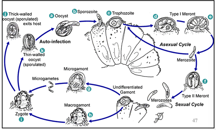
Cryptosporidium pavum pathogenesis
Ingestion of Oosysts (Infective form)
Oocyst releases sporozoite
Sporozoite attaches to enterocytes and becomes trophozoite
Trophozoite matures into Type I Meront
Type I Meront hatches and releases type I merozoites
Type I merozoite either reforms a trophozoite to release more type I merozoite (Asexual), OR, transforms into Type II Meront (Sexual)
Type II Meronts release Type II Merozoites
Type II Merozoites either become Microgamont (Male), or Macrogamont (Female)
The male Microgamont hatches, releasing microgametes that will fertilize the female Macrogamont → Zygote formation
The zygote either becomes a thin-walled oocyst that will progress the infection, OR, a thick-walled oocyst which will exit the host
Cryptosporidium outbreak causes
Close-contact with animals
Oocysts are robust and have low ID, and small
Oocysts resistant to chlorine
Nosocomial infection
Cryptosporidiosis lab diagnosis
Modified Kinyoun’s Acid-Fast stain (For all coccidians)
Iodine stain
Fluerescent antibodies, PCR
Trichomonas vaginalis habitat
Female lower genital tract / male urethra and prostate
Trichomonas infectious form
Tichomonas exist as trophozoites ECLUSIVELY
Trichomoniasis symptoms in males vs females
Males: Asymptomatic usually
Females: Purulent greenish discharge, dysuria & dyspareunia, itching, burining sensation
Trichomoniasis diagnosis
Demonstration of actively-motile trophozoites in wet films
Direct fluorescent antibodies (DFA)
Punctate cervical hemorrhages (Strawbeey cervix)
Trichomoniasis treatment
Metronidazole
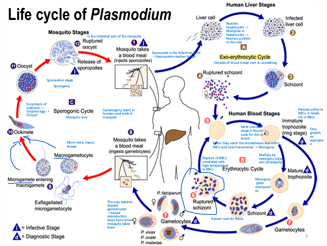
Explain the 3 parts of Plasmodium’s life cycle in details
Plasmodium uses mosquitos as their definitive hosts (Support sexual reproduction), and humans as their intermediate hosts (Support asexual reproduction and maturation)
Exo-erythrocytic Cycle:
Infected mosquito takes a blood meal and injects its plasmodium-infused saliva
The form injected into humans is the sporozoite form, it travels hematogenously to the liver, where it infects hepatocytes (Using Kupffer cells as an entry point)
The protozoan replicates inside the infected cells, eventually the parasite load will push the hepatocyte nucleus against the membrane, this cell form is known as a Schizont (Tissue Schizont)
Alternatively, the sporozoite could just stay dormant and would thus be known as a Hypnozoite
The schizont eventually ruptures, releasing Plasmodium Merozoites with RBC tropism
Erythrocytic Cycle:
Plasmodium merozoites infect RBCs in the circulation
Inside RBCs merozoites mature into Immature Trophozoites, which look like a wedding ring inside an RBC - Most commonly found stage in the blood
From this stage, we get divergent paths
A: Continuation of the Asexual Erythrocytic cycle through trophozoite maturation and schizont (Blood-stage schizont) formation then schizont rupture to give merozoites again which directly infect RBCs
B: Switch to gametocyte formation of male and female gametocytes in preparation for the Sporogonic Cycle
Sporogonic cycle:
When a mosquito takes a blood meal from an infected human, it uptakes the gametocytes (Other forms of the parasite die), and becomes infected itself
The drop in temperature inside the mosquito’s gut triggers the maturation of male and female gametocytes into micro and macro-gametes respectively
The microgamete fertilizes the macrogamete to give an Ookinete
The ookinete encysts and becomes an Oocyst
Rupture of the oocyst in the salivary glands of the mosquito releases the sporozoites, which can start the Exo-Erythrogenic cycle again
Sporogonic cycle
Exo-erythrocytic and Erythrocytic Cycle
What’s an important cause of Malaria Relapse
Persistence of Plasmodium parasites in the liver during the exoerythrocytic cycle
Name the different Plasmodium species and their different affinities of RBCs
P. falciparum → Infects RBCs of all ages, even reticulocytes in the BM
P. vivax and ovale → Prefer to invade younger RBCs
P. malariae → Affinity for older RBCs
How do malaria relapses occur?
During the initial sporozoite stage, some just stay dormant inside hepatocytes, and become known as hypnozoite. At some point, they can regain virulence.
*Note: Hypnozoites only in Po, Pv & Pm (None in Pf → no relapses in Pf)
*Note: Re-infection is possible due to a high degree of antigenic variation resulting from gene shuffeling (Applies to all Plasmodia but most clinically relavent for Pf)
How can people get non-vector acquired Malaria
It can be acquired:
Through contaminated blood transfusions
Congenitally - transplacentally, but only if there’s a placental defect cuz normal placenta is a good enough barrier
Needle sharing among drug addicts
*Note: In this case the infectious form is Trophozoites → Trophozoite-induced malaria
High-risk individuals for malaria
Pregnant women, babies, young children, elderly.
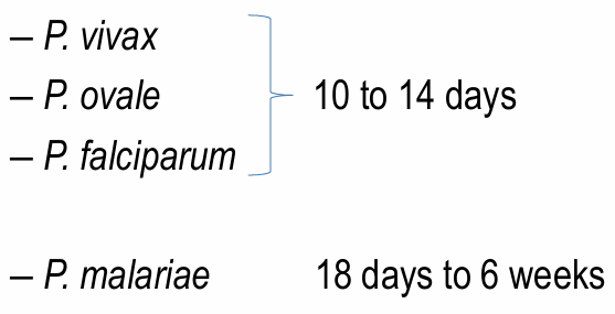
Plasmodium incubation period
Acute malaria symptoms
Malaria causes febrile paroxysms that are associated with the repture of erythrocytic schizonts in the circulation.
3 stages:
Cold stage: Intense cold, but patient is febrile (15-60 minutes)
Hot stage: Intense heat, febrile, headache, body & joint pain, vomitting & diarrhea (2-6 hours)
Perspiration stage: Profuse sweating, fall in temperature, exhaustion & weakness followed by sleep (2-4 hours not including the sleep period)
Other symptoms: Jaundice and Anemia (due to hemolysis, pigments from the parasite contribute to jaundice color), splenomegaly
Distinguish the fever patterns of Plasmodium based on the different species
It depends on what type of RBCs they infect
Pf: Any age, and even reticulocytes → Subtertian/Irregular episodes (Most severe)
Pv/o: Younger RBCs → Episode every other day - Every 48 hours
Pm: Older RBCs → Episode every 72 hours
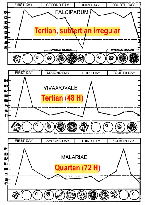
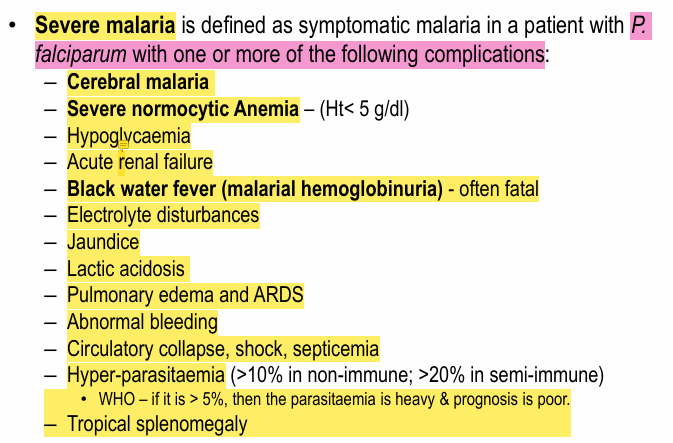
Severe malaria complications
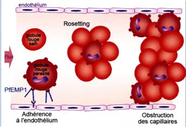
Cerebral Malaria
Complication of Pf infection.
→ Proteins of trophozoite-containing RBCs have receptors (PfEMP) on endothelial cells that result in their adhesion and aggregation of infected and non-infected RBCs (Rosetting) → Obstruction of capillaries
High TNF:
Fever
Confusion, seizures, and coma
Metabolic acidosis and RDS6
Black Water Fever
Pf complication
Due to presence of Hb in urine following a hemolytic attack → Dark red/ brown-black
In addition to fever, rigor, biious vomiting, icterus, circulatory collapse, acute renal failure.
Laboratory diagnosis of Malaria
Identification of Malarial parasites in peripheral blood films using Thick or Thin smears (Thick smears 30-40 times more sensitive)
*Note: RBCs may appear elongated
Giemsa stain (10% solution for 10 min)
Serology: HRP2 (His-rish protein) Ag / pLDH (parasite LDH)
→ Both produced by the parasite during their growth & differentiation in RBCs
Which forms are most reliable for the diagnosis of plasmodium
Immature trophozoites: Rings inside RBCs
Other forms are rarely seen
Malaria prophylaxis
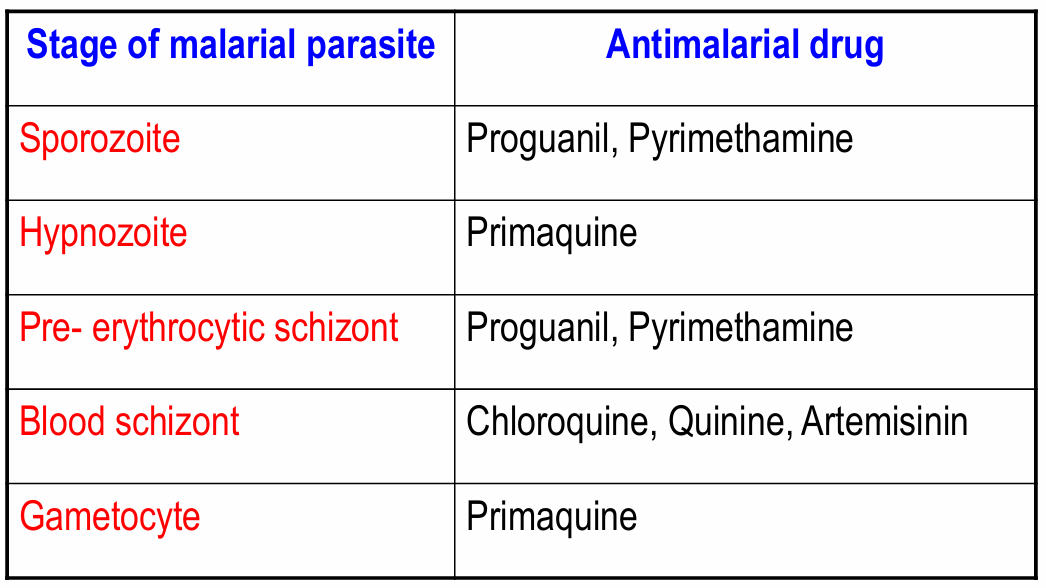
Causual Prophylaxis: Kill parasites in hepatocytes: Primaquine (++++) / Malarone (++)
Suppressive Prophylaxis: Kill asexual parasites in RBCs: Chloroquine/ malarone/ mefloquine/ Doxycyline (all ++++)
Gametocytoicidal Prophylaxis: Kill gameocytes: Primaquine (++++)
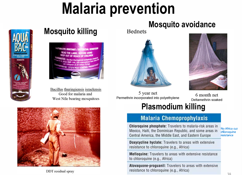
Antimalarial treatment
Babesiosis causative agents
Aka periplasmosis, caused by apicomplexan parasites of the Babesia genus
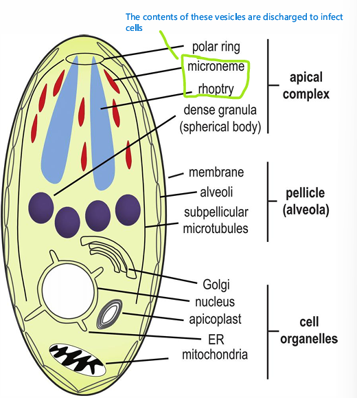
Babesia morphology
Babesia life cycle
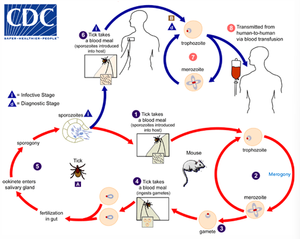
Babesia uses ticks as their definitive hosts and many mammals as their intermediate hosts.
Intemediate host: Tick blood meal causes sporozoite injections → RBC infection to obtain trophozoites (ring inside RBC) → Trophozoite undergoes merogony to give Merozoites
→ Merozoites either reinfect RBCs asexually or differentiate into gametes ready to be uptaken by ticks again
*Note: both babesia sporozoites and merozoites have RBC tropism
Definitive host: Microgamete fertilizes macrogamete → Ookinete enters salivary glands → Sporogony → Sporozoite release upon blood meal
Babiasis incubation period
1-4 Weeks
Babesiosis clinical manifestation
Fever
Chills and sweating
Myalgias
Fatigue
Hepatosplenomegaly
Hemolytic anemia.
When is Babesiosis more severe
Pathogen specie: B. divergens infection more severe than B. microti
Host: Immunosuppressed, splenecctomized, and elderly individuals
Laboratory diagnosis of Babesiosis
Blood smear stained with giemsa → Look for ring form or maltese cross inside RBCs
Serology
PCR
How can you morphologically differentiate between Plasmodium and Babesia-infected RBCs?
Babesia is pleomorphic
Babesia-infected RBCs may be vacuolated (In plasmodium, hepatocytes get vacuolated, but not RBCs)
Babesia does not produce any pigment
Babesiosis Treatment
Azithromycin, Clindamycin, Altovaquone, ELQs (Endochin-like quinones)
Toxoplasma gondii phylum
Phylum: Apicomplexa
Toxoplasma host types
Definitive: Felines
Intermediate: All warm-blooded animals
Toxoplasma localization in felines
It is an obligate IC parasite, and can be found in a cat’s intestinal epithelium
Describe toxoplasma tachyzoites
It is the infectious form of the parasite, and also the form that can travel trans-placentally
Crescent-shaped
Pointed anterior end and rounded exterior end
May be found single:
Free
IC trophozoites
Proliferative forms
Or may be found in masses called pseudocysts
Invasion: They invade cells from their apical side using AMA1 receptors to attach to RON2 host cell receptors to get endocytosed (Clathrin-mediated)
Describe toxoplasma tissue cysts
Cyst-like forms - an adaptation against immune responses
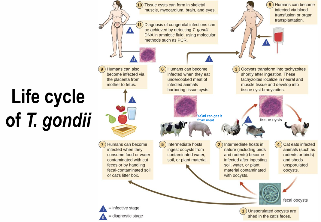
Describe toxoplasma’s life cycle
Cat - Sexual cycle:
Cat ingests tissue cysts in mice
Upon digestion by cat, the cyst wall will dissolve, releasing bradyzoites into the intestines
Bradyzoites invade cat enterocytes and start dividing by schizogony
Many divisions take place and, and eventually, the schizonts rupture to release Merozoites
Merozoites will infect more cells and keep dividing or form micro and macro gametes that may mate inside or outside the enterocytes
Fertilization will lead to the formation oocysts
Immature oocysts are excreted via feces
Maturation takes place in the soil by sporulation
1 Mature Oocyst will contain 2 sporocysts, and each sporocyst will contain 8 sporozoites - This is the infectious mature oocyst
Humans - Asexual:
Ingest Oocyst (cat feces-contaminated food)- > Sporozoite infectious form / Ingest cyst (Raw meat) → Bradyzoite infectious form
Both Sporozoites and Bradyzoites will infect enterocytes and give rise to Tachyzoites (Multiply quickly)
Tachyzoites will move from the intestines to other tissues - Acute phase of toxoplasmosis
Tachyzoites infect Macrophages, and replicate by endodyogeny
After a few divisions - > Rosette shape + Acidocalcisome accumulation
Eventually tachyzoites will be carried to the CNS hematogenously and will invade neurons and transform to bradyzoites intracellularly
Bradyzoites will secrete a cyst wall and form tissue cysts an immune evasion mechanism
Tissue cysts, can stay dormant for as long as the host is alive, and are the hallmark of toxoplasmosis’s chronic phase
Post-natally-acquired toxoplasmosis clinical presentation
Asymptomatic, subclinical, or mononucleosis-like (but unlike mono, heterophile antibody test will be negative)
Note: this is the acute form of the disease
Toxoplasmosis presentation in IC hosts
Systemic form
Almost always resulting from reactivation of latent infections in AIDS
Symptoms include Myocarditis, pneumonitis, chorioretinitis, and eventually brain abscesses and fatal encephalitis
*Note: Ring-enhancing lesions show up on MRI
Congenital Toxoplasmosis
Occurs in 1 in 1000 pregnancies
May cause damage or death of the fetus
Symptom Triad: Chorioretinitis, hydrocephalus, intracranial calcifications
Neonatal toxoplasmosis
The risk of vertical transmission to the fetus increases with the age of pregnancy, however the severity of the lesions decreases
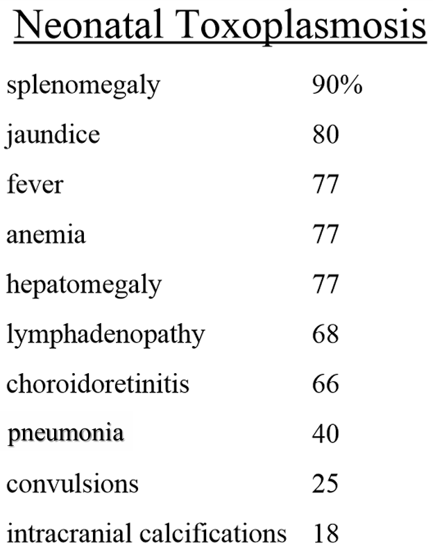
Toxoplasmosis diagnosis
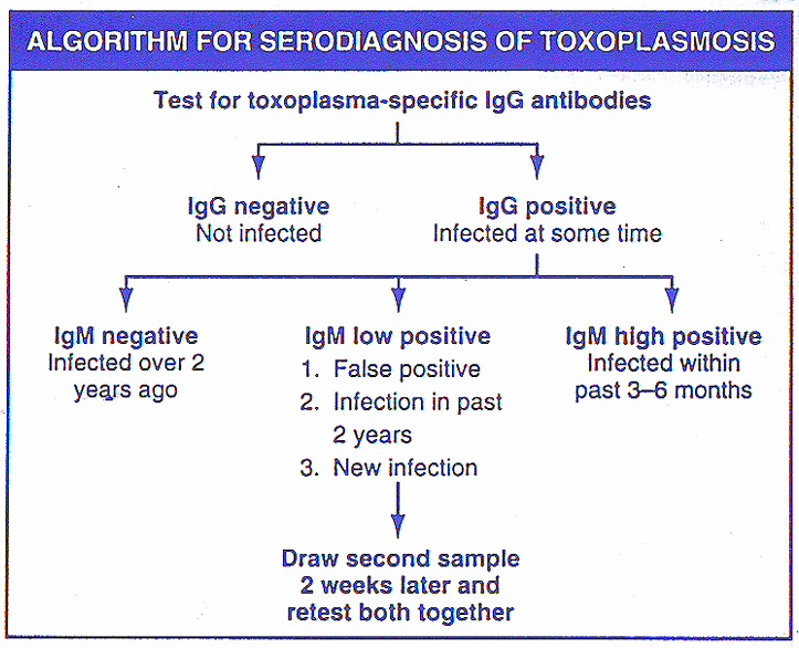
Toxoplasmosis serology interpretation
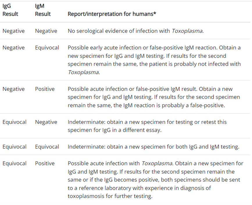
Laboratory diagnosis of toxoplasmosis
Take tissue biopsy for tachyzoite - Stain with Giemsa
PCR of amniotic fluid for possible intrauterine disease
Toxoplasmosis treatment & Prophylaxis
Recommended for pregnant women, IC patients, people with ocular diseases
Sufadiazine + Pyrimethamine (Potential teratogen, only use after 3rd trimester)
Spiramycin if pregnant woman is infected during 1st trimester
TMP-SMX: For AIDS with <100 cells/mm3 CD4+
Toxoplasmosis prevention
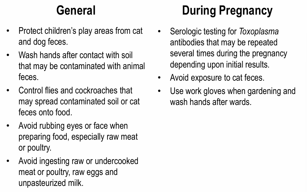
Causative agents of Amoebic Meningoenccephalitis
Naegleria fowleri & Acanthamoeba
Amoebic Meningoencephalitis Life cycles and treatments
Naegleria fowleri: Flagellated Trophozoites enter the olfactory neuroepithelium and cause amebic meningoencephalitis in healthy individuals
*Note: In healthy individuals
Acanthamoeba / Balamuthia mandrillaris: Cysts or trophozoites enter through LRT or through ulcerated/ broken skin → Granulomatous amebic Encephalitis
*Note: In IC patients
Brain-eating amoeba treatment
Amphotericin B or Rifampin