RBC Evaluation and Qualitative Assessment
 Call Kai
 Learn
Learn Practice Test
 Spaced Repetition
 Match
Match Flashcards
 Knowt Play
1/31

There's no tags or description
Looks like no tags are added yet.
Name | Mastery | Learn | Test | Matching | Spaced | Call with Kai | Chat |
|---|
No analytics yet
Send a link to your students to track their progress
32 Terms
What four CBC values measure circulating RBC mass?
RBC, HGB, HCT, PCV
What is a calculated value, expressed as (RBC x MCV)/10?
HCT
What is the percent of packed RBCs relative to whole blood?
PCV
What is measured via spectrophotometry after lysis of RBCs?
HGB
What is the automated concentration of erythrocytes in millions/microliter of whole blood?
RBC
True or False: Hyperchromasia is always an artifact.
True
What is the typical regenerative response pattern in anemia for dogs?
>80-100 K/uL Abs Retic
What is the typical regenerative response pattern in anemia for cats?
>60 K/uL Abs Retic
What is the typical regenerative response pattern in anemia for cattle?
>1% Retic %
What is the typical regenerative response pattern in anemia for horses?
cannot be assessed
What is the prefix “poikilo-” refer to?
shape variation
What is the prefix “aniso-” refer to?
size variation
What are the exceptions to the normal erythrocyte appearance?
Camelidae family: elliptocytes
White-tailed deer: artifactual sickle shape
Some goats: artifactual poikilocytes
Non-mammals: nucleated elliptocytes
What is Rouleaux?
linear association or “coinstacking” appearance
What can be reversed by saline dilution?
rouleaux; but agglutination is NOT
What is normal about RBC appearence in horses and cats due to having a low zeta potential/magnetic charge?
rouleaux
What is agglutination?
antibody-mediated binding of RBCs, looks like random clumping
What shape variation is shown in the image?
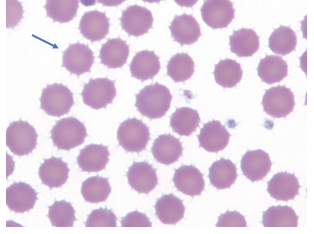
Echinocytes
What shape variation is shown in the image?
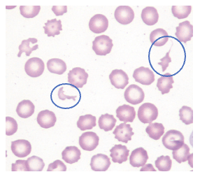
Schistocytes
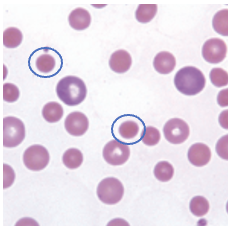
What shape variation is shown in the image?
Spherocytes
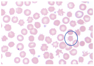
What shape variation is shown in the image?
Acanthocyes
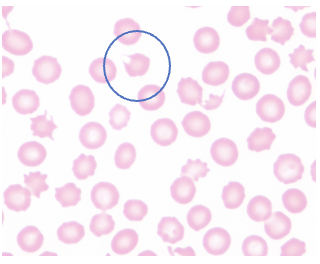
What shape variation is shown in the image?
Keratocytes
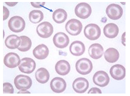
What shape variation is shown in the image?
Codocytes
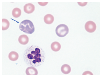
What shape variation is shown in the image?
Polychromasia
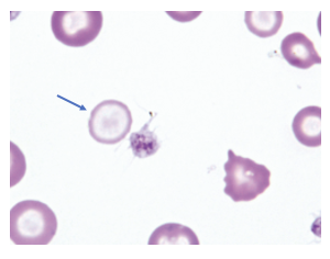
What shape variation is shown in the image?
Hypochromasia
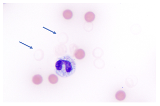
What shape variation is shown in the image?
Ghost Erythrocyte
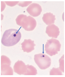
What erythrocyte inclusion is shown in the image?
Howell-Jolly Bodies
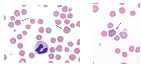
What erythrocyte inclusion is shown in the image?
Basophilic Stippling
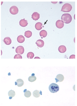
What erythrocyte inclusion is shown in the image?
Heinz Bodies
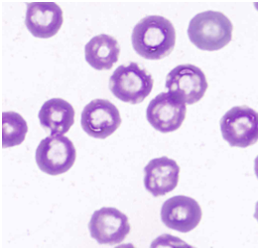
What erythrocyte inclusion is shown in the image?
Water Artifact
How does a hematology analyzer classify a nucleated erythrocyte?
WBCs
What is an increase in RBCs is termed as?
rubricytosis