Biology (Science 10)
1/90
Earn XP
Description and Tags
Biology concepts from science 10!
Name | Mastery | Learn | Test | Matching | Spaced | Call with Kai |
|---|
No analytics yet
Send a link to your students to track their progress
91 Terms
Life
The condition that distinguishes plants and animals from inorganic matter, including the capacity for movement, homeostasis, sensitivity, reproduction, nutrition, excretion, and growth preceding death.
The three tenets of cell theory
All organisms are made up of cells
The cell is the basic unit of life
All cells come from pre-existing cells
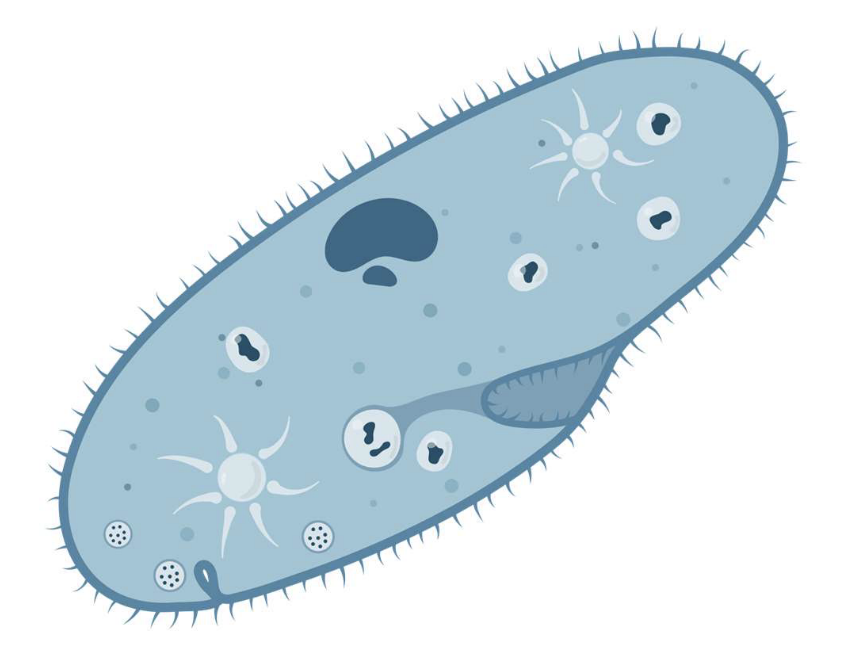
Cell
The smallest unit of life that can live on its own. It makes up all living organisms
Organism
Any living individual entity, from the smallest bacteria to the largest whale, that is capable of carrying out its own life processes.
Unicelluar
Organisms made up of one cell
Multicellular
Organisms made up of more than one cell
Homeostasis
The process by which organism maintain a stable internal environment (i.e, body temperature)
Zacharias Janssen
1590, he invented the first microscope.
Robert Hooke
1665, he observed thin slices of cork under the microscope and coined the term ‘cells.’
Francesco Redi
1668, he conducted the meat jar experiment and was the first to challenge and disprove the theory of spontaneous generation.
Anton Van Leeuwenheok
1673, he was the first to observe microscopic organisms in pond water and called them ‘animacules.’
Matthias Schleiden
1838, he concluded that all living plants are made up of cells. One of the two people to be credited for the first two tenets of cell theory.
Theodore Schwann
1839, he concluded that all living animals are made up of cells. One of the two people to be credited for the first two tenets of cell theory.
Robert Remak
1852, he discovered that all cells come from pre-existing cells when observing chicken embryos and frog spawn. He had his work stolen by another scientist.
Rudolph Virchow
1855, he concluded that ‘where a cell exists, there must have been a pre-existing cell.” Stole his work from another scientist, but is credited for the third tenet of cell theory.
Louis Pasteur
1862, he was the last person who helped disprove the theory of spontaneous generation through his swan necked flask experiment. He also helped develop sterilization and pasteurization techniques.
Manipulated variable
The aspect or factor of the experiment that is purposely changed
Controlled variable
The factor of an experiment that stays fixed/it does not change.
Responding variable
The resulting change from the manipulated variable.
Robert Brown
1831, he discovered and named the plant cell nucleus.
Light microscopes
Light is passed through a visible specimen, then focused by a glass lens. Light microscopes can observe both living and dead specimen. It has a magnification of around 1000x, and a resolution of 200 nanometers. They are best used for observing external structure.
Magnification
How many times the image has been scaled up/how zoomed in the image is
Resolution
How clear and defined an image appears, aka the smallest distance between objects that a microscope can see.
Confocal Florescence Microscope
Confocal florescence microscopes utilize staining techniques to determine differences in structure. They can technically observe both dead and alive specimen, but the vast majority is dead. These microscopes will never scan a full animal, only cells. It has a magnification of around 2000x and a resolution of 250 nanometers. They are best used for observing internal cell structure.
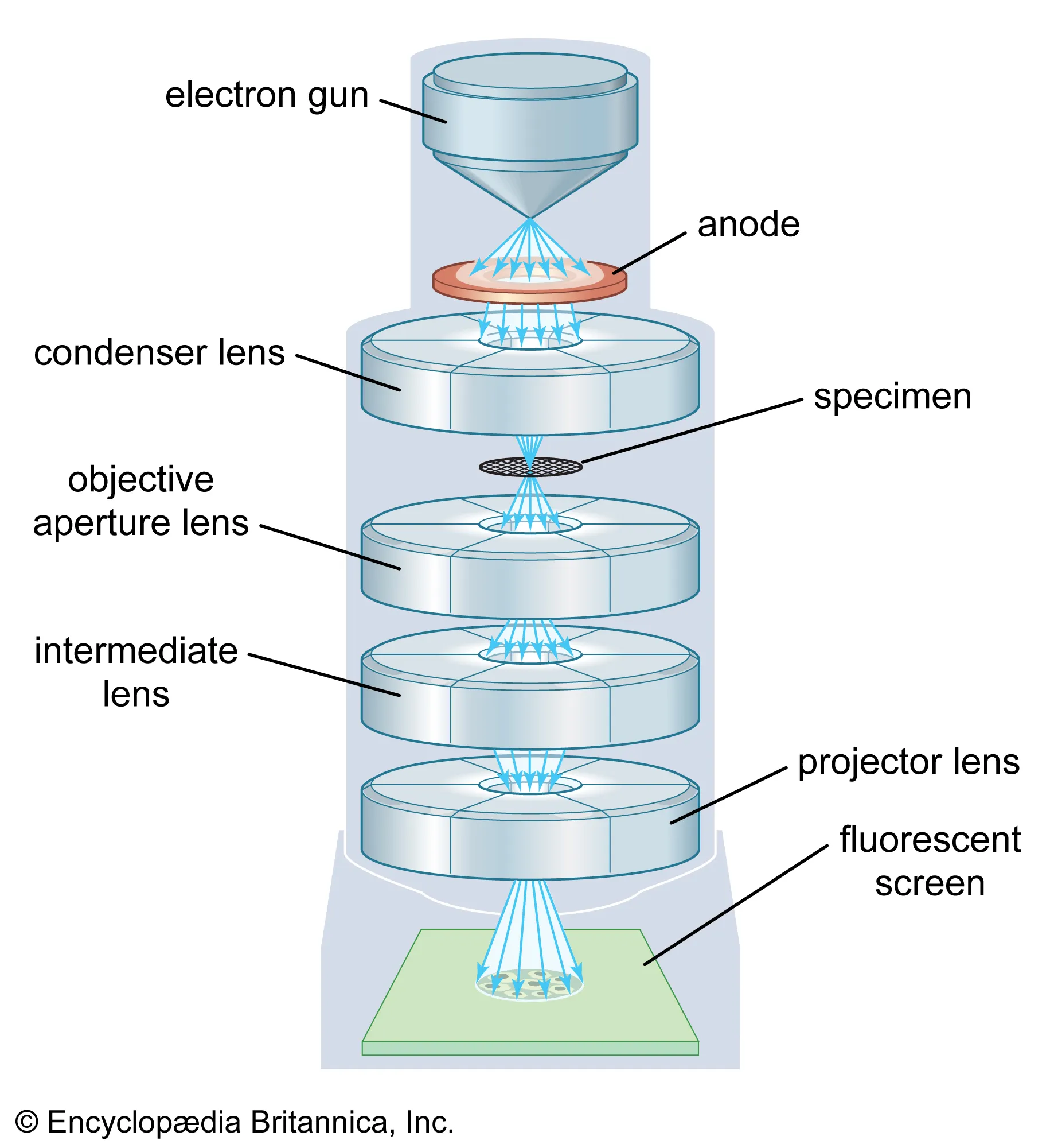
Transmission electron microscope
Electrons are passed through the specimen, then focused by a magnetic lens. TEM’s will only ever operate on dead specimen, and are best for viewing internal structure. They have a magnification of around 1,000,000 (million)x, and a resolution of 0.02 nanometers.
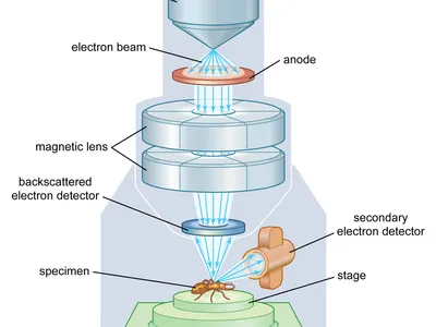
Scanning electron microscope
Specimen are coated in a thin layer of metal to prevent charging, which affects the image quality. Electrons are beamed across the surface of the dead specimen. They are best for viewing a 3D external structure. Magnification is around 10-3 million X and has a resolution of 1-20 nanometers.
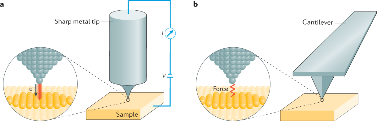
Scanning probe microscope
This microscope has very sharp probes that pass over the specimen. It can scan both alive and dead specimen, and is best for viewing external structure. It has a magnification of up to 100 million X, and a resolution of 0.01-10 nanometers.
Bright field
When the surrounding field of view is bright, but the specimen is dark in contrast.
Dark field
when the surrounding field of view is dark, but the specimen is light in contrast.
Two lens of a microscope
Eyepiece lens (typically 10x mag) and Objective lens
Meters → Millimetres → Micrometers → Nanometers
1 Meter = 1000 millimetres, 1 mm = 1000 micrometers, 1 micrometer = 1000 nm.
Which microscope produces a 3d image?
Why do electron microscopes have a higher resolution than light microscopes?
Which microscopes can observing living specimen?
Light microscope, Scanning probe microscope, and sometimes confocal fluorescence microscope.
Why are staining techniques useful?
They help visualize metabolic processes, differentiate cell components, and distinguish between live and dead cells.
Simple stain
A basic smear stain with a single dye. It is used to highlight microbes and illustrate cellular shapes and arrangements. Organisms are only stained one color.
Negative stain
Smear stain mixed with Nigrosin and spread onto thin film. This makes the specimen appear light with a dark contrasting background.
Gram stain
Primary stain: crystal violet is applied to the film and then treated with iodine, alcohol and then counter stained with safranin. This stain gives the microbes a dark appearance on a light contrasting background. It characterizes bacteria into two groups; gram positive(purple) or gram negative(pink).
Acid fast
Film is stained with hot Z.N.C.F decolourized (acid-alcohol) and counter stained with methylene blue. It helps separate non-acid-fast and acid-fast bacteria (depending on whether or not the cell membrane resists decolourization)
Why is gram staining used on bacteria?
It can help seperate bacteria groups from one another. Different types of bacteria will be different colors depending on their cell wall properties.
Basis for which stains work off?
Properties of the cell wall.
Passive transport
The movement of substances across a membrane without expending energy
Active transport
The movement of substances across a membrane that requires energy
Diffusion
The movement of substances from a high concentration to a low concentration until it reaches an equilibrium. Diffusion can occur through gases, liquids, solids, and semi-permable membranes. The movement of particles along a concentration gradient is known as diffusion.
Osmosis
The movement of water molecules through a semi-permable membrane from an area of high concentration to low concentration until it equalizes.
Concentration gradient
Substances will move down the gradient from a high concentration to a low concentration. A concentration gradient is the difference in concentration of a substance; the larger the difference, the steeper the gradient.
Equilibrium
The balancing of opposing two forces.
Dynamic diffusion
When substances on opposing sides are balanced, they move randomly along the membrane.
Facilitated diffusion
The passive transport of molecules across the cell membrane with the help of transport proteins, or channels.
Open system
A system in which both matter and energy can enter and exit
Closed system
A system in which only energy can enter and exit
Isolated system
A system where energy and matter cannot enter or exit
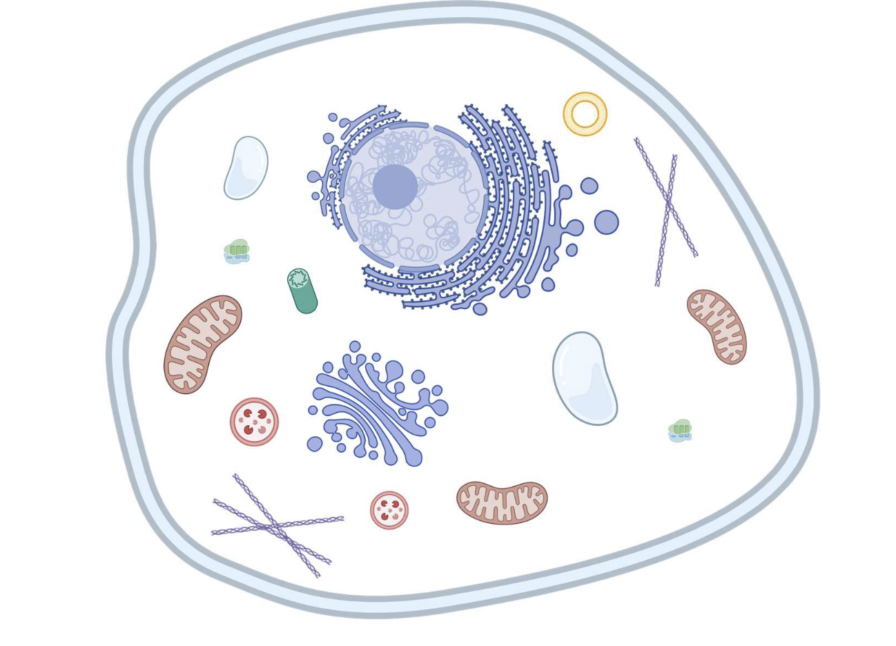
Nucleus
Contains and protects genetic material. Site of ribosome production.
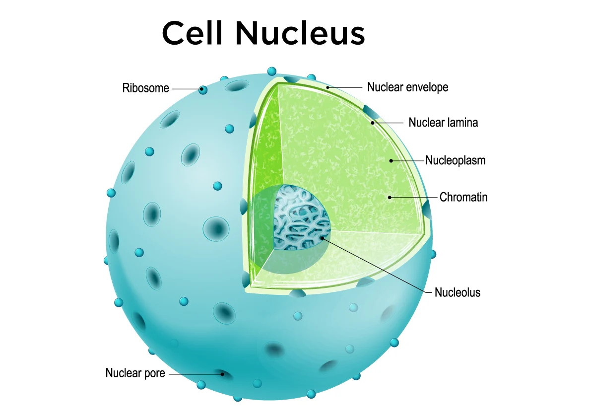
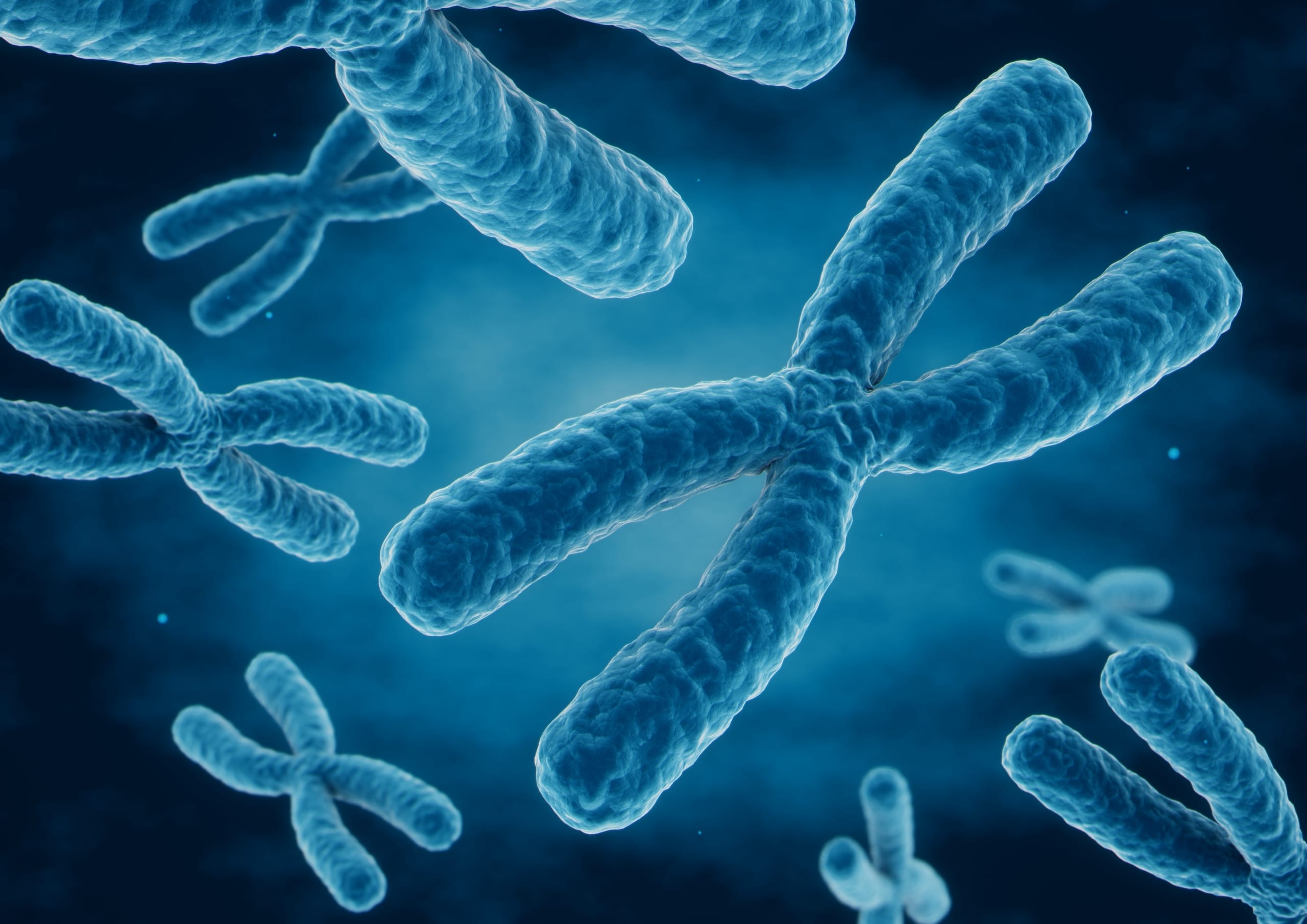
DNA/chromosomes
Provides i2nformation for making proteins. Found in the nucleus
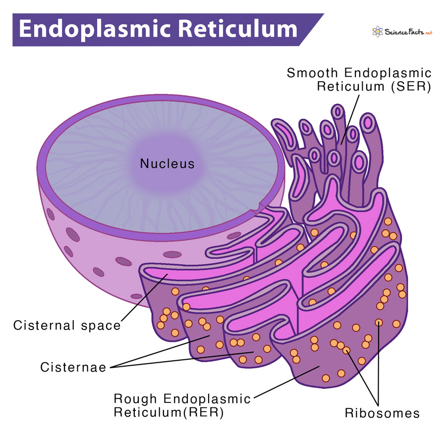
Ribosomes
Makes proteins. Found in the rough endoplasmic reticulum or free floating.
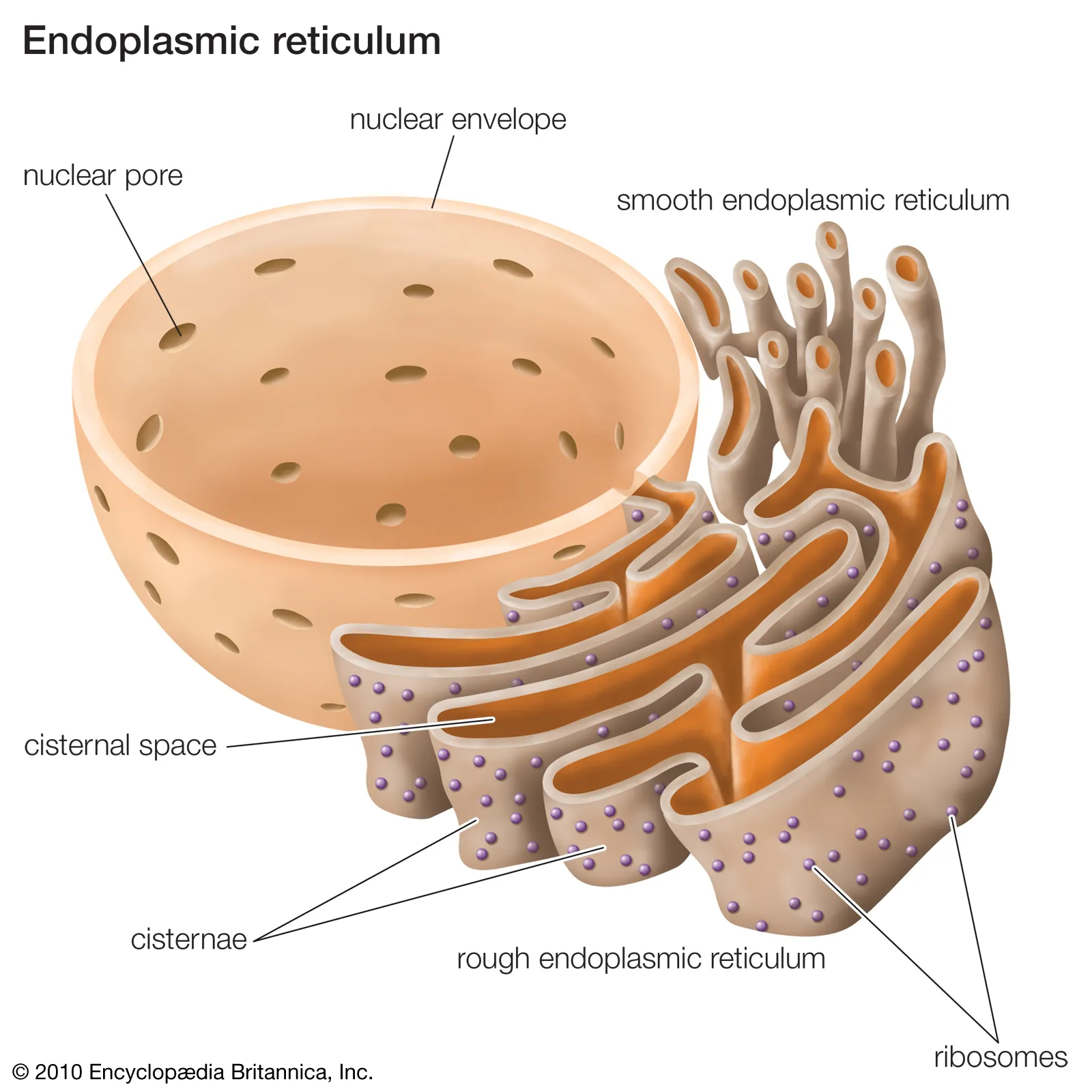
Endoplasmic reticulum
Transports and finishes proteins and other molecules. Smooth is responsible for lipid synthesis, while rough is for protein/ribosome synthesis.
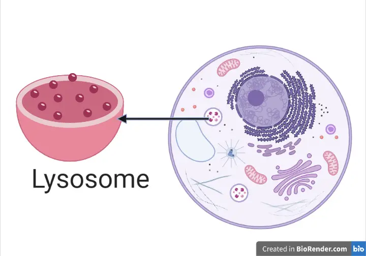
Lysosomes
Cleans waste. Only found in animal cells
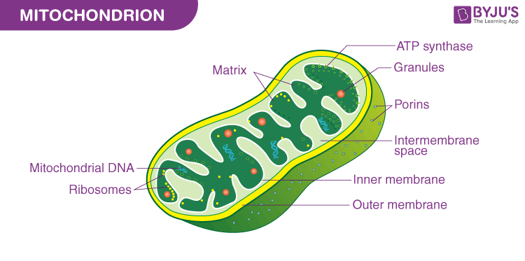
Mitochondria
Powerhouse of the cell, generates energy (ATP).
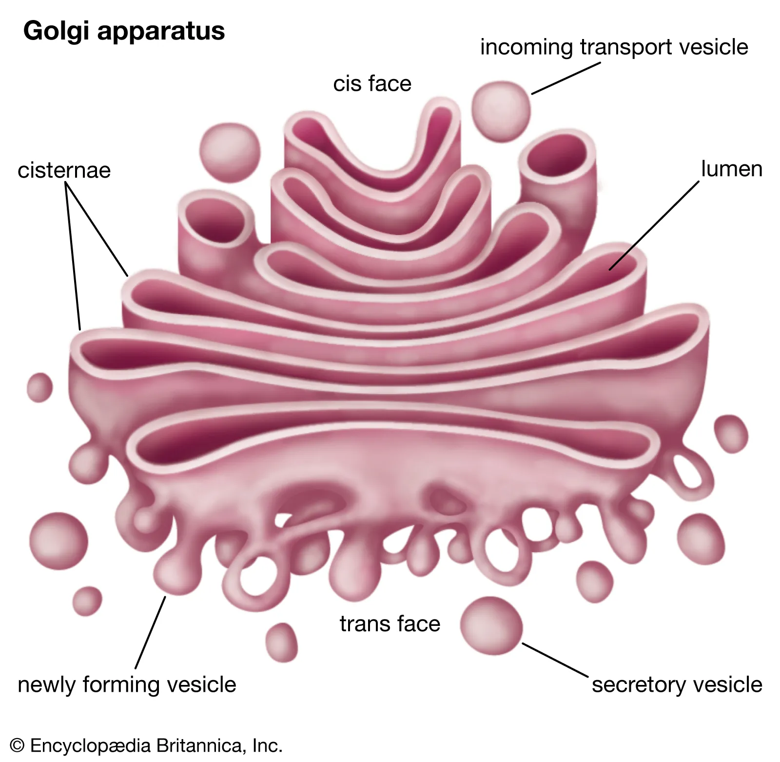
Golgi apparatus
Transports nutrients
Cytoplasm
Gel-like substance that supports the cell structure and protects the organelles.
Vesicles
Small sacs used for the transportation and storage of materials like proteins and lipids. Also facilitates cell-cell communication

Vacuole
An organelle that stores water, nutrients, and waste products. Plant vacuoles are typically one large sac and help with waste disposal, while animals have many small ones.
Pores/Gated channels
Points of entry and exit for materials
Cell membrane
Contains/protects organelles. Only found in animal cells.
Cell wall
Rigid layer containing and protecting plant cell organelles.

Chloroplasts
The organelle where photosynthesis occurs. They contain the pigment chlorophyll, which gives them their green pigment.
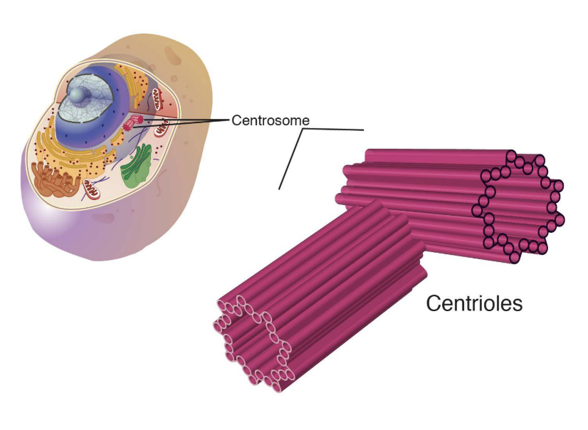
Centrioles
Helps organize microtubules to form centrosomes. Comprised of protein tubulin. Not found in fungi or flowering plants.
Eukaryotic cell
Organisms whose cells have a membrane-bound nucleus and other organelles.
Turgor pressure
The pressure exerted by the cells fluid content on the cell wall

Cytoskeleton
A network of protein filaments that help provide structural support, maintain cell shape, cell movement and chromosome segregation
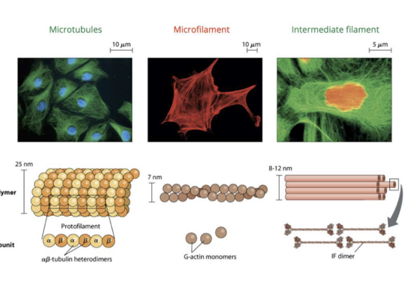
Microtubules
Found inside the cytoskeleton. They help provide structural support.
Cilia
Short, hair like projections along the cell membrane. Used for sensing and moving around. Only in animal cells.
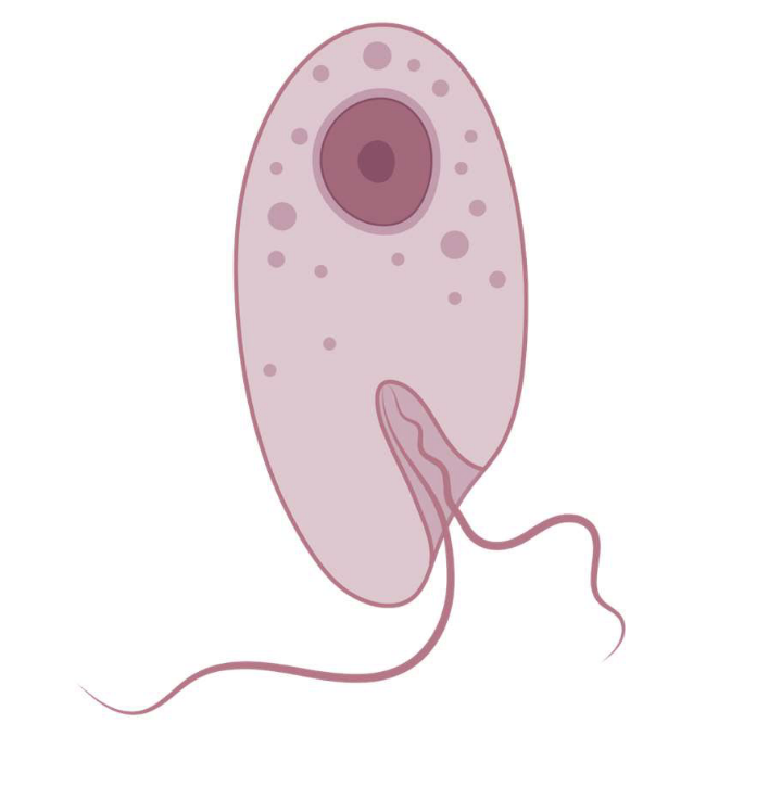
Flagellum
A whip like appendage used for movement in microorganisms. Only in animal cells.
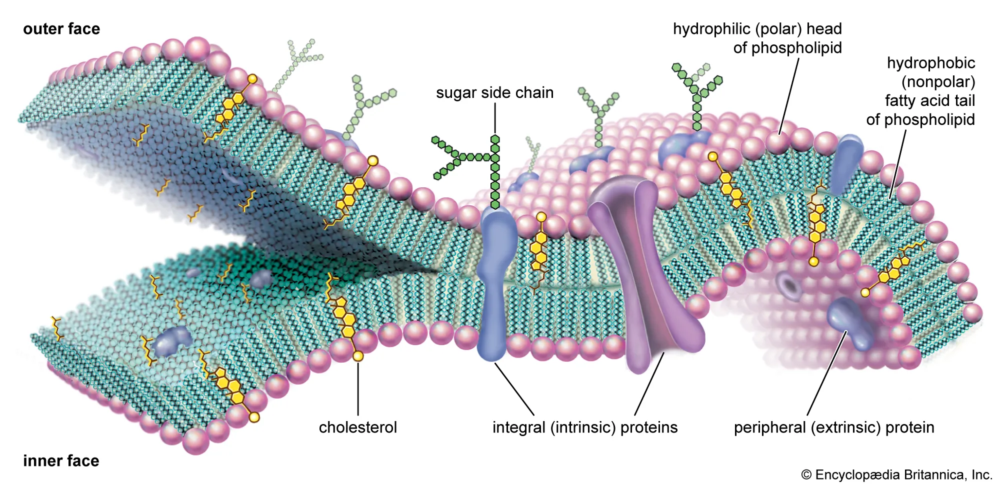
Phospholipid bilayer
The substance that makes up the cell membrane. The phospholipids have a hydrophobic tail and a hydrophilic head.
Diffusion
The movement of particles from a high-low concentration in a solution, gas, or through a membrane.
Osmosis
The movement of water particles through a semi-permable membrane to help balance out solute concentrations.
Facilitated diffusion
Diffusion, but the molecules move in and out of a membrane through a protein channel or carrier proteins.
Channel protein
Integral proteins which contain a pore through which ions may cross a membrane. These proteins are ion-specific.
Carrier Protein
Integral glycoproteins which bind a solute and undergo a structural change to translocate the solute across the membrane.
Active transport
The movement of molecules against the concentration gradient that requires the use of ATP (energy). Uses channel proteins to move molecules. Often results in a structural change of the protein.
Endocytosis
The process by which large (or large amounts of small substances) substances enter the cell without crossing the cell membrane
Pinocytosis
The taking in of fluids, or the 'drinking’ of cells.
Phagocytosis
The process by which a cell takes in large particles, or cell ‘eating.’
Receptor-mediated endocytosis
When a substance enters a cell after binding to a receptor.
Exocytosis
The process by which large substances (or large amounts of small substances) exit the cell without crossing the cell membrane.
Role of surface area in cells
Allows for more nutrients, waste, products, etc. to be absorbed and expelled more efficiently.
Role of volume in cells
Higher volume makes it harder for nutrients to get to the nucleus, and for waste to leave. This can lead to the cell ‘suffocating’ itself.
High SA:Volume ratio
Means the largest difference between surface area and volume. The larger the difference, the more efficient the cell is.
Specialized cell
A cell that has adapted to meet a specific function; skin cells, heart cells, root hair cells, etc.
Levels of organization
(Lowest to highest) Atom, molecule (proteins are very large molecules), organelle, cell, tissue, organ, organ system, organism, population, community, ecosystem
Plant organ systems
Root system and shoot system