M7 Van- Containment: from lipids to membranes

0.0(0)
studied byStudied by 0 people
0.0(0)
full-widthCall Kai
learnLearn
examPractice Test
spaced repetitionSpaced Repetition
heart puzzleMatch
flashcardsFlashcards
GameKnowt Play
Card Sorting

1/10

encourage image

There's no tags or description

Looks like no tags are added yet.

Study Analytics
Name
Mastery
Learn
Test
Matching
Spaced

No study sessions yet.

11 Terms

1
New cards

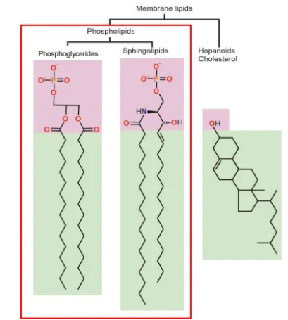
what are the three main types of membrane lipids?

  • phosphoglycerides (‘typical’ phospholipid)- glycerol linker, phosphate group + two fatty acids, all joined by ester bonds

  • sphingolipids (another phospholipid)- sphingosine linker, phosphate group, joined by an ester bond, + two fatty acids, joined by amide bonds

  • hopanoids + cholesterol- flat, hydrophobic molecules with saturated rings that intercalate into the bilayer and increase membrane stiffness

    • hopanoids are pentacyclic compounds, found in prokaryotic membranes

    • cholesterol is a tetracyclic compound, found in eukaryotic membranes

<ul><li><p><strong>phosphoglycerides </strong>(‘typical’ phospholipid)- <strong>glycerol </strong>linker, phosphate group + two fatty acids, all joined by <strong>ester </strong>bonds</p></li><li><p><strong>sphingolipids </strong>(another phospholipid)- <strong>sphingosine </strong>linker, phosphate group, joined by an <strong>ester </strong>bond, + two fatty acids, joined by <strong>amide </strong>bonds</p></li><li><p><strong>hopanoids + cholesterol</strong>- flat, hydrophobic molecules with saturated rings that intercalate into the bilayer and <strong>increase </strong>membrane stiffness</p><ul><li><p>hopanoids are <strong>pentacyclic </strong>compounds, found in <strong>prokaryotic </strong>membranes</p></li><li><p>cholesterol is a <strong>tetracyclic </strong>compound, found in <strong>eukaryotic </strong>membranes</p></li></ul></li></ul><p></p>
2
New cards

how can phospholipids vary?

variation in the tails:

  • tail length (longer = less fluid)

  • fatty acid saturation, normally only in one tail- cis double bonds are common, trans are rare (unsaturated = less tightly packed + more fluid)

variation in the heads:

  • head groups attached to the phosphate are involved in protein interactions, signalling and recognition

  • eg. glycerol, serine, glucose, choline, ethanolamine, inositol

3
New cards

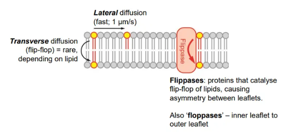
compare diffusion of lipids within and between membrane leaflets

  • diffusion within leaflets is very fast

  • diffusion between leaflets is rare, because it is difficult to get the hydrophilic head group past the hydrophobic tails, but can be catalysed by flippases

    • this causes asymmetry in the membrane, because there will be more phospholipids in one leaflet than the other

<ul><li><p>diffusion <strong>within </strong>leaflets is very <strong>fast</strong></p></li><li><p>diffusion <strong>between</strong> leaflets is <strong>rare</strong>, because it is difficult to get the hydrophilic head group past the hydrophobic tails, but can be catalysed by <strong>flippases</strong></p><ul><li><p>this causes <strong>asymmetry </strong>in the membrane, because there will be more phospholipids in one leaflet than the other</p></li></ul></li></ul><p></p>
4
New cards

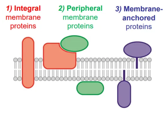
what are the three types of membrane protein?

  • integral membrane proteins have transmembrane domains (alpha helices, helical bundles and beta barrels) which have many hydrophobic residues that interact with the fatty acids

  • peripheral membrane proteins associate with membrane lipids and proteins via polar interactions, so they can be dirsputed by high salt concentrations

  • membrane-anchored proteins have lipid tails that are added post-translationally to interact with the fatty acids

<ul><li><p><strong>integral membrane proteins</strong> have transmembrane domains (alpha helices, helical bundles and beta barrels) which have many hydrophobic residues that interact with the fatty acids</p></li><li><p><strong>peripheral membrane proteins</strong> associate with membrane lipids and proteins via polar interactions, so they can be dirsputed by high salt concentrations</p></li><li><p><strong>membrane-anchored proteins</strong> have lipid tails that are added post-translationally to interact with the fatty acids</p></li></ul><p></p>
5
New cards

in what three ways are membrane-anchored cytoplasmic proteins lipidated?

  • S-acylation- post-translational, reversible modification on cysteine residue by a thioester bond

  • N-myristoylation- post-translational or co-translational (during translation), irreversible modification of an N-terminal glycine residue (once methionine removed) by an amide bond

  • prenylation- post-translational, irreversible modification of cysteine residue near the C-terminus by a thioether bond

6
New cards

in what two ways are membrane-anchored extracellular proteins lipidated?

in prokaryotes:

  • lipoprotein- post-translational modification on N-terminal cysteine after a signal peptide has been removed

in eukaryotes:

  • GPI anchor- co-translational modification at C-terminus

7
New cards

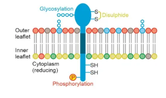
how and why are membranes asymmetrical?

  • different lipids aren’t evenly distributed between leaflets due to flippase action

  • proteins are asymmetrical and can’t flip

  • different PTMs are found on the cytoplasmic and extracellular sides of proteins (eg. outer environment is more oxidative, so disulphide bridges mostly form outside the cell)

<ul><li><p>different lipids aren’t evenly distributed between leaflets due to flippase action </p></li><li><p>proteins are asymmetrical and can’t flip</p></li><li><p>different PTMs are found on the cytoplasmic and extracellular sides of proteins (eg. outer environment is more oxidative, so disulphide bridges mostly form outside the cell)</p></li></ul><p></p>
8
New cards

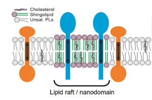
what are lipid nanodomains?

  • lipid nanodomains/rafts are localised membrane regions with distinct lipid compositions that can attract different proteins

  • these robust regions arrange membrane proteins into functional clusters, and alter local membrane rigidity

<ul><li><p>lipid nanodomains/rafts are localised membrane regions with distinct lipid compositions that can attract different proteins </p></li><li><p>these robust regions arrange membrane proteins into functional clusters, and alter local membrane rigidity</p></li></ul><p></p>
9
New cards

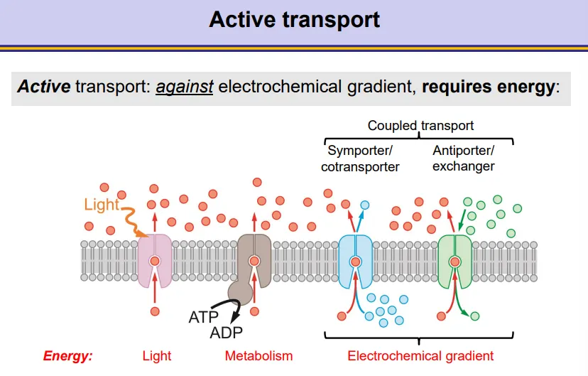
what are the different sources of energy for active transport membrane proteins?

  • light

  • ATP from metabolism

  • the electrochemical gradient of another molecule- coupled transport

    • symporters/cotransporters move the two molecules in the same direction

    • antiporters/exchangers move the two molecules in opposite directions across the membrane

<ul><li><p><strong>light</strong></p></li><li><p><strong>ATP </strong>from metabolism</p></li><li><p>the <strong>electrochemical gradient </strong>of another molecule- coupled transport</p><ul><li><p>symporters/cotransporters move the two molecules in the same direction</p></li><li><p>antiporters/exchangers move the two molecules in opposite directions across the membrane</p></li></ul></li></ul><p></p>
10
New cards

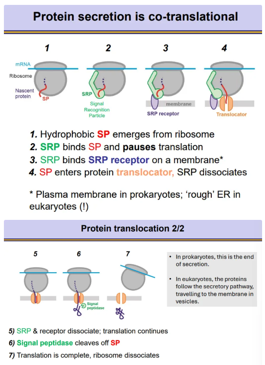
how are proteins secreted co-translationally?

  • proteins to be secreted contain a signal peptide sequence at the N-terminus

  • as the ribosome translates the protein, once the signal peptide emerges, a signal recognition particle (SRP) will bind, which pauses translation

  • the SRP binds to an SRP receptor on a membrane, so that the signal peptide can go through a translocator across the membrane

  • SRP dissociates so translation can continue

  • the signal peptidase enzyme cleaves the signal peptide off at the end

    • in prokaryotes, the membrane is the plasma membrane, so the protein is excreted straight out the cell

    • in eukaryotes, the membrane is the rough ER, so the proteins must go through the secretory pathway for excretion via vesicles

<ul><li><p>proteins to be secreted contain a <strong>signal peptide sequence</strong> at the <strong>N-terminus</strong></p></li><li><p>as the ribosome translates the protein, once the signal peptide emerges, a <strong>signal recognition particle</strong> (SRP) will <strong>bind</strong>, which <strong>pauses </strong>translation </p></li><li><p>the SRP binds to an<strong> SRP receptor </strong>on a membrane, so that the signal peptide can go through a <strong>translocator across </strong>the membrane</p></li><li><p>SRP <strong>dissociates </strong>so translation can <strong>continue</strong></p></li><li><p>the <strong>signal peptidase</strong> enzyme <strong>cleaves </strong>the signal peptide off at the end</p><ul><li><p>in <strong>prokaryotes</strong>, the membrane is the <strong>plasma membrane,</strong> so the protein is excreted straight out the cell</p></li><li><p>in <strong>eukaryotes</strong>, the membrane is the <strong>rough ER</strong>, so the proteins must go through the<strong> secretory pathway </strong>for excretion via vesicles</p></li></ul></li></ul><p></p>
11
New cards

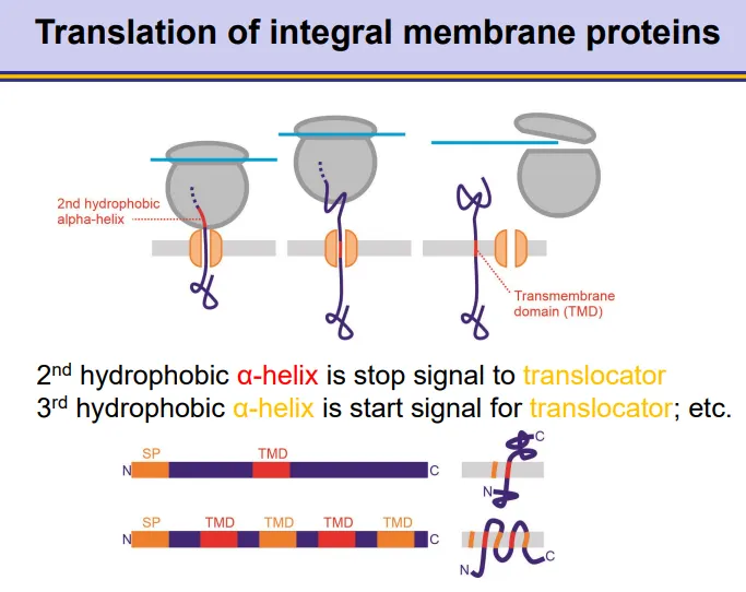
how are membrane proteins integrated co-translationally?

  • integral membrane proteins contain a signal peptide sequence at the N-terminus

  • as the ribosome translates the protein, once the signal peptide emerges, a signal recognition particle (SRP) will bind, which pauses translation

  • the SRP binds to an SRP receptor on the plasma membrane, so that the signal peptide can begin to go through a translocator across the membrane

  • SRP dissociates so translation can continue

  • when hydrophobic alpha helices are translated, these will get recognised as a transmembrane domain

  • the signal peptidase enzyme cleaves the signal peptide off at the end

<ul><li><p>integral membrane proteins contain a <strong>signal peptide sequence</strong> at the <strong>N-terminus</strong></p></li><li><p>as the ribosome translates the protein, once the signal peptide emerges, a <strong>signal recognition particle</strong> (SRP) will <strong>bind</strong>, which <strong>pauses </strong>translation </p></li><li><p>the SRP binds to an<strong> SRP receptor </strong>on the plasma membrane, so that the signal peptide can begin to go through a <strong>translocator across </strong>the membrane</p></li><li><p>SRP <strong>dissociates </strong>so translation can <strong>continue</strong></p></li><li><p>when hydrophobic alpha helices are translated, these will get recognised as a transmembrane domain</p></li><li><p>the <strong>signal peptidase</strong> enzyme <strong>cleaves </strong>the signal peptide off at the end</p></li></ul><p></p>