OMFPR 6: Advanced Imaging Modalities - CT, CBCT Technique and Application
 Call Kai
 Learn
Learn Practice Test
 Spaced Repetition
 Match
Match Flashcards
 Knowt Play
1/56

There's no tags or description
Looks like no tags are added yet.
Name | Mastery | Learn | Test | Matching | Spaced | Call with Kai | Chat |
|---|
No analytics yet
Send a link to your students to track their progress
57 Terms
False
T/F: Advanced imaging (3D) is routine form of imaging.
Tomographic Imaging
3D imaging techniques make a __________ image.
Tomographic image
Thin slice through tissue rather than a simple projection image
Projection image (PA) vs. tomographic image
Left vs. right
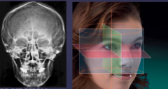
Axial
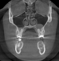
Coronal
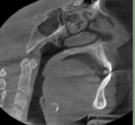
Sagittal
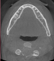
3 spatial planes of tomographic images
Axial
Coronal
Sagittal
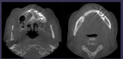
CBCT (cone beam computed tomography)
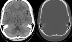
CT (computed tomography)
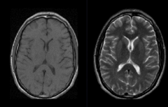
MRI
CT - fan shaped x-ray beam
Which imaging modality:
X-ray tube emits a fan shaped beam
Beam directed through a patient
Series of detectors arranged around the patient in a ring
Detectors measure the # of photons that exit the patient
Tissue density
For image display, each voxel is assigned a CT number representing _____ ______.
Attenuation of x-rays caused by the material by the material within that "voxel"
The CT number is proportional to the degree of...
+ 1000
Hounsfield number / CT number for bone
0
Hounsfield number / CT number for water
- 1000
Hounsfield number / CT number for air
Density of the anatomic structure in the image
Hounsfield number / CT number depends on...
CT scan
Which scan shows bone window + soft tissue window?
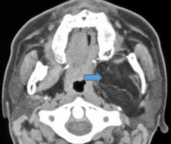
Soft tissue window
Lipoma: axial view
Uniform magnification
Lateral, coronal, and axial views available
Post acquisition reformatting may provide additional views
CT advantages
Limited availability
Expensive
Results in relatively high dose of radiation
Radiologist required for acquisition and interpretation
CT disadvantages
CT
What would be used for investigation of severe trauma?
CT
What would be used for evaluation of neoplasia?
CT
What would be used for assessment of chronic inflammation or infection?
CBCT (cone beam computed tomography)
3D imaging modality that uses x-ray beam that is "cone shaped" as opposed to "fan shaped" x-ray beam of traditional multi-slice CT
Hardware + software
Components of CBCT system
Rotating platform/gantry is fixed to x ray source and image detector
X-ray beam is cone shaped
X-ray source and detector rotate around a fulcrum fixed within the center of ROI (region of interest)
How does CBCT image acquisition work?
CT
CT or CBCT:
Fan shaped
CBCT
CT or CBCT:
Cone shaped
CT
CT or CBCT:
Multiple rotations of the gantry
CBCT
CT or CBCT:
Single rotation of the gantry
CT
CT or CBCT:
More dose
CBCT
CT or CBCT:
Poor contrast resolution - poor soft tissue detail
CBCT
CT or CBCT:
More scatter, due to a wide conical beam
CT
CT or CBCT:
More cost
CBCT
CT or CBCT:
Lower radiation exposure, higher spatial resolution, limited contrast resolution
CT
CT or CBCT:
Lower spatial resolution, superior contrast resolution
Voxels
Building blocks of 3D image volume
Voxel size
What is spatial resolution primarily dependent on?
Higher
Smaller voxel = ______ resolution
MPR
Axial, coronal, and sagittal is ___ view.
Ray rum vs. MIP
Left vs. right
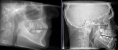
Ray sum
The slice of orthogonal or MPR images can be "thickened" by increasing the # of adjacent voxels included in the display
Increasing the # of adjacent voxels included in the display
The slice of orthogonal or MPR images can be "thickened" by...
MIP (maximum intensity projection)
Achieved by evaluating each voxel volume along an imaginary projection ray from the observer's eyes within a particular volume of interest and representing only the highest value at the display value
Volume rendering
Allows visualization of volumetric data by selective display of voxels within a dataset
Image fusion
Fusion of CBCT data with photographic image sets
Metallic beam hardening
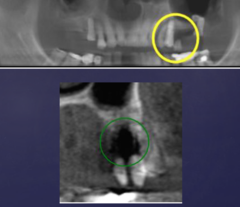
Patient motion
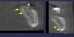
Floor of maxillary sinus
Nasopalatine canal
Inferior alveolar nerve canal (IAN)
Mental neurovascular canal and foramen
Landmarks that need to be avoided/prevented injury of in CBCT
CBCT
Useful for assessment of osseous components of TMJ
False; MRI
T/F: CBCT is useful for assessment of articular disk related issues.
MRI
Recommended for evaluation of soft tissue structures including articular disk of TMJs
CBCT
Useful in detecting missed canal during endodontic treatment
CBCT
Accessory neurovascular canals, anterior extension - these are best visualized with ____ images.
Labial undercut
Smooth depressions on the facial surface of the anterior maxilla that dental implants could perforate through the undercut during placement