Overview of heart and blood
 Call Kai
 Learn
Learn Practice Test
 Spaced Repetition
 Match
Match Flashcards
 Knowt Play
1/71

Earn XP
Description and Tags
Bio 211
Name | Mastery | Learn | Test | Matching | Spaced | Call with Kai | Chat |
|---|
No analytics yet
Send a link to your students to track their progress
72 Terms
Blood
A connective tissue with a liquid matrix called plasma and three kinds of formed elements: erythrocytes, leukocytes, thrombocyte
Blood functions
Transportation, Defense, Maintenance of homeostasis, Body temperature, Body pH, Fluid Balance/Water content of cells
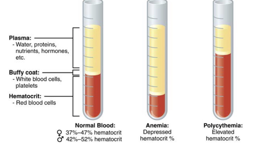
blood composition and volume
Measured by a Hematocrit (percent of erythrocyte in the blood) Consists of, from bottom to top, a layer of erythrocytes, a buffy coat, and a liquid portion known as plasma, Average values vary depending on gender and pathology
blood plasma composition
Water, Proteins, Electrolytes/Ions
albumins- protein blood plasma
Function to transport solutes and increase blood volume by osmosis
globulins- protein blood plasma
Alpha and beta function as transport proteins; gamma globulins function as antibodies
fibrinogen- protein blood plasma
Functions in blood clotting
How would liver disease affect the osmotic pressure of the blood?
Liver disease lowers the colloid osmotic pressure (oncotic pressure) of the blood. Because a damaged liver cannot make enough of the protein albumin, there are fewer proteins left in the blood vessels to pull and hold water. This drop in pressure causes fluid to leak out of the blood and into body tissues.
Sites of hematopoiesis in the embryo
Yolk sac, fetal liver, fetal spleen, lymphatic tissue, bone marrow
Sites of hematopoiesis post-birth through adulthood
Red bone marrow in most bones, including the diaphyses
Sites of hematopoiesis from adulthood
Epiphyses of long bones, sternum, skull bones, ribs, vertebrae, os coxae
hematopoiesis
the biological process by which the body creates new blood cells and platelets
Red blood cell function
Function to carry respiratory gases usually oxygen
red blood cell development
When they enter the bloodstream, they are immature and must develop. As they develop they eject their nucleus and other organelles (includin mitochondria which means they engage only in anerobic respiration
red blood cell proteins
Hemoglobin (carrying gases) and Spectrin (allows for bending)
hemoglobin
Also abbreviated Hg or Hgb, Hemoglobin is a 4-polypeptide protein, It consists of the 4
polypeptides of amino acids, 4 “heme groups” (an organic molecule called porphyrin), and 4
iron ions. The color of blood comes from the red pigment of the heme
anemia
A generic term for several forms of insufficient erythrocytes counts
polycythemia
Overproduction of erythrocytes (which can lead to a more viscous blood. The heart has to work
harder to deliver the same amount of oxygen)
low hemoglobin
A test called a “percent saturation” (or simply “percent sat”) indicates how many hemoglobin
binding sites are occupied by oxygen. 95-100% is normal. Lower values = not enough oxygen delivered to tissues
altitude hypoxemia
Decreased partial pressure of oxygen at high altitudes leads to less oxygen available to bind to the hemoglobin sites. Fatigue, breathlessness, and headache result. Causes EPO release from the kidneys in response
erythrocyte lifespan
120 days
erythrocyte rate of production
2 million every second
erythrocyte nutrients production
Glucose, lipids, amino acids, Vitamin B12, the B vitamin Folate, Trace amounts of Iron, Copper, and Zinc
destruction of erythrocytes
Occurs mostly in the spleen. The breakdown products include the Globin protein’s amino acids, which are recycled by the liver, the irons, which are recycled by the liver, and the heme molecule, which is repurposed by the liver
heme group breakdown
The liver converts the organic heme group to biliverdin (a green pigment responsible for some of the greenish tint to bruises as they develop). Biliverdin is converted into bilirubin (a yellow pigment
responsible for the yellowish tint to bruises as they heal, or jaundice, a liver disease). Bilirubin is used by the liver to make bile (a necessary substance for lipid digestion/metabolism). Bilirubin is then broken apart from the bile and converted to urobilinogen (responsible for the yellow color to urine), and then some to stercobilin (responsible for the brown color of feces)

anemias caused by bloodloss
Wounds, Hemorrhoids, Cancer, NSAIDS, Aspirin, Childbirth, Menstruation
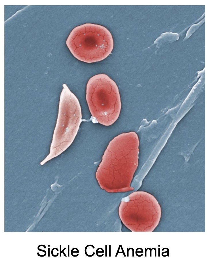
anemia caused by faulty erythrocytes
Sickle Cell, Iron-deficiency, Vitamin deficiency, Megaloblastic, Pernicious, Celiac
Why are arteries thicker than veins?
Veins bring blood to the heart, which is a smaller muscle. Arteries bring blood to the rest of the body which needs more blood, and also has more urgency.
ANEMIAS CAUSED BY EXCESSIVE DESTRUCTION OF ERYTHROCYTES
Aplastic (Stem Cell Destruction), Thalassemia, Lead Exposure

charcterstics of leukocytes
Number: 5-9K/mm3, Function: Immunity/defense; guard against microorganisms
and foreign DNA; cellular “janitors” (clean up cellular debris), Shape – spherical with large nuclei
leukocytosis
An increased production of leukocytes due to infection or trauma
margination
Leukocytes will stick to blood vessel walls at the sites of injury
diapedisis
the movement of leukocytes to the sites of infection or injury
chemotaxis
the reason the leukocytes are drawn to those sites of injury. Pathogens leave chemical trails
that leukocytes “follow
granular leukocytes
Histologically, appear to have very grainy cytoplasm, Stain blueish (basic – basophils), reddish (acidic eosinophils), or lavender (neutral neutrophils), depending on the type Have multi-lobed nuclei
agranular leukocytes
Histologically, appear to have very few grains in the cytoplasm, Have large, rounded nuclei
Basophils- granular
Contain histamine granules that increase inflammation, Contain and release heparin, which inhibits
blood clotting Increase in number with allergies and parasitic infections
Eosinophils- granular
Produce anti-histamines to counteract inflammation Produce molecules that
are toxic to parasitic worms Increase with allergies, parasitic worm infestation, and some autoimmune disorders
neutrophils- granular
First responders to infection (produce our innate immunity), Phagocytize bacteria (contain lysozymes – enzymes that break down bacterial cell walls), Increase in number with generic bacterial
infections, burns, and even stress
lymphocytes types - agranular
NK (Natural Killer) Cells
• B-Cells
• T-Cells
NK/ natural killer lymphocytes
attack cells that do not express “self” on their plasma membranes (are seen by the body as being
foreign). They increase during cancer and when viruses infect cells. They provide non-specific immunity.
B-cells lymphocytes
produce antibodies; provide humoral (bodily fluids) immunity; retain memory of previous infections in
case of repeat infection; are produced in and mature in the red bone marrow.
T- cells lymphocytes
directly attack infected cells; produced in the bone marrow but mature in the thymus.
monocytes - agranular
Exist as monocytes when in the blood but become MACROPHAGES when they move to tissue to
take up residence. Phagocytize dead cells, cellular debris, large wastes, pathogens, dead
erythrocytes, damaged cells
differential count
A differential count determines both the number and types of leukocytes in a sample of blood. Gives an indication of potential issues with elevated or reduced numbers as a whole, and of certain types of
cells
leukocytes lifespan
few hours to a few days
leukocytes production
Most lymphocytes can undergo mitosis which leaves ‘memory’ to certain foreign pathogens Production occurs in response to leukocyte hematopoietic factors; monocytes take the myeloid line
and lymphocytes take the lymphoid line
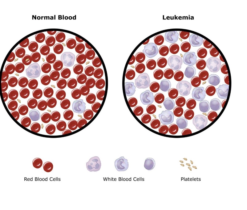
leukemia
leukocyte disorder cancer that produces non-functional cells
lymphoma
leukocyte disorder that is a cancer of lymph organs that assist with immunity
leukocytosis
leukocyte disorder that has a high count of non-functional leukocytes
leukopenia
leukocyte disorder that has insufficient numbers of leukocytes
thrombocytes/ platelets
Their name is a misnomer; they never were cells. Number: 350K-500K. Develop via the myeloid
stem cell line as broken fragments of megakaryocytes. Function to help stop blood loss in damaged vessels and produce factors that promote tissue healing. 1/3rd of them are stored in the spleen “if needed”
thrombocytosis
platelet disorder with too high numbers of platelets; leads to excessive blood clotting
thrombocytopenia
platelet disorder that has too low numbers of platelets; leads to excessive bleeding and inability to
clot
hemostasis
the multi-step process of stopping blood loss from damaged vessels
- Occurs in three stages:
• Vascular spasm
• Platelet Plug Formation
• Coagulation
vascular spasm
vasoconstriction limits blood flow to the damaged area
platelet plug formation
The platelets begin to stick to the damaged endothelial wall of the broken vessel. They form a temporary dam, limiting the blood leaking through the wall of the vessel
coagulation
This third step involves a series of chemical reactions involving “clotting factors”. Clotting factors are 12 different molecules that react together to ultimately produce the insoluble protein ‘fibrin’. Fibrin forms the permanent clot to seal the vessel wall so the healing process can begin. Occurs via an intrinsic (internal vascular damage) or an extrinsic (external vascular damage) pathway until
these join to form a single pathway
clot removal
Fibrinolysis: the gradual degradation of the clot. Plasma Anticoagulants: those substances naturally
found in plasma that assist with fibrinolysis. Thrombolytic Agents: administered agents that
break up clots. Anticoagulants: prolong the time it takes for coagulation to occur. Antithrombins: prevent formation of thrombin
thrombophillia
platelets tend to spontaneously stick to each other; can be caused by Lupus, heparin sensitivity, polycythemia, sickle cell disease, pregnancy, obesity
causes of clotting
• Vitamin K deficiency
• Calcium deficiency
• Hormones
• Stagnant body position
• Smoking
blood antigens
Antigens are proteins embedded in the plasma membrane of erythrocytes. Because there are other
antigens attached to cells, these particular ones are called “blood” antigens. The most common are
named A and B (forming the ABO System), and Rh (or D) (forming the Rh System
blood antibodies
Antibodies are proteins found in the plasma of the blood (*not* attached to the cells). Antibodies are generally referred to as “Anti-A Antibody” or “Anti-B Antibody” so as not to confuse them with “A
Antigen” or “B Antigen”. While Anti-Rh Antibodies can be made they are normally not present in blood; Under special situations, they can appear
Rh blood type
A different type of antigen may be present on erythrocytes, named the Rh antigen. It is inherited differently from the A and B antigens Therefore it is standard practice to state a letter (A, B, AB or O) and a symbol (positive or negative) when giving a blood type, to give a full type.
Rh antibodies
Unlike Anti-A and Anti-B Antibodies, which are present from birth, even with no exposure to foreign blood, the Rh negative body does not contain antibodies to the Rh antigen unless it has been exposed to Rh positive blood. These might happen in transfusions with Rh positive blood or when an Rh negative mother bears an Rh positive child.