Lecture 1 – UMN & LMN Part 1
 Call Kai
 Learn
Learn Practice Test
 Spaced Repetition
 Match
Match Flashcards
 Knowt Play
1/133

There's no tags or description
Looks like no tags are added yet.
Name | Mastery | Learn | Test | Matching | Spaced | Call with Kai | Chat |
|---|
No analytics yet
Send a link to your students to track their progress
134 Terms
Why is understanding UMN vs LMN important?
UMN and LMN lesions create different clinical signs, helping clinicians locate where nervous system damage occurred
What are the two halves of the somatic motor system?
Upper motor neurons
Lower motor neurons
What is the basic job of an upper motor neuron?
It carries motor signals from the brain → spinal cord
What is the basic job of a lower motor neuron?
It carries motor signals from the spinal cord → muscle or target organ
What is another name for the lower motor neuron?
The final common pathway
Where do upper motor neurons begin?
In the primary motor cortex, located in the precentral gyrus just anterior to the central sulcus
What is the main goal of the upper motor neuron?
To provide precise control of movement, especially contralateral movement
Does the UMN directly affect the target organ?
No, the UMN communicates with the LMN, which then acts on the target organ
Why can’t the UMN create movement by itself?
Because it only initiates the motor signal
Movement requires the LMN to activate muscle
Where do UMNs terminate?
In the ventral horn of the spinal cord, where they communicate with LMNs
What is the UMN’s only target?
The alpha motor neuron
What does the LMN communicate with?
The muscle or target organ
What determines the final output from the LMN to muscle?
Interactions between sensory input and descending UMN signals
What major structures adjust descending UMN activity?
The cerebellum and basal ganglia
How do the cerebellum and basal ganglia influence movement?
They adjust movement by influencing excitation or inhibition of LMN activity
What type of information adjusts descending motor data throughout the CNS?
Ascending sensory information
What brain areas give rise to UMNs?
Premotor cortex
Primary motor cortex
Supplementary motor area
Somatosensory area
Superior parietal lobe
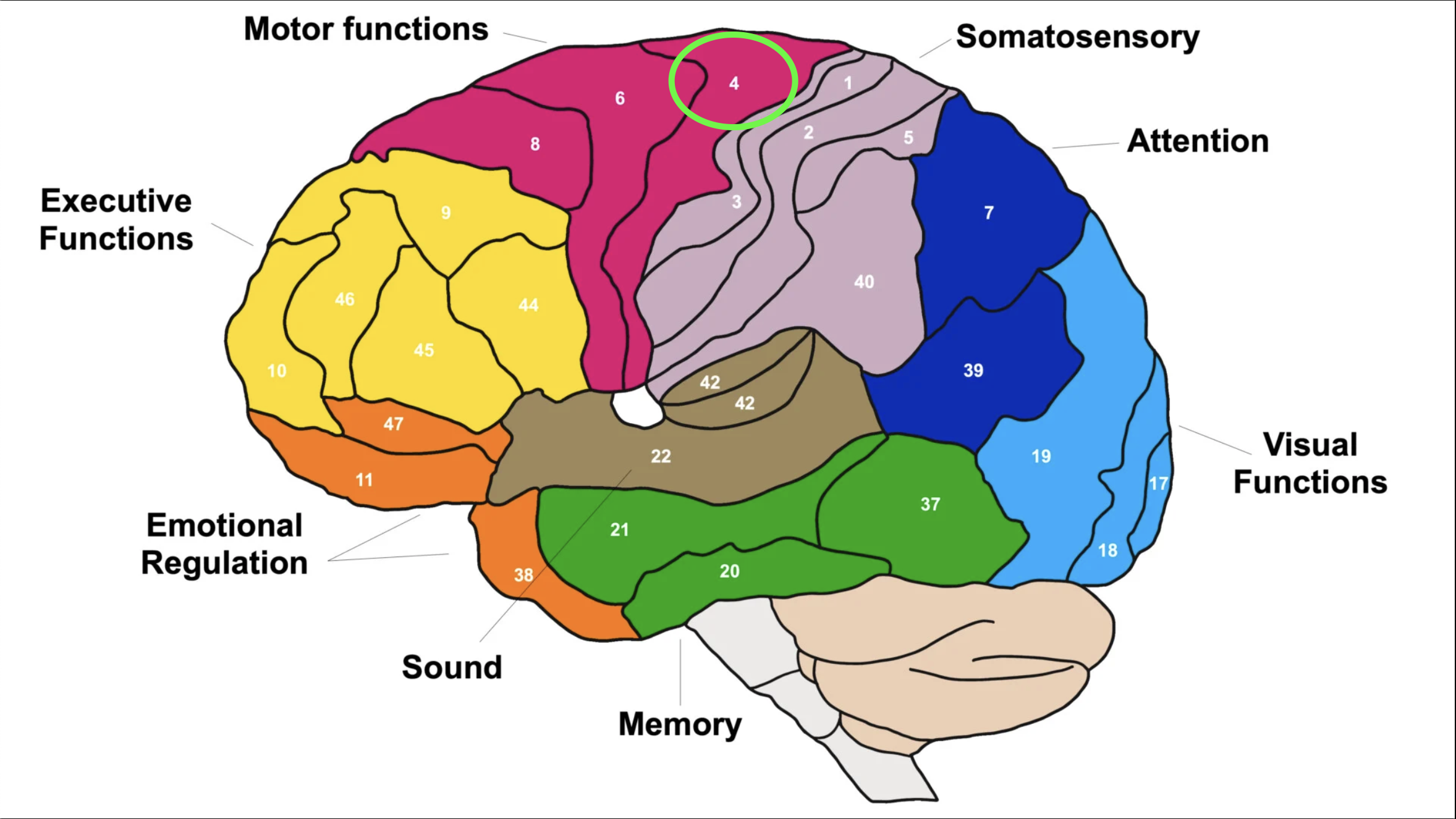
Which Brodmann area is the primary motor cortex?
Brodmann area 4
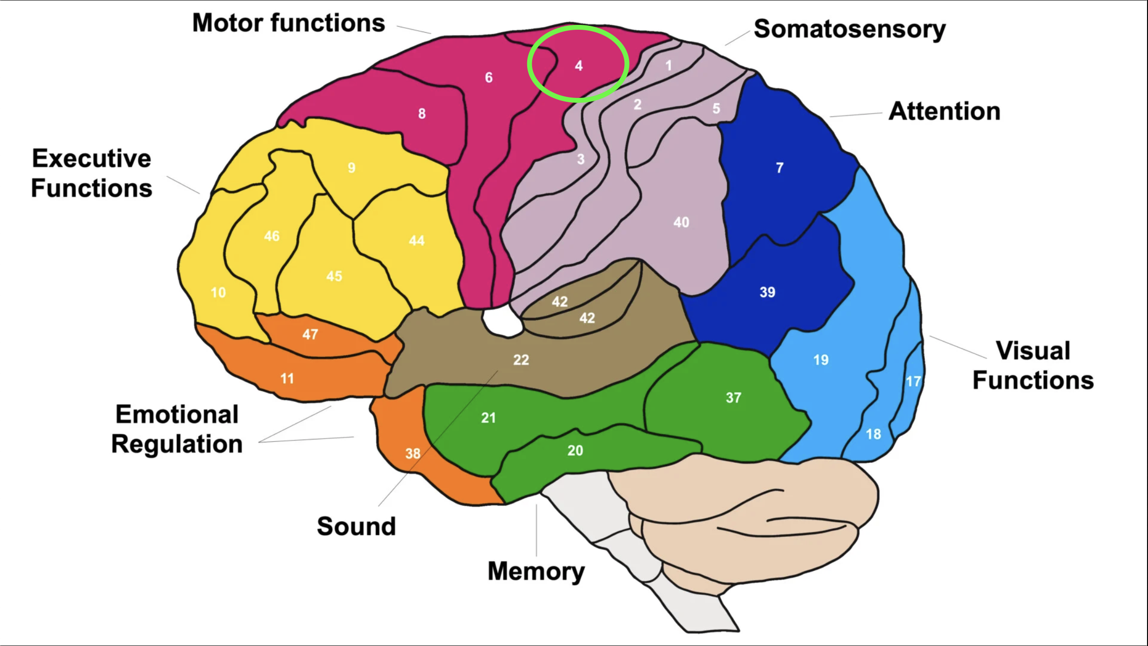
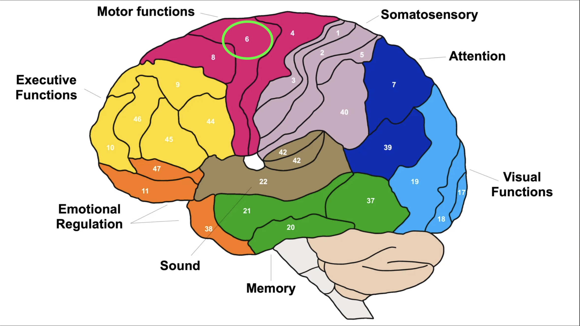
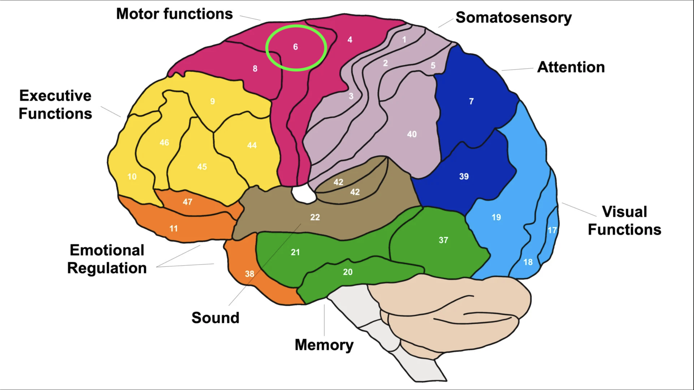
Which Brodmann area is the premotor area?
Brodmann area 6
Where is Brodmann area 4 located?
In the precentral motor cortex
Where is Brodmann area 6 located?
In the premotor cortex
Why are Brodmann areas clinically important?
They help identify functional cortical regions involved in conditions such as stroke
What are Brodmann areas based on?
Histological or cellular architecture of the cerebral cortex
What do Brodmann areas form?
A functional map of the brain
What are Brodmann areas 44 and 45?
Broca’s areas
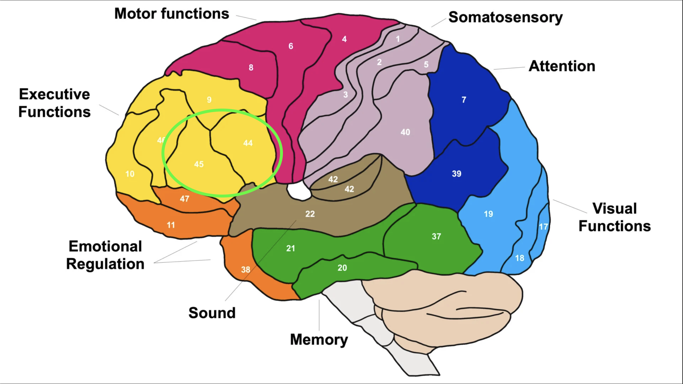
What is Broca’s area involved in?
Producing coherent speech
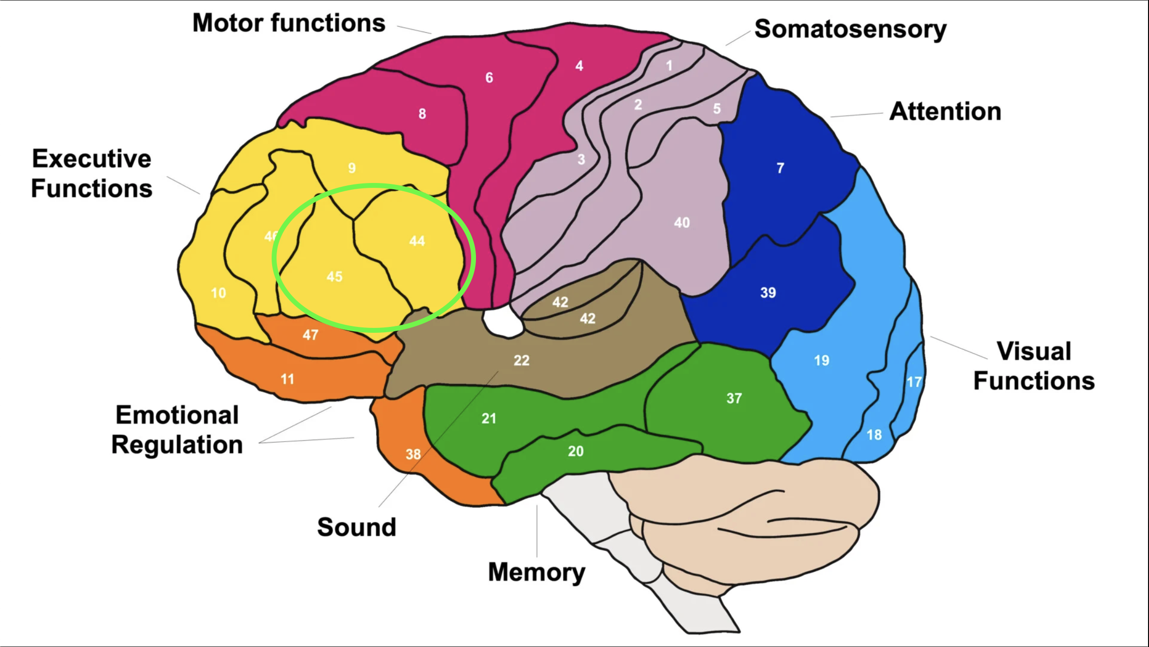
What type of stroke commonly affects Broca’s area?
Middle cerebral artery bleed or infarct
What is Broca’s aphasia?
The patient understands language but has difficulty producing speech
Why can Broca’s aphasia be frustrating for patients?
They know what they want to say but cannot get the words out effectively
Which Brodmann area is Wernicke’s area?
Brodmann area 22
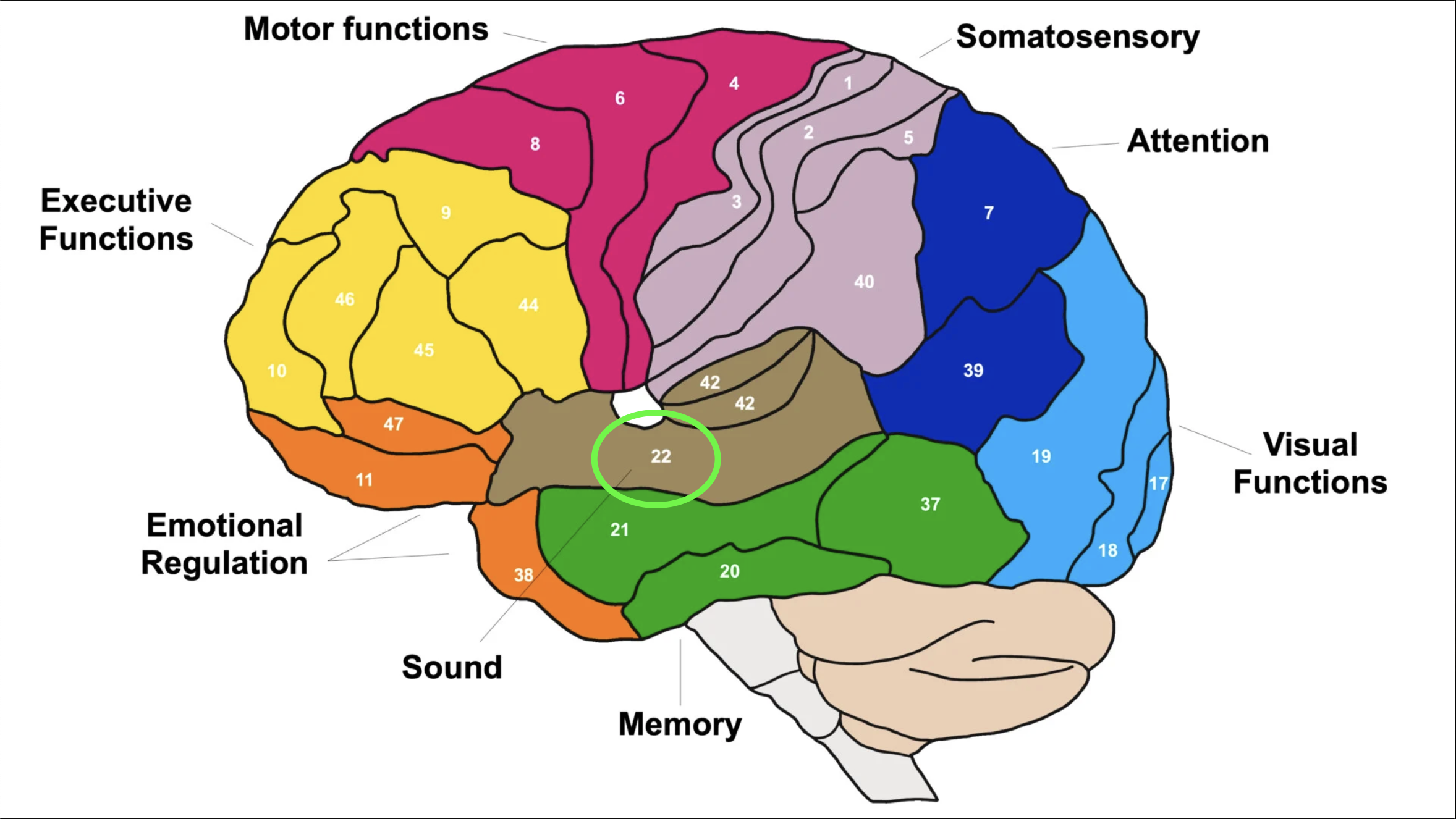
What is Wernicke’s area involved in?
Processing speech and understanding language
Where is Wernicke’s area generally located?
In the temporal-parietal region
What is Wernicke’s aphasia?
The patient may speak fluently, but the words do not make meaningful sense and comprehension may be impaired
What is “word salad” associated with?
Wernicke’s aphasia
How is Broca’s aphasia different from Wernicke’s aphasia?
Broca’s = understands but cannot produce speech well
Wernicke’s = fluent speech that does not make sense and poor comprehension
Why might patient history be unreliable with Wernicke’s aphasia?
Because the patient may not understand questions or produce meaningful answers
What structures can contain cranial nerve nuclei and relay stations?
Cortex
Cerebellum
Brainstem
Midbrain
Pons
Medulla
What are tracts?
Bundles of long axons within white matter
How are tracts usually named?
By their origin and termination
What does the first part of a tract name usually indicate?
Where the tract begins
What does the second part of a tract name usually indicate?
Where the tract ends
Why is the spinothalamic tract an ascending tract?
Starts in the spinal cord → ends in the thalamus
Why is the corticospinal tract a descending tract?
Starts in the cerebral cortex → ends in the spinal cord
What do descending tracts do?
They carry motor signals from the brain toward LMNs or effector organs
What are the two main UMN pathway categories?
Pyramidal and extrapyramidal systems
What does “pyramidal system” mean?
Pathways that travel through the pyramids of the medulla
Which tracts are pyramidal tracts?
Corticospinal tract
Lateral and Anterior
Corticobulbar tract
What are the divisions of the corticospinal tract?
Lateral corticospinal tract
Anterior corticospinal tract
What is the main movement system?
The corticospinal tract
What does the corticobulbar tract control?
Movements of the head and neck
What does “extrapyramidal” mean?
Outside the pyramids of the medulla
What are extrapyramidal tracts mainly involved in?
Involuntary control and fine-tuning of movement
Do extrapyramidal tracts travel through typical corticospinal tract channels?
No
Which tracts are UMN extrapyramidal tracts?
Reticulospinal tracts
Rubrospinal tracts
Vestibulospinal tracts
Tectospinal tracts
What three things should you know about descending tracts?
Spinal cord location
Whether they cross
Their function
What is the lateral corticospinal tract especially important for?
Fine, skilled limb movement
Where does the lateral corticospinal tract cross?
In the medulla at the medullary pyramids
Why is the lateral corticospinal tract part of the pyramidal system?
Because it crosses/travels through the medullary pyramids
What is the anterior corticospinal tract more responsible for?
Control of the upper extremity, head, neck, and upper trunk
Does the anterior corticospinal tract cross the same way as the lateral corticospinal tract?
Not necessarily
Some fibers combine with the lateral corticospinal tract
What is the tectospinal tract?
A descending extrapyramidal tract involved in reflexive head turning
Where does the tectospinal tract cross?
In the midbrain
What does the tectospinal tract respond to?
Visual and auditory stimulation
What is the function of the tectospinal tract?
Reflexive head turning while helping maintain postural control
Do the lateral and medial reticulospinal tracts cross?
No
What are the reticulospinal tracts responsible for?
Posture
Balance
Pain awareness regulation
Tone
Postural preparation
What are the vestibulospinal tracts responsible for?
Posture
Balance
Maintaining head position when the base of support changes
What are the two vestibulospinal tracts?
Lateral vestibulospinal tract
Medial vestibulospinal tract
What neurotransmitter is key at the neuromuscular junction?
Acetylcholine (ACh)
What other neurotransmitter can influence motor pathways?
Glutamate
What is a motor neuron made of?
Soma/cell body
Axon
Dendrites
Neuromuscular junction/motor end plate
What is the neuromuscular junction also called?
The motor end plate
What happens at the motor end plate?
The neuron attaches to and communicates with the muscle
What is included in the lower motor neuron system?
Alpha motor neuron, gamma motor neuron, neuromuscular junction, motor end plate, muscle spindle, Golgi tendon organ, free nerve endings, and muscle.
Where is the Golgi tendon organ located?
In the tendinous portion of muscle.
Where is the muscle spindle located?
In the central portion of muscle.
Where do alpha and gamma motor neurons synapse?
In the ventral horn of the spinal cord.
What sensory fibers are included in the spinal nerve?
Ia and Ib fibers.
What is a motor unit?
An alpha motor neuron and all the muscle fibers it innervates.
What happens when an alpha motor neuron is activated?
Acetylcholine is released at all its neuromuscular junctions, causing all innervated muscle fibers to contract.
What type of muscle fibers are common in postural muscles?
Slow-twitch fibers.
Why do postural muscles have many slow-twitch fibers?
They are important for postural control and slow, sustained contractions.
What principle relates to motor unit recruitment and muscle fiber activation?
Henneman’s size principle.
What are C fibers mainly related to?
Pain signaling.
Do all motor units innervate the same number of muscle fibers?
No, motor units vary depending on the precision required.
How many muscle fibers may one alpha motor neuron innervate in the gastrocnemius-soleus complex?
About 2,000 muscle fibers.
What is another name for the gastrocnemius-soleus complex?
Triceps surae.
How many muscle fibers may one alpha motor neuron innervate in eye muscles?
About 2 muscle fibers.
Why do eye muscles have fewer muscle fibers per alpha motor neuron?
To allow very fine motor control.
What is alpha-gamma coactivation?
Simultaneous activity of alpha and gamma motor neurons to coordinate muscle contraction and spindle sensitivity.
What does the alpha motor neuron activate?
Extrafusal skeletal muscle fibers.
What does the gamma motor neuron activate?
Intrafusal skeletal muscle fibers through the muscle spindle.
Is the alpha motor neuron excitatory?
Yes.
What does the gamma motor neuron regulate?
Muscle spindle sensitivity and tone-related adjustments.
Where does the gamma motor neuron go?
Directly to the muscle spindle.
What does the gamma motor neuron adjust muscle activity for?
Tonic or phasic activity.
What forms the spinal nerve?
Sensory information from the dorsal root plus motor information from the ventral root.
Where are alpha motor neuron cell bodies located?
In the ventral horn of the spinal cord.
Where do alpha motor neuron axons project?
To a single muscle.
What are motor pools?
Clusters of alpha motor neurons that innervate muscles.