3. Kinsesology 2Y03 Embrology and Development - Module 3
 Call Kai
 Learn
Learn Practice Test
 Spaced Repetition
 Match
Match Flashcards
 Knowt Play
1/26

There's no tags or description
Looks like no tags are added yet.
Name | Mastery | Learn | Test | Matching | Spaced | Call with Kai | Chat |
|---|
No analytics yet
Send a link to your students to track their progress
27 Terms
Hapliod cells
Gamates
Half sex cells come from males
Half sex cells come from females
Gonads
Organs that produce gamates
Female: Ovaries
In each menstrual cycle, about 14 days after ovulation begins
Process of producting Secondary oocyte
One ovary matures a female gamete cell + preps to release (Once released called seconary oocyte)
The time of release is known as ovulation
At this point, the oocyte has not finished meiosis (Secondary)
Only finished the first meiotic division
Oocyte enters uterine tube/Fallopian tube
Via Finger like projectors
Passes funnel area
Enters widened region known as AMPULLA
Ampulla is where fertilization will occur (SECONDARY OOCYTE DOES NOT MOVE FROM HERE)

Uterus
Pear shape
Muscular structure
Implantation of embryo happens here
Development + Growth of fetus occurs here
it connects to Vagina Through opening known as the cervix
Vagina: opening to the outside of the body + entry point for male gamete cell (Sperm)
2 uterine tubes connect to the UTERUS
Sperm Killing
During intercourse, 20 million sperm are deposited into vagine
ONLY 1% make it to the uterus
Only 100-200 reach the oocyte in the ampulla
WHY????
Some sperm don’t have good motility (can’t swim well)
pH in secretions of the female reproductive system kills off many sperm
Overall Goal of Process
Take male genetic material and combine with female genetic material
Sperm path to Oocyte
Initially when sperm enter the vagina:
They are self-propelled by flagella (tail)
Once inside uterus and uterine tube
Muscular contractions of the walls of structure help push sperm along
Underline: Some are caused by hormones released during intercourse (e.g., oxytocin)
The system also has prostaglandins from liquid secretions that support sperm (semen)
Also stimulate small contractions

Sex cell life span
Secondary oocyte only stay fertilizable for aproxx 24 hours
Sperm can live up to 6 days
Most die within 24 hours
Window for fertilization
6 days before ovulation
1 day after
Germinal Period
first two weeks of development
Formation of primitive germ layers (cells multiply)
Become body tissue
Embryonic Period
Week 3 to end of 8th week organs systems develop
Fetal period
9th week until birth; organ systems grow + mature (aproxx 38 weeks - embryonical age)
Clinical/ Medical Events (date set up)
Date since last menstrual period (clinical age of unborn)
Embryologists (date set up)
Post ovulatory age (aproxx 14 days less than clinical age)
oocyte Info
chromosomes held within the nucleus
females sex cells don’t divide equally during meiosis
big cell (oocyte)
small cell (polar body
no fucntion in fertilization excepting removing extar genetic material from steps of meiosis
Degrade overtime
Oocyte Layers
Around its plasma membrane it has
Clear glyoprotein layer
zona pellucida
loosely packed cells
corona radiata
AKA crown of oocyte
made of follicular cells
Maturation of secondary oocyte + release happens in a follicle
follicle: Cells surrounding oocyte
Supporting them
Some of these loose cells stay stuck onto zona pellucida (which is the corona radiata)
Fertilization
Once sperm reaches secondary oocyte
Penetrates corona radiata
Zona pellucia has a ZP3 glycoprotein (sperm receptor)
Sperm must bind to get through
ZP3 is species specific
NO MAN-HORSE
Once sperm acrosome (at head of sperm tip is called acrosome → contains digestive enzymes)
Acrosomal Reaction starts (activation of digestive enzymes)
Zona pellucida starts to break down
Many sperm must bind to break zona pellucida
to allow only one to go through
Once sperm is into the oocyte, it binds with the plasma membrane of the oocyte
Contains receptor “Integrin α6β1”
As soon as one sperm binds to it, it causes fast depolarization
electrical charge in the plasma membrane of the oocyte
shocks all other sperm trying to reach the plasma membrane
blocks other sperm (fast block to polyspermy)
Head of sperm is into Oocyte!!!
Female nucleus does 2nd meiotic division
Ovum Formed
2nd Polar body
Once inside, sperm tail disintegrates (only head left)
Nucleus undergoes 2nd meiotic division (on one side of cell)
Now cells contain:
Female pronucleus
male pronucleus (sperm head)
Fusion of two hybrid pronuclei produces a single nucleus (zygote) - diploid

fast block to polyspermy
Once sperm is in the oocyte, it binds to the plasma membrane of the oocyte
Contains the receptor “Integrin α6β1”
As soon as one sperm binds to it, it causes fast depolarization
electrical charge in the plasma membrane of the oocyte
shocks all other sperm trying to reach the plasma membrane
blocks other sperm
Slow Block to Polyspermy
Once sperm enters the oocyte
Release of Ca2+ Intracellularly
Ca2+ moves out of the cell, taking water with it
Fluid and ions make their way between the zona pellucida and plasma membrane
Causes the oocyte to shrink
Zona pellucida denatures along with ZP3 → inactive receptor
Space now between zona pellucida and membrane
Formation of Blastocyst
Zygote undergo clevage
Cleavage: Series of cellular divisions
Very first division
takes the longest
Usually happens in the first 24 hours
Can take 6-36 hours to occur
Divisions become faster
2 cells in 24 hours
4 cells in 48 hours
Solid ball of cells (12 + cells)
Up until this point, all cells are totipotent
One single cell can divide + differentiate into every cell type in body (up-until Morula)
Morula Formed (day 5)
Size of morula is the same size as the original cell
Cells get smaller with each cleavage
Denatured zona pellucida still exists around the cells
At this point, all cells are now pluripotent
Started to Differentiate
Cant form all cells but still can differentiate into many
Hatching
Morula breaks free of the zona pellucida
Creates fluid which pushes cells to the side to create a fluid-filled cavity
Blastocyst Born (day 6)
Outer layer (single layer of cells (trophoblast)
Blastocele
Inner cell mass → Becomes Embryo
Cells that become embryo proper
Everything else → some extra embryonic membrane

While Blastocyst is forming….
As the disviosn occur
cells move closer to uterine cavity
By the time it is a blastocyst, it is in the uterine cavity
undergoes implantation into the wall of uterine cavity
Implantation (8-12 days)
Until now:
Cells survided on nutrients that were part of originals cells cytosol
+ secretions by walls of uterus
Uterine milk
glycogen rich
As cells get bigger, diffusion of nutrients won’t work
needs connection to mother’s circulatory system
IMPLANTATION and formation of the placenta (8-12 days)
Blastocyst reaches Uterine Endometrium
When the blastocyst reaches this layer, it is very thick and vascular
Large blood supply
Blastocyst orients region of inner cell mass towards wall of endometrium
Starts this on DAY 7
Made of both the embryo proper + trophoblast
Trophoblast differentiate into two cell types
trophoblast Differentiations:
Syncytiotrophoblasts (away from embryo proper)
Invade into uterine wall
Digest Blood vessels in that area
Aim for the maternal Ateriole so they can break down walls
Get blood connection
These are multinucleated
Lose plasma membrane
one mass with many nuclei
They release hormones
Human chorionic gonadotropin into the blood supply
Circulates in blood
Once in sufficient concentration can be detected in the urine of women
Used for pregnancy test
Helps maintain the thickened vascular wall of endometrium
Stops the menstrual cycle
Cytotrophoblast (close to Embryo proper)
Layer of separation for embryo proper
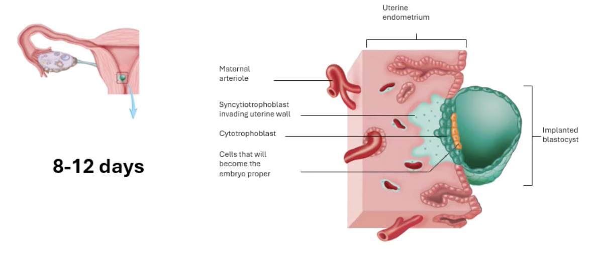
Uterine endometrium
Wall that is closest to the cavity of the uterus
Changes in thickness with each monthly cycle (menstrual)
Implantation and formation of the placenta (14-20 days)
Syncytiotrophoblast invades further into the wall
Changes in Embryo proper
Connection made
Connection stalk develops
Which later becomes umbillical cord
Blastocyst made its way into wall
Cytotrophoblast:
Continues to expand and form finger-like structures
Syncytiotrophoblast:
further invade
As they digest more arterioles, they form pools of blood
called lacuna
will become suplly of nutrients + oxygen for embryo

Implantation and formation of the placenta (14-20 days)
More lacune
Cytotrophoblast cord forms
surrounds the syncytiotrophoblast + lacunae on the maternal side
creates a protective layer for
maternal blood
new embryonic blood
they dont like to mix
embryo makes its own blood + blood vessels
Mesoderm cells:
Extar embryonic mesoderm cells
not part of embryo
builiding own blood + vessels

Fetus
Cytotrophoblast chords form chorionic villi
Fill with blood vessels from the fetus
ONE BIG LACUNA surrounding chorionic villi
Materal aterioles bring fresh blood
Venules take away used blood
Chorionic villi connected via umbelical chord
NOTE:
Umbilical arteries carry deoxygenated blood away from the fetus (arteries move blood away from the heart)
in this case the oxygen comes from the maternal blood supply and goes into the heart of the baby (veins of baby), and then the deoxygenated blood is carried away from the heart by ateries back to refuel.

Chorion
barrier created by chorionic villi
made of
Syncytiotrophoblast (remaining cells)
Basement membrane (thin)
Made of protein fibres
Chorion is the final division between
maternal blood supply
Embryo blood supply
Cytotrophoblast cells disintegrated by this stage.
