Cell structure
1/171
There's no tags or description
Looks like no tags are added yet.
Name | Mastery | Learn | Test | Matching | Spaced | Call with Kai |
|---|
No analytics yet
Send a link to your students to track their progress
172 Terms
What does cell fractionation allow scientists to do?
Study the functions of organelles
What do we do during the first stage of cell fractionation?
We take a sample of tissue containing the cells and homogenise the tissue.
What does ‘homogenising’ a cell mean?
It means to break up the tissue and break open the cells,
What are 2 appliances that can be use to homogenise a cell?
1) Blender
2) Homogeniser
How can we homogenise a cell using a homogeniser?
Place the tissue sample into the glass tube and cover it with buffer solution.
Why do we need to use buffer solution whilst using a homogeniser?
To keep the pH constant
Why is it important that the pH remains constant whilst homogenising?
Because a shift in pH could lead the enzymes in the cell’s organelles to denature
1) What do we do after homogenising the tissue in cell fractionation?
2) Why do we have to do this?
Place the homogeniser on ice
Because cooling the sample means that enzymes work more slowly, preventing any destructive enzymes from damaging the organelles.
1) What do we do after placing the homogeniser on ice?
2) What does this step produce?
1) Push the homogeniser’s plunger up and down to break open cells and disrupt tissue
2) A cell homogenate
What does the cell homogenate (produced from pushing the homogeniser’s plunger up and down) contain?
It contains all the organelles that we find in the cell.
State the largest to smallest of these 4 organelles: Nucleus, Ribosomes, Lysosomes and Mitochondria
1) Nucleus
2) Mitochondria
3) Lysosomes
4) Ribosomes
What is the term used to describe separating the different organelles in a cell?
Fractionation
What machine do we use to carry out fractionation?
Centrifuge
How can we use the centrifuge to carry out cell fractionation?
Place tube containing cell homogenate into the sample holder.
What happens when the centrifuge spins the sample? (Note the different sizes of the organelles)
The organelles are flung towards the bottom of the tube.
Larger organelles (i.e nucleus) experience a greater force and move towards the bottom of the tube faster than smaller organelles.
How does the speed of the centrifuge vary throughout the fractionation process?
First we start with a low speed spin.
What can we see right after the centrifuge spins at a low speed for the first time with the sample inside?
The larger organisms such as the nuclei are flung to the bottom of the tube forming a pellet
What is the ‘pellet’ and ‘supernatant’ in the fractionation process?
Pellet- larger organelles that settled at the bottle of the tube
Supernatant- remaining organelles suspended in the liquid.
What do we do after the pellet and supernatant have formed in fractionation?
We transfer the supernatant into a new tube and centrifuge for a second time at a higher speed spin.
After this spin the pellet would contain mitochondria.
What do we do after the mitochondria has formed in the pellet, made during ultracentrifugation?
Transfer supernatant to new tube and centrifuge again at a higher speed
This time the pellet contains lysosomes
What do we do after the lysosomes have formed in the pellet, made during ultracentrifugation?
Take supernatant for the last time and centrifuge again at the highest speed
The pellet will contain ribosomes, the smallest organelle
How many millilitres is 1m divided into? (1m=xmm)
1000mm
How many micrometres is 1mm divided into (1mm=x micrometres)
1000 micrometres
How many nanometres is 1 micrometre divided into? (1 micrometre= xnanometres)
1000 nanometres
What 3 features do the early light microscope and modern light microscope have in common?
Eyepiece lens
Stage for specimen
Focusing dial
What is the specific difference in magnification between the early light microscope and modern light microscope?
Early light microscope- 300x
Modern light microscope- 1000x
1) What big advantage do light microscopes have over other types of microscopes?
2) What can this advantage allow us to explore
Light microscopes can be used on living cells
Which allows us to explore processes such as (active) cell division and movement of cells
What is the main disadvantage of light microscopes and why does it occur?
It has a low resolution
Because visible light has a longer wavelength
Define the resolution of an object
It is defined as:
The minimum distance between two objects where they can still be seen as two separate objects
What is the limit of resolution for a standard light microscope?
200 nm.
What is the range of wavelength of visible light?
400nm to 700nm
1) Why can we not see two objects that are closer than 200 nm in light microscopes?
2) Which microscope would we use instead?
1) Because that is the limit of resolution in standard light microscopes
2) An electron microscope
How can we calculate the diameter of a cell using a scale bar?
Measure length of scale bar with ruler in millimetres
Measure length of cell using ruler in millimetres
Divide length of cell by length of scale bar and multiply by the value on the original scale bar
1) State the formula for magnification
2) Unit used for image and actual size?
Image size/Actual size
Micrometres- p.s Always keep units the same!!!
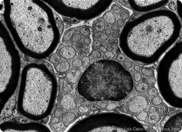
What do electron microscopes use instead of light?
Electrons
Why do electron microscopes have a higher resolution than light microscopes?
Because the beam of electrons have a much shorter wavelength than light
Enabling them to resolve much finer detail.
How much times better is the resolution of an electron microscope compared to a light one?
2000x better
How do transmission electron microscopes work?
They start with an electron gun which produces a beam of electrons
These electrons pass down the microscope.
Why can the electrons in a transmission electron microscope pass through it without bouncing off the molecules in air?
Because the inside of an electron microscope contains a vacuum.
What can we use to focus the electron beams in transmission electron microscopes?
Electromagnetic lenses
How can electromagnetic lenses focus on the specimen in transmission electron microscopes?
Specimen is placed in path of electron beam
Electrons pass through the specimens (through some parts more easily than others)
Final image is displayed on a fluorescent screen
How far can we resolve with an electron microscope?
Up to 0.1nm (under good conditions)
What is one major disadvantage of electron microscopes and why is this the case?
We cannot view living specimens inside it
Because the interior of an electron microscope is a vacuum
Besides the fact that we can’t view living specimens, what is another disadvantage of electron microscopes? (not cost)
It requires careful staining of the specimen and it has to be thin.
What unwanted result can electron microscopes sometimes display and how do they occur?
Artefacts (false images)
due to the staining process
or conditions inside electron microscope
Name the two different types of electron microscope
Transmission electron microscope
Scanning electron microscope
What kind of images does the transmission electron microscope produce?
Flat, black and white 2D shapes
Why do transmission electron microscopes always need thin slices of specimen?
So that the electron beam can pass completely through the specimen
And display an image on the fluorescent screen
How does the scanning electron microscope work?
Electrons are scattered from the surface of the specimen and detected.
What type of images does the scanning electron microscope produce>
3D images which do not need specimen to be thinly sliced
Why does the scanning electron microscope have a lower resolution than the transmission electron microscope?
Because they use lower energy electrons that only scan the surface of specimens instead of passing through them
1) Why do scanning electron microscopes require the specimen to be coated with a metal?
2) What unwanted result can this lead to?
1) So that the specimen can become electrically conductive and thus prevents a build up of charge from the electron beam
2) This can lead to artefacts.
What are the two categories that living organisms are divided into?
Eukaryotes and prokaryotes
Where is the DNA contained in all eukaryotic cells?
A membrane-bound nucleus
What is the DNA of eukaryotic cells tightly wrapped around?
Proteins called histones
What do the DNA and histone proteins combine to form?
Chromosomes
How can eukaryotic cells pack lots of DNA into their nucleus?
By tightly coiling their DNA into chromosomes
Why is eukaryotic DNA considered a linear molecule?
Because the ends of the DNA molecule in a chromosome do not join together to form a loop
What are 2 membrane-bound organelles that are found in plant cells but not animal cells?
Chloroplasts and a vacuole
What is an example of a non-membrane bound organelle in eukaryotic cells?
Ribosomes
What is the difference in size between ribosomes in eukaryotic and prokaryotic cells?
Eukaryotic cells contain 80s ribosomes
Prokaryotic cells contain 70s ribosomes
What does the ‘S’ represent in 80s and 70s ribosomes’?
It represents how quickly organelles move in a centrifuge
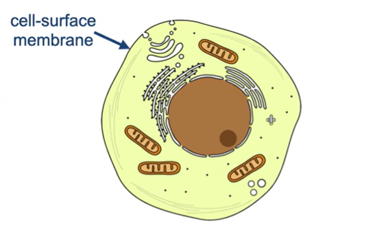
What role does the cell-surface membrane have in a eukaryotic cell?
It helps to control the molecules that can pass in and out of the cell
What is the function of the cell wall in plants?
Maintain the structure of these cells
What is the cell wall made out of in plant cells vs fungal cells?
Plant cell wall- The polysaccharide cellulose
Fungal cell wall- The polysaccharide chitin
Why are prokaryotic cells smaller than eukaryotic cells?
Because prokaryotic cells contain no membrane-bound organelles.
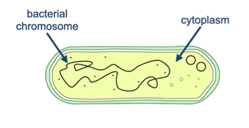
Where is the DNA found in prokaryotic cells?
The cytoplasm
How is the DNA arranged in prokaryotic cells?
Is the DNA bound to any proteins?
The DNA is arranged into a circular chromosome with no free ends
Not bounded to any proteins
What do plasmids in bacterial cells refer to?
Small loops of DNA
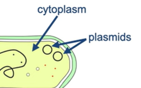
What sort of genes do plasmids contain in bacterial cells?
Genes which make the bacterium antibiotic resistant
What are prokaryotic cells surrounded by?
A cell wall
What is the bacteria cell wall made out of?
Peptidoglycan (also referred to as murein)
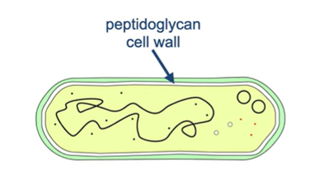
What is peptidoglycan?
Its a polymer formed between peptides and polysaccharide molecules
What use would a bacterial cell wall have if water moved into the cell via osmosis?
It would prevent the cell from bursting.
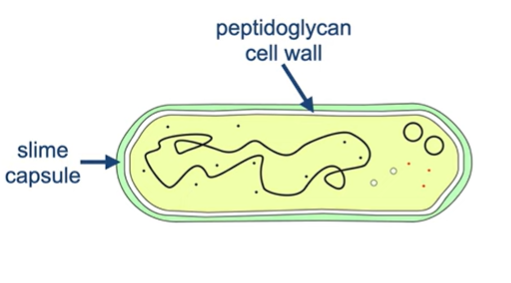
What does the slime capsule produced by some bacteria outside the cell wall help with?
It helps protect the bacteria from phagocytosis by white blood cells
What does the flagellum in prokaryotic cells help with?
Movement
1) What are the fine protein strands on the surface of bacteria celled?
2) What is its function?
1) Pili
2) Pili helps bacteria to attach to surfaces and other bacteria
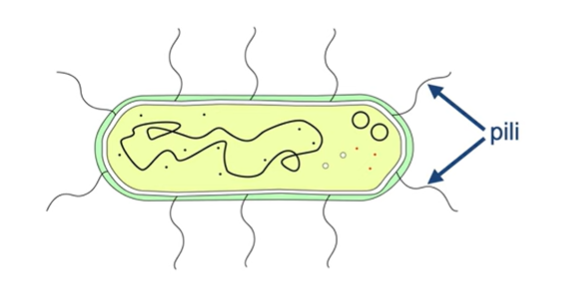
When can DNA be transferred from one bacterium to another?
When two bacterium are attached due to pili
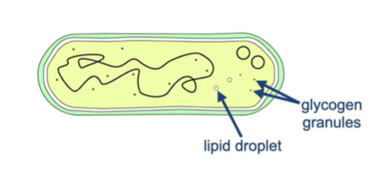
What is the function of lipid droplets and glycogen granules in bacterial cells?
They act as nutrient stores for the bacterial cell
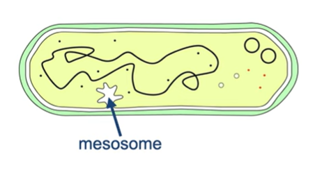
1) What are mesosomes?
2) What did scientists initially think their function was and what are they actually related to?
1) They are infoldings of the membrane of prokaryotic cells
2) Initial suggestion- they played a role in respirati on
What they actually are- Artefacts that are created when bacterial cells are prepared for electron microscopy
What is the general shape of viruses?
Spherical
What is the range in size of virus particles?
Ranges from the smallest with 20nm to the largest with a length of around 300 nm
Why can’t viruses reproduce independently?
Because they can only reproduce inside a host cell
How does a virus reproduce inside a host cell?
Virus particle attaches to surface of host cell
Virus enters host cell
Virus uses host cell’s enzymes to produce copies of itself
Virus leaves host cell (and goes on to infect new host cells)
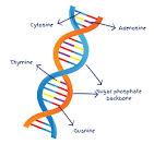
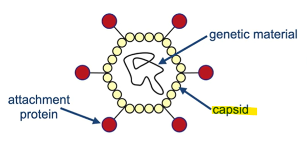
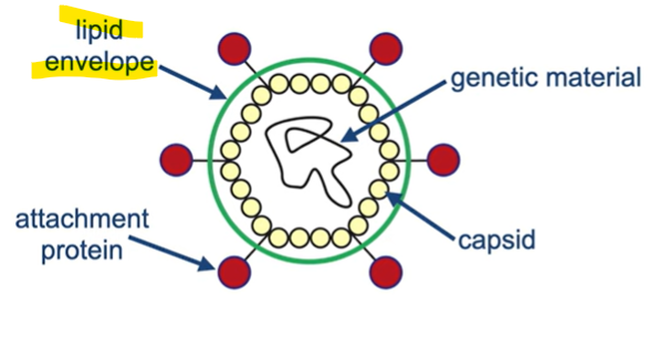
What 3 things do all viruses contain?
Genetic material (such as DNA or RNA)
Capsid (contains genetic material)
Attachment proteins
Where is the genetic material contained in a virus?
It’s contained inside a protein structure called a capsid
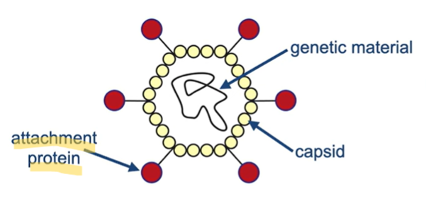
1) What do we find on the surface of viruses?
2) What do they allow the virus particle to do?
Attachment proteins
Allows it to attach to and enter the host cell
1) What is the capsid surrounded by in some viruses?
2) What is it formed from?
A lipid envelope
Formed from the host cell membrane
Why aren’t viruses considered living organisms?
Because viruses cannot reproduce on their own
What are the three main uses of proteins for our cells?
Enzymes for chemical reactions
Structure (eg. organelle movement)
Transport (eg. haemoglobin which transports oxygen)
What is contained within the gene of a protein?
The instructions for encoding its amino acid sequence.
Name the first step of protein synthesis and describe what occurs during this
Transcription
The genetic info encoded by that gene is converted to messenger RNA (mRNA).

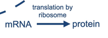
1) What happens after transcription in protein synthesis?
2) What is this process called?
The mRNA leaves the nucleus
Ribosome reads the information contained in the mRNA and synthesises the protein molecule
This is referred to as translation
1) When does translation take place on a free ribosome?
2) Give an example of a protein that this occurs with.
1) It takes place on a free ribosome if the protein remains in the cytoplasm after being synthesised.
2) Cellular enzymes because they stay within the cell.
Where are proteins that are secreted from the cell translated? (proteins that leave the cell)
They’re translated on a ribosome attached to the rough endoplasmic reticulum
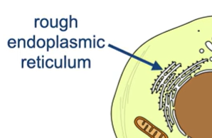
Where do the proteins that secrete the cell go after being translated by ribosomes attached to the ER? (hint: it’s two organelles)
They make their way through the rough ER and the Golgi apparatus before leaving the cell.
What is the nucleoplasm & what does it contain?
A material contained within the nucleus
Contains molecules such as nucleotides and enzymes which are needed for DNA and RNA synthesis.
What is the nuclear envelope & what does it consist of?
It is the double membrane that surrounds the nucleus.
Consists of two phospholipid bilayers.
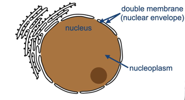
1) Besides the phospholipid bilayers, what is found within the nuclear envelope?
2) What is its function?
1) Nuclear pores
2) It allows molecules to enter and leave the nucleus.
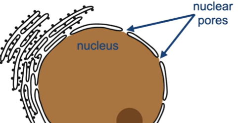
1) How do RNA nucleotides enter the nucleus?
2) What are these nucleotides used for?
1) Through nuclear pores from the cytoplasm.
2) They are used in the nucleus to synthesise mRNA.