Veinous Supply & Lymphatics of Head and Neck
 Call Kai
 Learn
Learn Practice Test
 Spaced Repetition
 Match
Match Flashcards
 Knowt Play
1/48

There's no tags or description
Looks like no tags are added yet.
Name | Mastery | Learn | Test | Matching | Spaced | Call with Kai | Chat |
|---|
No analytics yet
Send a link to your students to track their progress
49 Terms
What are the boundaries of the scalp? anterior, posterior and laterally
anterior: superior orbital margin
posterior: superior nuchal lines (occipital bones)
laterally: zygomatic arch on each side
What does each letter of scalp stand for?
skin, connective tissue, aponeurosis, loose areolar tissue (allows the aponeurosis to glide over the pericranium), pericranium
What is found in the skin layer?
hair, sebaceous glands and sweat glands
What may occur if there is any issues with the sebaceous glands? (2)
dandruff and skin dryness
What is found in the connective tissue layer of the scalp?
what is it?
what does it connect?
contains (2)
close network of fibro-fatty tissue
connects the overlying skin with the under lying aponeurosis
contains large blood vessels and nerves
the connective tissue is stretched
What can injury to a scalp cause?
bleeding (vessels can’t retract) and pain (when a haematoma forms)
What is the aponeurosis?
role?
the layer underneath?
acts as a tendon, connects the frontal belly and occipital belly of the occipito-frontalis muscle
tightly attached to the connective tissue
the loose subaponeurotic layer - loose areolar tissue
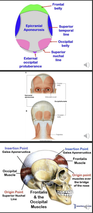
What is the pericranium?
the periosteum of the skull
continuous with endosteum at the sutures
What are the 3 cerebral meninges?
dura mater
arachnoid mater
pia mater
What is the dura mater?
what are the 2 layers?
its attachments?
dense inelastic membrane
meningeal - inner
endosteal - outer
loose contact with calvaria
strongly adherent to the base of the skull
What does the dura mater have?
reflections that are essentially fold of the dura mater,
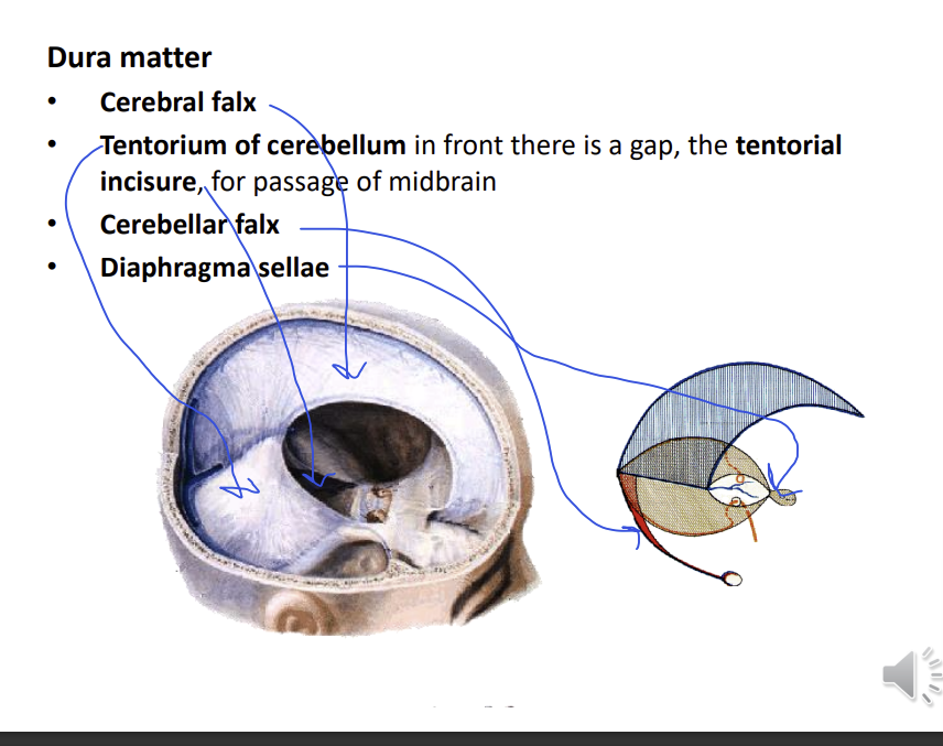
What are the 4 folds of the dura amter
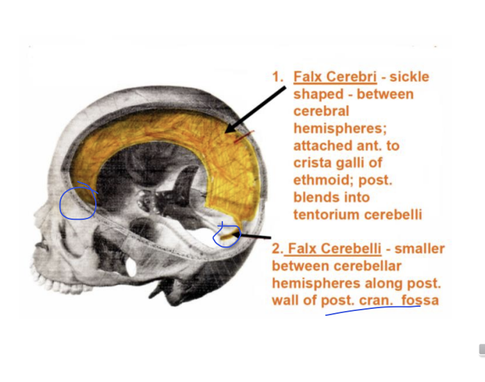
cerebral falx - lies between the 2 cerebral hemispheres
tentorium of cerebellum - separates the cerebellum from the cerebral hemispheres - roof of the cerebellum
tentorial incisure - for passage of the midbrains
cerebellar falx
diaphragma sellae - in contact with the pituitary gland - on the sella turcica
What is the cerebral falx shape and attachments?
sickle shaped
attached anteriorly to the crista galli of ethmoid,
posteriorly it blends into tentorium of the cerebellum
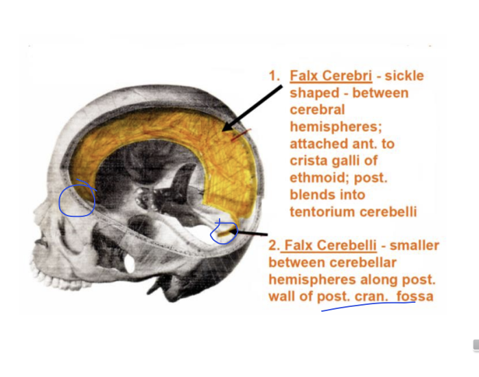
What is the cerebellar falx?
smaller
between cerebellar hemispheres along the posterior wall of the posterior cranial fossa
What is the role of the tentorium of the cerebellum?
forms the roof of posterior cranial fossa
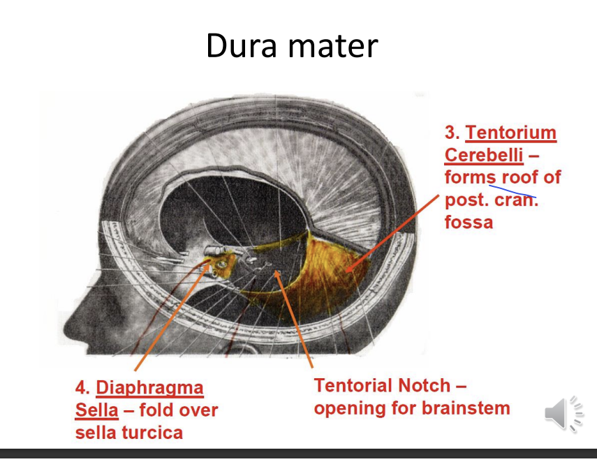
What does the tentorial notch allow for?
opening for the brain stem
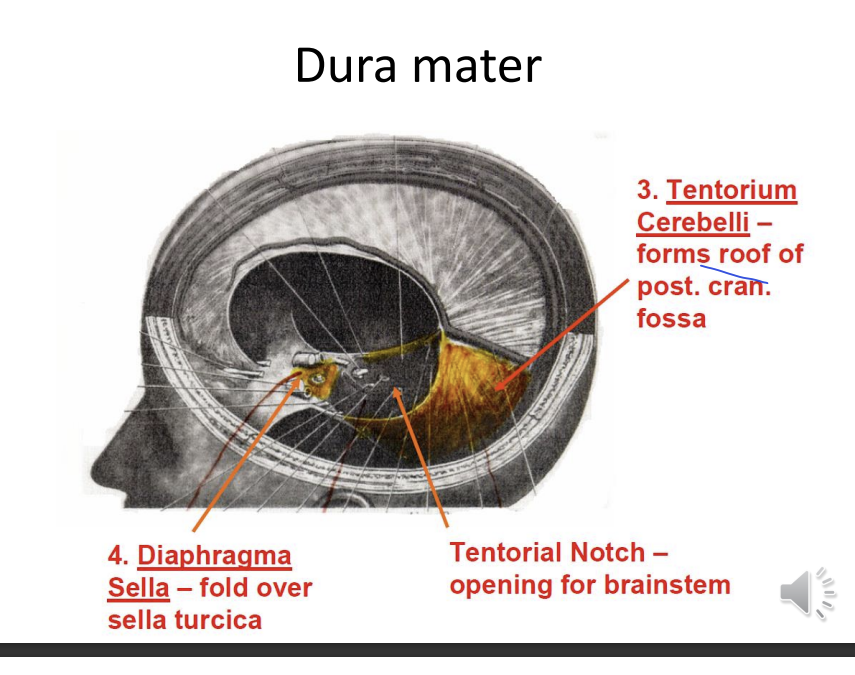
What is the diaphragma sella?
fold over the sella turcica
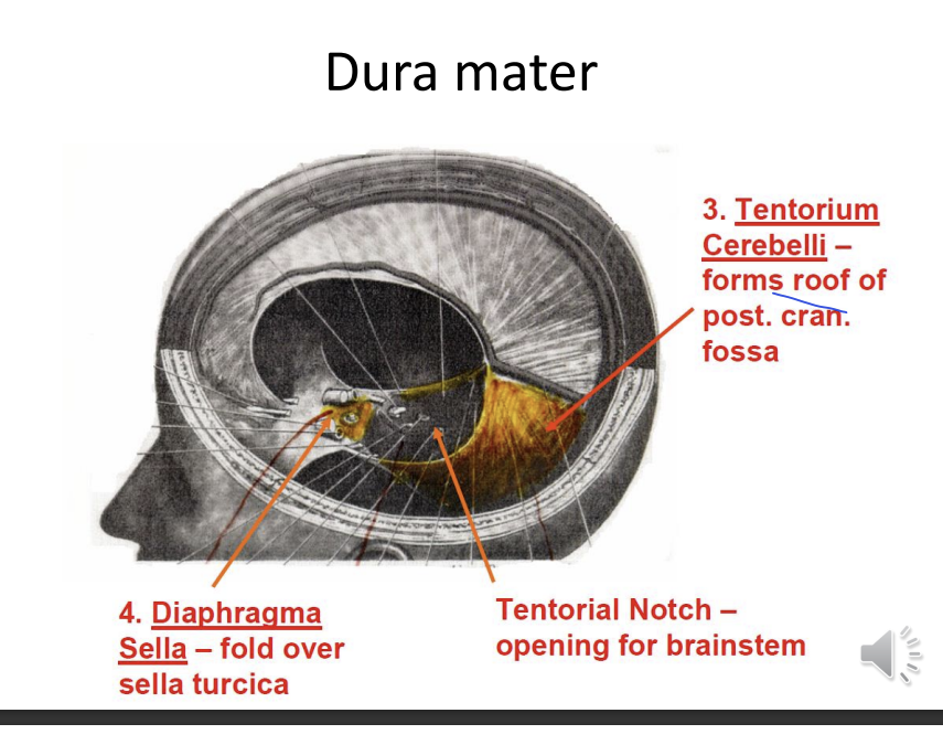
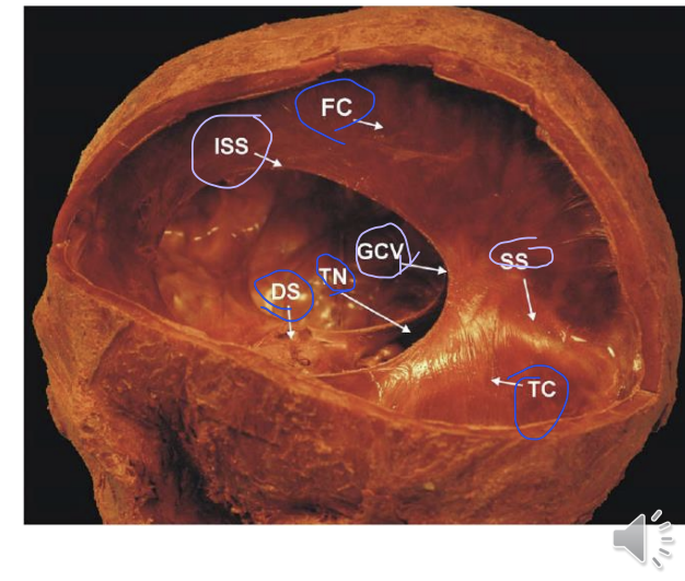
What are these sections?
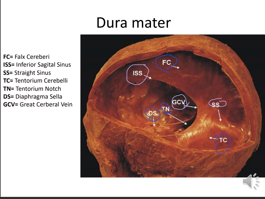
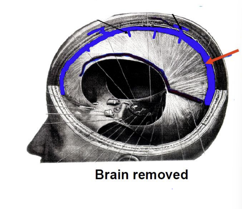
What is this sinus called?
superior sagittal sinus
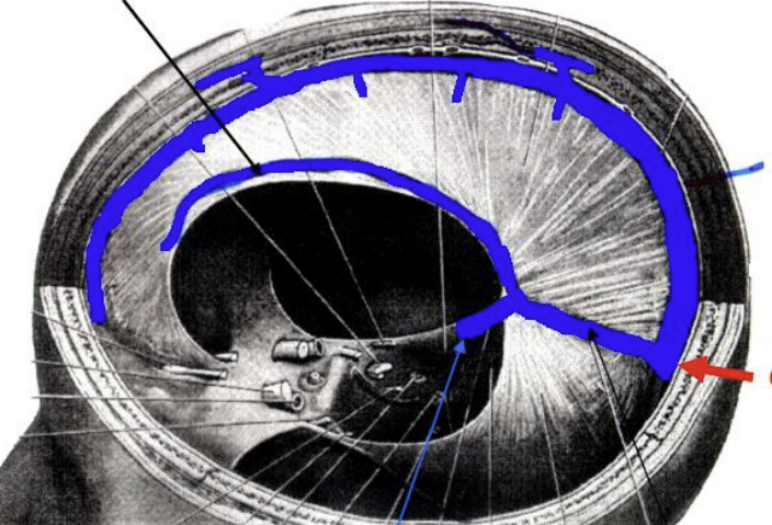
What are the other 2 sinuses?
what is the red arrow?
straight sinus
inferior sagittal sinus
confluence - where the superior sagittal connects with the straight sinus
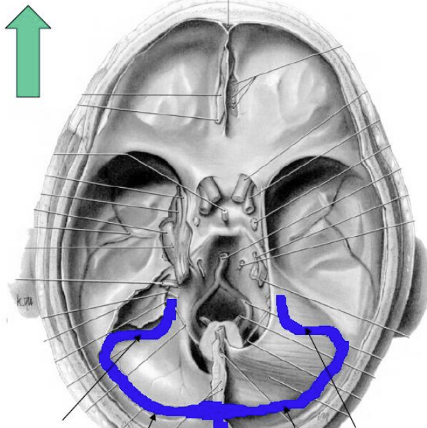
What are these two sinuses?
sigmoid sinus - connects to the jugular foramen and drains venous blood into the jugular vein
transverse sinus
they are both connected to the confluence
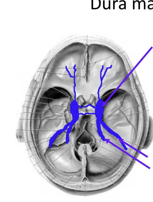
What are these 2 sinuses?
where are they found?
cavernous sinus - in middle cranial fossa, on the side of the body of the sphenoid bone
superior and inferior petrosal sinuses - petrous part of the temporal bone
superior is more lateral on the image
the superior petrosal sinuses are connected to the transverse sinus
inferior petrosal sinus drains directly into the jugular vein through the jugular foramen
What structures are found in the cavernous sinus?
internal carotid artery and abducent nerve (C6) - run through the sinus
oculomotor (C3), trochlear (C4)and ophthalmic and maxillary divisions of trigeminal (C5) nerve lie in the lateral walls of the sinus
orange - pituitary gland
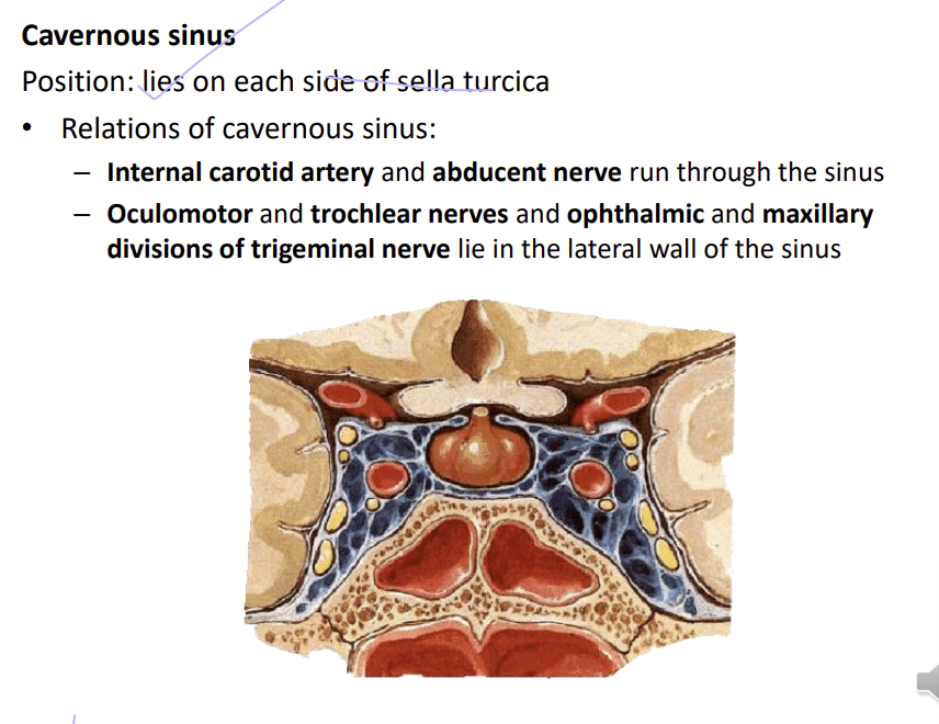
How is the flow of the blood in the Dural sinuses (Flow chart)
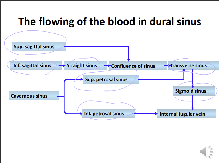
The venous drainage from the head and neck and brain terminates where?
internal jugular vein
the internal jugular vein drains where?
superior vena cava
what is the course of the internal jugular vein? (4)
emerges from the jugular foramen
passes through the carotid sheath
joins the subclavian vein to form the brachiocephalic vein
drain into the superior vena cava - the largest vein in the body
Name 4 other veins in the head and neck?
facial, pterygoid plexus. retromandibular vein, maxillary vein
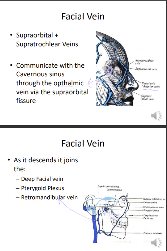
Describe the facial vein
supraorbital and supratrochlear veins join it
communicates with the cavernous sinus through the ophthalmic vein via the supraorbital fissures
as it descends, it joins the deep facial vein (brings deoxygenated blood from the pterygoid plexus), pterygoid plexus and retromandibular vein
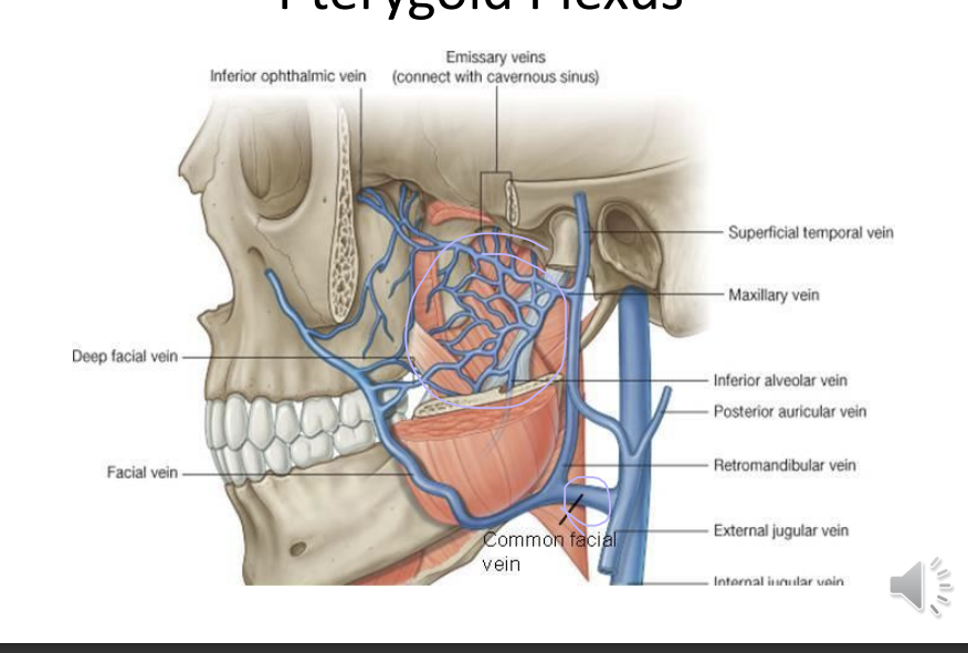
What i this image showing?
pterygoid plexus
PP drains into the deep facial vein join the facial vein
facial vein joins the retromandibular vein
all join the common facial vein
this will then join the internal jugular vein
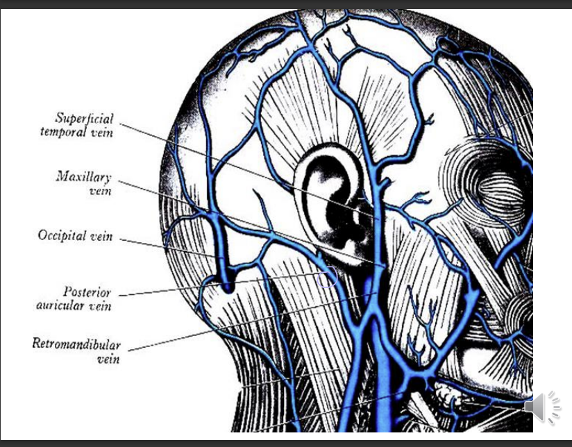
Describe the retromandibular vein
formed by?
passes through
two divisions of exit?
formed by superficial temporal vein and maxillary vein
passes through the parotid gland
anterior - join the facial vein
posterior it joins the posterior auricular vein
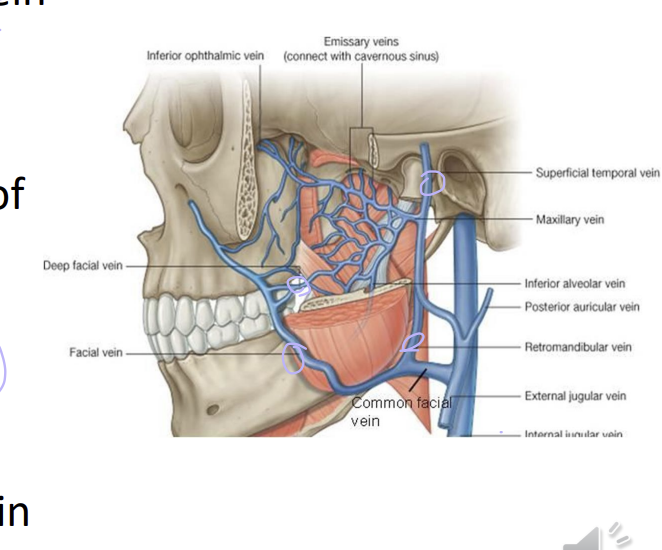
Describe the maxillary vein
what can drain into it?
what does it pass between?
what does it join with
pterygoid plexus can drain into the maxillary veins
sphenomandibular ligament and the neck of the mandible
joins the superficial temporal vein
joins with the superficial temporal vein - forms the retromandibular veins
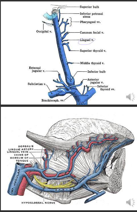
Give an example of a vein that also drains into the internal jugular vein?
lingual vein - drains the floor of the mouth and tongue
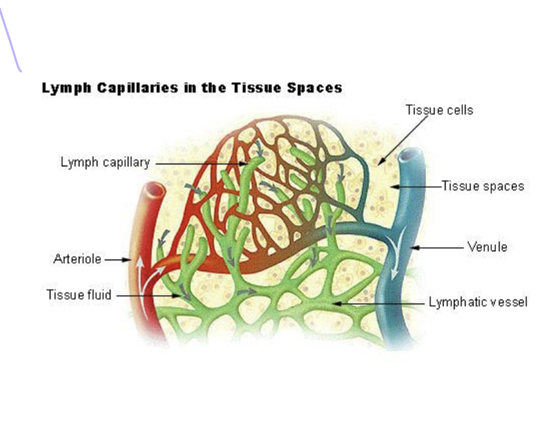
How is lymph formed and what is it composed of?
fluid within the blood is - plasma
plasma is filtered through blood capillaries into tissue space - interstitial fluid
interstitial fluid passes into the lymph vessels - lymph
lymph is composed of interstitial fluid and lymphocytes
What are the 3 functions of the lymphatic system?
1) draining excess interstitial fluid
2) carrying out immune response
3) transporting dietary lipids and lipid soluble vitamins such as A D E K
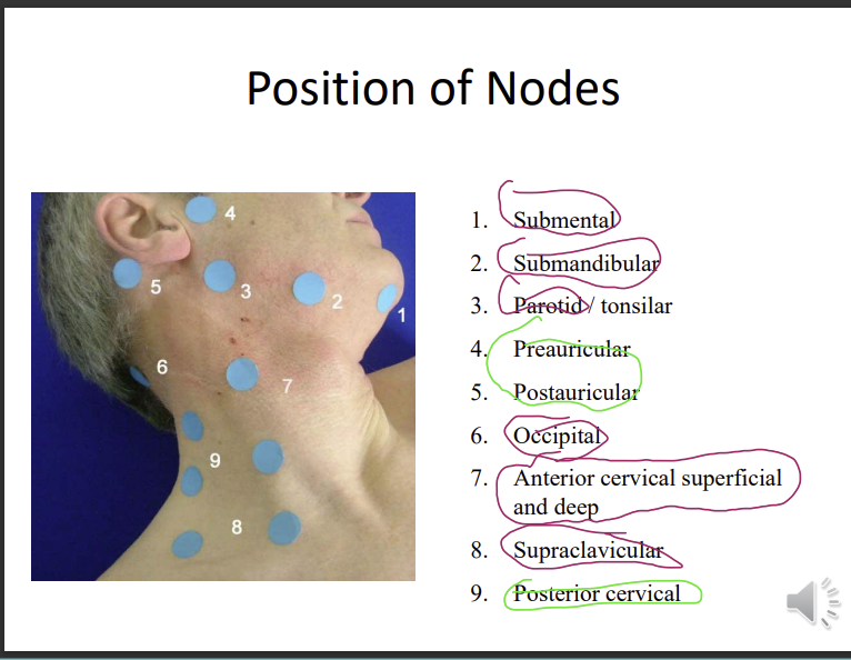
What are regional groups of lymph nodes? 5 regions
2, 2, 2, 2, 3
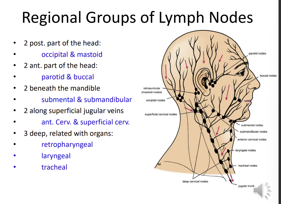
What do all of those lymph noes drain into?
drain into the deep cervical lymph nodes, which then enter the jugular vein
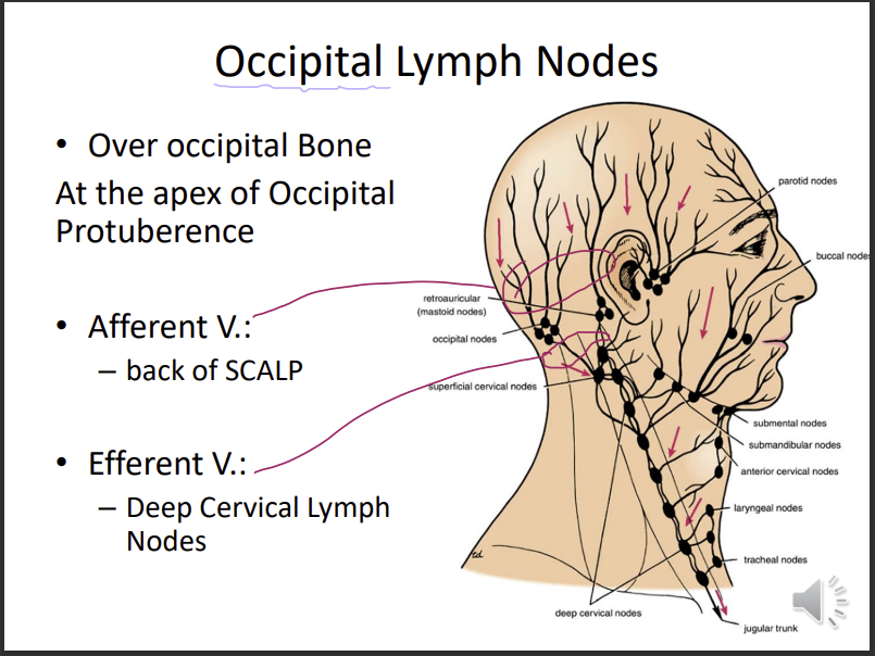
describe ethe occipital lymph nodes?
location
over the occipital bone - apex of the occipital protuberance
afferent vessels - back of the scalp (take in lymph)
efferent vessels - release lymph into the deep cervical lymph nodes
The other name for the mastoid lymph nodes?
location
retroauricular
over the lateral surface of the mastoid process
afferent vessel:
scalp above the auricle
posterior medial auricle
posterior wall of the external auditory meatus
efferent vessel:
deep cervical lymph nodes
auricle - ear
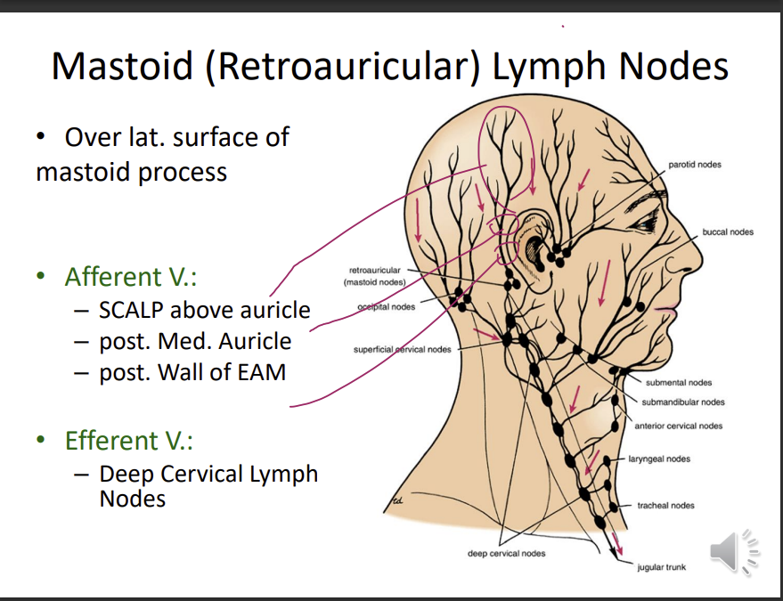
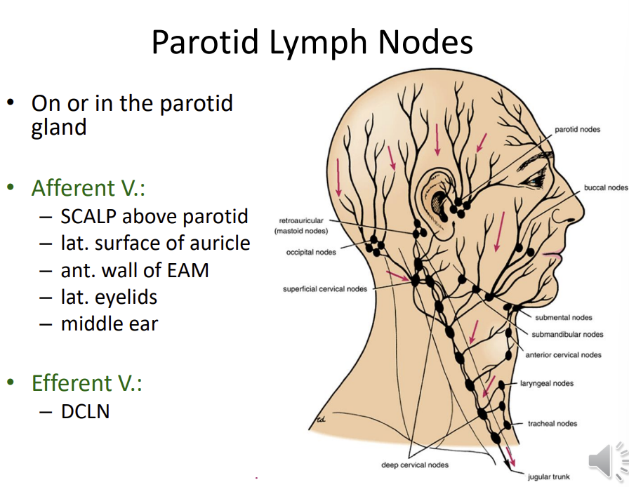
Describe the parotid lymph nodes
location?
on or in the parotid gland
afferent vessels:
scalp above the parotid
lateral surface of the auricle
anterior wall of the EAM
lateral eyelids
middle ear
efferent:
DCLN
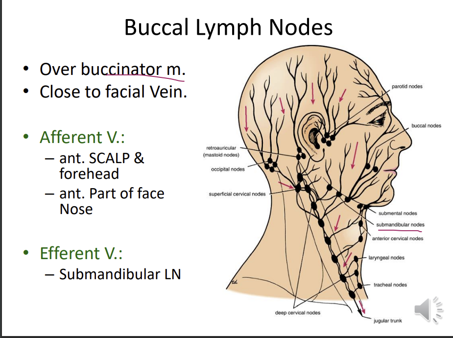
describe the buccal lymph nodes
location
found over the buccinator muscle, close to the facial vein
afferent vessels:
anterior scalp and forehead
anterior part of the face sides of the nose
efferent:
submandibular lymph nodes
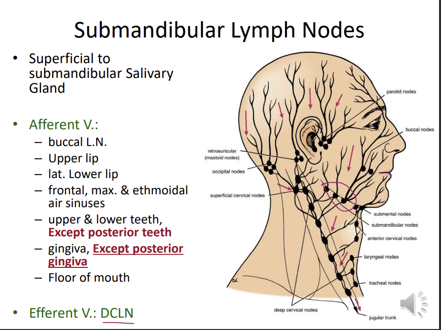
Describe the submandibular lymph nodes
location
superficial to submandibular salivary glands
afferent:
buccal lymph nodes
upper lip
lateral lower lip
frontal, max, ethmoidal, air sinuses
upper and lower teeth except the posterior teeth
gingiva, except for the posterior gingiva
efferent:
floor of the mouth
What are the efferent and afferent drainage of submental lymph nodes?
afferent vessels:
tip of the tongue
lower incisors and associated gum
central lower lip
Efferent:
submandibular ln
Describe the anterior CLN
location
along the course of anterior jugular vein
afferent - anterior neck
efferent : DCLN
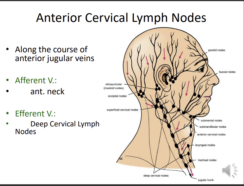
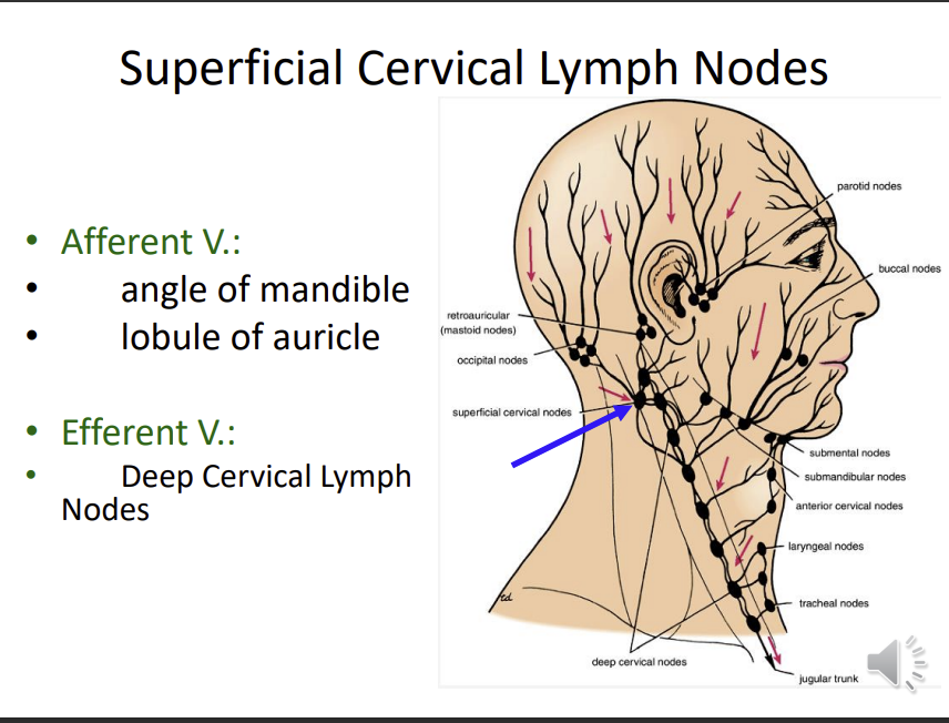
Describe the superficial cervical lymph nodes
afferent and efferent?
afferent vessels:
angle of the mandible
lobule of auricle
efferent lymph nodes:
DCLN
What are the DCLN?
location
efferent
afferent
a chain of lymph nodes located along the course of the internal jugular vein mainly on its lateral aspect
extends from the base of the skull to the root of the neck
inside the carotid sheath
afferent - receives lymph from all lymphatic structures in the head and neck
efferent - join to form the jugular lymph trunk
What does the jugular lymph trunk join to?
right lymphatic duct or thoracic duct
venous system (vena cava)
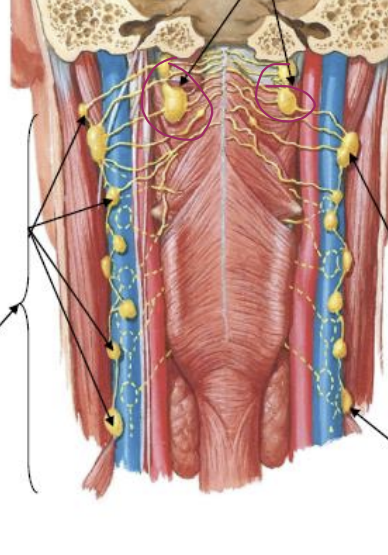
where are the retropharyngeal lymph nodes in thi image?
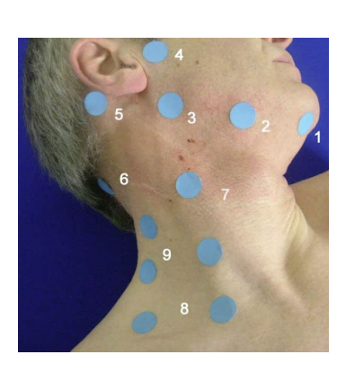
name the lymph nodes