Fall NMT: GI
 Call Kai
 Learn
Learn Practice Test
 Spaced Repetition
 Match
Match Flashcards
 Knowt Play
1/121

There's no tags or description
Looks like no tags are added yet.
Name | Mastery | Learn | Test | Matching | Spaced | Call with Kai | Chat |
|---|
No analytics yet
Send a link to your students to track their progress
122 Terms
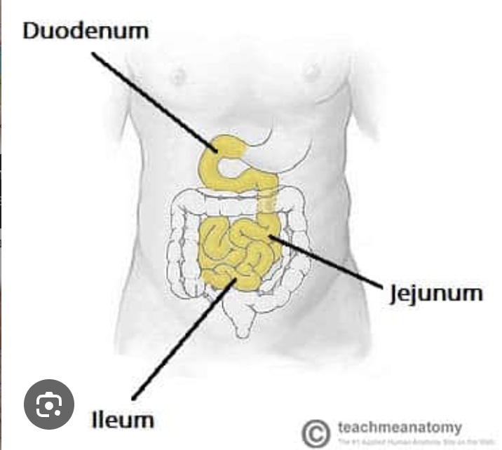
Are these parts apart of the small or large intestine
Duodenum
jejunum
ileum
small intestine

Are these parts apart of the small or large intestine
cecum Ascending and descending colon
rectum
anal canal
large intestine
small intestine function
digestion and absorption of nutrients
large intestine function
reabsorption of water (formation of fecal material), some vitamin absorption
Localization techniques for upper GI bleeds
endoscopy
Localization techniques for lower gi bleeds
Superior anatomical visualization
must be active
require IV contrast
Angiography CTA
Localization techniques where higher blood flow= better chances to indetify a bleed
Radionuclide test
specifically tagged RBCs
T or F: ACUTE GI bleeding is potentiall life threatening
true
what are the two radionuclide techniques to find a GI Bleed when imaging
Tc- 99m Sulfur Colloid
Tc-99m tagged RBCs
what is the preffered method of radionuclide techniques to find a GI Bleed when imaging
Tc-99m tagged RBCs
These are characteristics of which radionuclide for a Gi Bleed
Simpler process/ procedure
less sensitive
high-target-to -background ratio
increased spleen, liver, and bone marrow uptake
must have an active GI bleed at injection time
false negatives
possible under abdomen sites can be missed
higher radiation doses to liver and spleen
99m-Tc sulfur colloid
when immaging for a GI bleed using sulfuf colloid you should block what area and why
small portion of the liver and spleen since their is a large amount of uptake so the area can look intense
when using Tc-99m sulfur colloid for a Gi Bleed what is the critical organ
liver and spleen
what position and area should be imaged for GI bleed procedure
Supine including the liver to the pubis symphysis
what is the dose range for sulfur colloid GI bleeds
7-10 mCi 99m-Tc Sulfur Colloid through an IV bolus
what imaging is used for a SC GI bleed study
ANT flow for 2-3 min
ANT static every 1-2 min for 20-30 min
obliques, Lats, and POST as necessary
results of a SC GI bleed
active bleeding OR bleeding most likely seen after 5min of imaging
appears focal area of inc. activity then becomes more intense ad background decreases
IF YOU HAVE TO RESCAN YOU HAVE TO RE-INJECT
Higher bleeds can be missed
These are characteristics of which radionuclide for a GI bleed
Agent of choice for intermittent bleeding
Stays in the system for over 36hrs allowing delayed images
Lower radiation dose to liver and spleen
Better detection of GI bleed in upper abdomen
Tc-99m RBCs
what imaging is used for a Tagged RBC GI bleed study
ANT flow for 2-3 min
ANT Dynamic 10-60sec/60-90min
Obliq, lats,POST, SPECT as necessary
Delayed: 10-60sec/ 30-60min
what is the dose range for tagged RBC GI bleeds
15-25mCi Tc-99m Tagged RBC through IV bolus
when using tagged RBC for GI bleeds wha do you have to watch out for
neck images “bad tags”
reuslts of a Tc-99m tagged RBC GI bleed
bleeding may be seen on flow since tagged
Pattern of focal tracer activity that becomes more intense and changes position with time and is not related to normal structures
SMALL Bowel: central location moves rapidly
LARGE Bowel: peripheral, curvilinear pattern
Delayed (+): hard to pinpoint site of bleed
reuslts of a Tc-99m tagged RBC GI bleed
SMALL Bowel:
central location moves rapidly
reuslts of a Tc-99m tagged RBC GI bleed
large bowel
peripheral, curvilinear pattern
normal abdominal strucures not related to GI bleed for tagged rbc
liver, spleen, abdominal vessels, kidneys, bladder, stomach, genitals
reuslts of a Tc-99m tagged RBC GI bleed
delayed (+)
hard to pinpoint site of bleeding
where the blood and tc-99m exit in a GI bleed
through the bowel witht he pt having stool in their blood

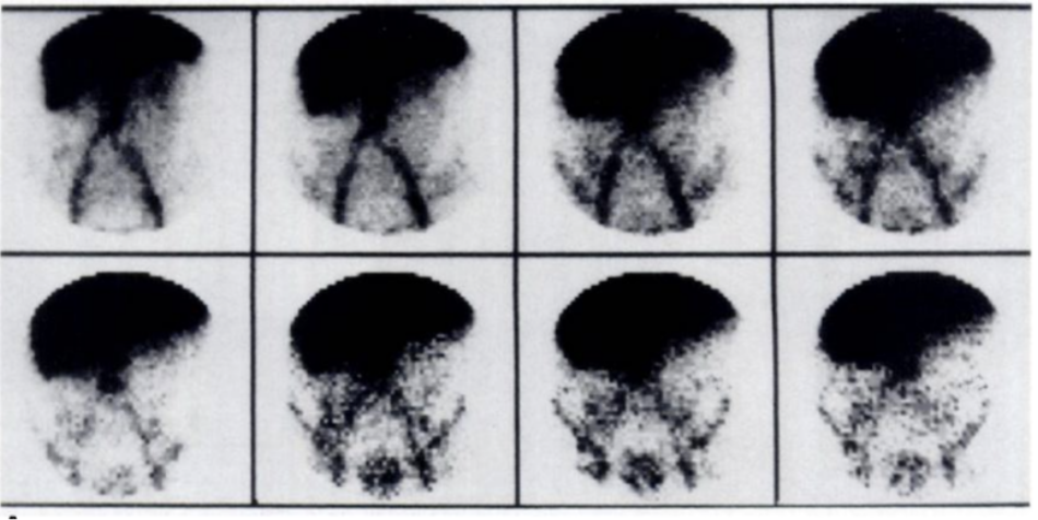
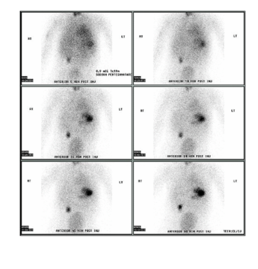
is this a negative Sulfur Colloid or tagged RBC
Negative Sulfur Colloid

is this a negative Sulfur Colloid or tagged RBC
Negative Tagged RBC

is this a positive or negative GI bleed
postive (centrally located)

is this a positive or negative GI bleed
positive (small intestine)

is this a positive or negative GI bleed
positive (large intestine)
Cells of the stomach mucosa
parietal cells
secrete hydrochloric acid
Cells of the stomach mucosa
Chief cells
secrete pepsinogen and intrinsic factor
Cells of the stomach mucosa
G cells
secrete gastrin
Cells of the stomach mucosa
columnar mucin-secreting epithelial cells
alkaline secretions
ectopic/ heteropic gastric mucosa
located anywhere in sbdomen most freq. in the RLQ
gastric mucosal secretion can cuase peptic ulcerations
what are the 2s used to describe Meckel’s Diverticulum
present in Approx. 2% of population
under the age of 2
Approx. 2cm in size
within 2 feet of the ileocecal

Meckel’s Diverticulum is more present in males or females
males
T or F: imaging for Meckels is mainly preformed in adults
false kids
cause of meckel’s
failure of closure of an embryonic duct
What radionuclide is used for meckel’s imaging
Tc-99m pertechnetate
prep for a meckel’s test
NPO (makes stomach smaller)
No laxitives or barium for 3 days prior (irritates bowels mucosa)
NO potassium perchlorate (blocks Tc uptake)
Pretreatment for a meckel’s test that enhances visualization
Pretreatment for a meckel’s test that enhances visualization
pentagastrin (ENHANCES)
stimulates gastric uptake of Tc
Removed from the market bc of side effects
Pretreatment for a meckel’s test that enhances visualization
Cimetidine (blocks and holds Tc)
decreases release of Tc into bowel
Pretreatment for a meckel’s test that enhances visualization
Glucagon (increases)
relaxes smooth muscles, decreases peristalsis, increases persistence in gastric mucosa
positioning for meckels imaging with LEHR or LEAP collimator
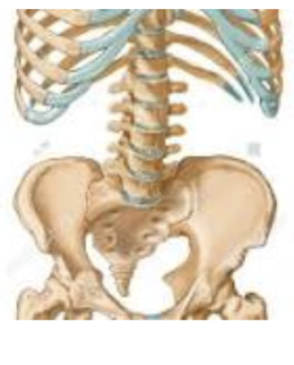
Supine including the xiphoid to the symphysis pubis
dose range for meckel’s imaging
5-15mCi pertechnetate FOR ADULTS*** given through iv bolus
Must caluclate a dose for kids
childs dose calculation
(kids wt) x (adult dose)/ 150lbs
Imaging set up for Meckel’s study
ANT flow 1-5sec/frame for 1 min
ANY dynamic 30-60 sec/for 30 mi.
ANT static 5 min intervals for 30 min.
Delay: Obliq., Lats., POST. for eliminating areas of increased activity
Postviod image-moves activity in renal structures
results of a meckes (+)
focal area with increased uptake
normally in RLQ
seen within 5-10 min. of injection
continuous incr. intensity with imaging
reasons for a false neg. in a meckels study
Failing to image the entire abdomen, small gastric mucosa, removal of Tc by bowel secretions
reasons for a false pos. in a meckel’s study
Intussusception, volvulus, tumor or cyst, renal structures
Intussusception
telescoping of the bowel
Intussusception can be seen as a flase pos for meckels how can it be seen as a false neg.
if blood flow becomes restricted
Volvulus
twisting of the bowel

pos or neg for meckel’s
positive
normal diffuse uptake areas areas seen for meckels
stomach, bladder, kidney

major salivary glands
parotid
serous cells
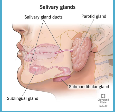
3 major salivary glands
sublingual
mucous glands

3 major salivary glands
submandibular
mix of serous and mucous cells
indications for salivary imaging
warthin’s tumor
differation from other malignant/ begign salivary tumors
Indications for salivary imaging
Xerostomia
evaluation of mouth dryness or other oral cavity / mucous membrane conditions
indications for salivary imaging
inflammation
of the salivary glands
indications for salivary imaging
Sjogren’s syndrome
a long-term autoimmune disease where the immune system attacks the glands that make tears and saliva
dose range and radionuclide for salivary imaging
5-10mCi Tc-99m through IV
method of localization for salivary imaging
active transport
psotioning for a salivary study
pt supine with a pillow under the shoulders wl the neck extended with camera ANT over calse and neck as close as possible
imaging foe a salivary study
Flow in water position (chin up): 1-2 sec image
Dynamic: 1-2 min/ frame 20-60 min or Static: 5-15 (ANT, RLAT, LLAT)
Lemon juice to stimulate saliva secretion while imaging
limits to salivary imaging
not very specific
poor indicator of anatomy
useful role in evaluating certain diseases
salivary normal results
glands have homogenous distribution of Tc
localization is symmetrical in all glands
rapid, symmetrical, and profound dec. of radiotracer loalization after lemon juice
waters position does what
prevents super inposition of the glands (imaging on top of eachother)
the lemon juice should do what
cause saliva to releas the Tc equally

is this normal or irregular results for a salivary
normal (neg) wash out is seen equal after lemon juice (arrow)
Abnormal results that could be an indicator of what…
vasular or connective diseases causing little or no uptake in the salivary glands (dry mouth)
asymmetric arival in glands or delayed compared to thyroid
delayed images showing bilateral decreased uptake (little to no repsonse to lemon juice)
Sjogren’s Syndrome
cold (no uptake) spots in a salivary study can mean
metastatic tumor, cyst, abscess, enlarged lymph nodes
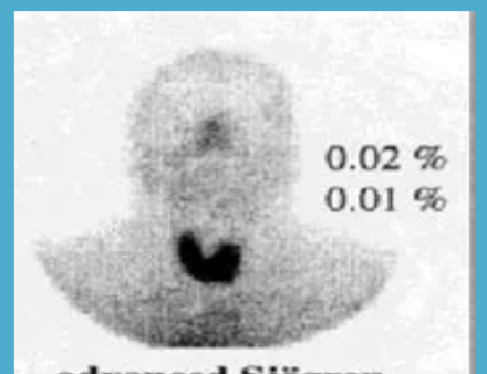
what could this salivary test indicate
no salivary glands seen
Sjogern’s syndrome

what could this salivary test indicate
cold spot warthins tumor
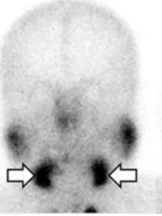
what could this salivary test indicate
normal salivary secretion
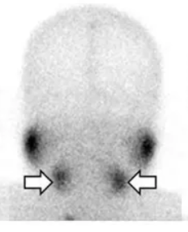
what does this salivary test indicate
hypofunction (since the glands are not evenly hot)
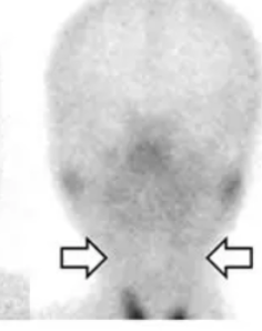
what does this salivary test indicate
no function m
a benign mass of tangled up blood vessels /cells that can be small large, or multiply
hemangioma

there are no signs or symptoms to liver hemangiomas, so how are they found
on accident when other imaging tests are being done like CT, ultrasounds, or MRIs
why do they avoid biopsy’s for liver hemangiomas
bc tumors are in high vascular areas so a biopsy can cause bleeding, invasive, and are unecessary
which scan would be better for visualizing a hemangioma, Planar or Spect
SPECT 88% while planar is 55%
what are the 2 types of radionuclides that can be used for a SPEct Hamangioma prodcedure
20-30 mCi Tc-99m Tagged RBC and Tagged RBC PYP (pyrophosphate)
while both 20-30 mCi Tc-99m Tagged RBCand Tagged RBC PYP (pyrophosphate) can be used for a liver SPECT hemangioma what is preffered
20-30 mCi Tc-99m Tagged RBC
method of localization for Tc-99m Tagged RBCand Tagged RBC PYP (pyrophosphate)
compartmental (meaning tagged to and circulating with the blood)
Positioning for a liver SPECT hemangioma
supine
imaging for a liver SPECT hemangioma
Flow: based on prior imaging
blood pool static: ANT marker, ANT, RAO, RLAT, POST
Delay/SPECT: 1-2 hrs later
normal areas of uptake for a liver SPECT hemangioma
Heart, great vessels, spleen and kidneys
liver SPECT hemangioma uptake (delay)
heterogenous uptake of liver parenchyma with the hepatic vessels prominent in various slices
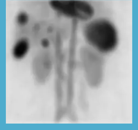
positive liver SPECT hemangioma image
initially hypovascular with filling over time
cold during blood pool with liver activity being greater than hemangioma
during delay: hemgioma activity greater and the rest of the liver and heart and spleen are equal
T or F: only hemangiomas retain RBC until the 2 hour interval
true
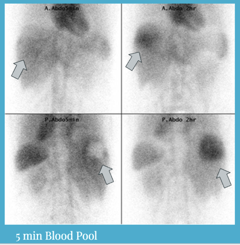
is this a positive or neg liver hamangioma
positive
what does the portahepatis consit of
hepatic artery
portal vein
bile ducts
75% of blood supply for the liver is via the
portal vein
25% of blood supply for the liver is via the
hepatic artery
venous blood draining from the intestines carried to liver; carries nutrients
absorbed from intestines to liver for processing by hepatocytes
portal vein
arterial oxygenated blood; branch of abdominal aorta
hepatic artery