Hematology Solo 3 Erythrocytes Lecture 1 Erythropoiesis, Maturation, Membrane Characteristics, and Metabolic Activities
 Call Kai
 Learn
Learn Practice Test
 Spaced Repetition
 Match
Match Flashcards
 Knowt Play
1/43

Earn XP
Description and Tags
Name | Mastery | Learn | Test | Matching | Spaced | Call with Kai | Chat |
|---|
No analytics yet
Send a link to your students to track their progress
44 Terms
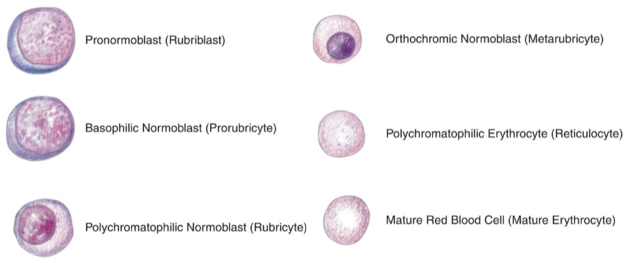
Erythropoiesis Nomenclature
Pronormoblast (Rubriblast)
Basophilic Normoblast (Prorubricyte)
Polychromatophilic Normoblast (Rubricyte)
Orthochromic Normoblast (Metarubricyte)
Polychromatophilic Erythrocyte (Reticulocyte)
Mature Red Blood Cell (Mature Erythrocyte)
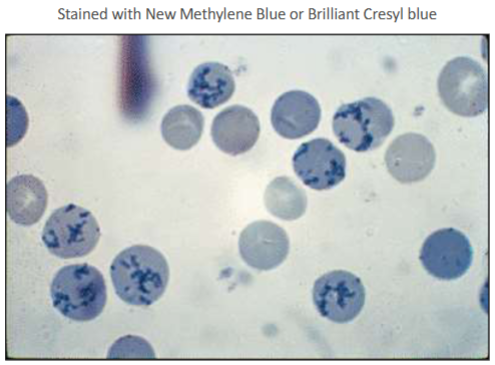
Reticulocytes are only classified as such when stained with new methylene blue revealing organelles in a granulofilamentous arrangement
Erythropoiesis
The production of erythrocytes in the bone marrow.
This occurs in erythropoietic islands of medullary bone marrow over a period of about 5 days.
The average life span of a maturing red blood cell is 120 days

Erythropoietic Islands
Distinct anatomic units that consist of a macrophage surrounded by a cluster of maturing erythroblasts
Hgb synthesis occurs
Arrow is pointing to a macrophage

Erythropoiesis
Begins with a pluripotential stem cell followed by a progenitor cell, which is stimulated by Erythropoietin (EPO) for erythrocyte production
EPO is a hormone produced by the kidney that maintains a constant red cell mass
1. Renal sensors sense a decrease in the body’s oxygen level
2. EPO production is increased
3. This stimulates the bone marrow erythroid precursors to proliferate and increase the production of red blood cells
Disorders Related to Erythrocyte Maturation and Production
Disorders of Erythropoietin
Polycythemia is the term used to refer to an increased concentration of erythrocytes (erythrocytosis) in the circulating blood that is above normal for gender and age
Secondary, or absolute, polycythemias reflect an increase in erythropoietin production and should not be confused with polycythemia vera or relative polycythemias
Mechanisms that can produce secondary polycythemia include the presence of high oxygen affinity hemoglobin, chronic lung disease, smoking, and dwelling in high altitudes
RBC Maturation: Key Points
The stage of maturation is determined by
1. Cell size
2. Nuclear size compared to the rest of the cell: Nucleus to cytoplasm (N:C) ratio
3. Nuclear chromatin: Texture, density, homogeneity
4. Presence or absence of nucleoli
5. Color of cytoplasm
Nucleus is round in all maturation phases
The primary characteristic to evaluate when determining maturation is Nuclear Chromatin

Erythropoiesis
The following changes occur in the developmental stages of RBC maturation
1. Cell volume decreases
2. N:C ratio decreases
3. Nuclear chromatin goes from fine to becoming condensed
4. Nucleoli disappear
5. Cytoplasm color changes from blue to pink as RNA decreases and hemoglobin synthesis increases

Select the correct maturation sequence of erythropoiesis
Rubriblast, Prorubricyte, Rubricyte, Metarubricyte, Diffusely basophilic erythrocyte, Erythrocyte
As erythroid cells mature with each stage
Amount of RNA in the cytoplasm decreases

Rubriblast
Also known as pronormoblast or proerythroblast
Earliest and largest cell in the erythroid series
Cell size is 14-24 micrometers
Nucleus
Nucleus is round (almost spherical) with distinct, linear chromatin strands
Nuclear to cytoplasmic ratio is high (8:1 to 6:1)
Nucleus stains reddish-blue with 0-5 nucleoli visible
Chromatin: blue, fine, diffuse, loose, no evidence of clumping
Cytoplasm is deep royal blue; scant amount with no granules
Reference range: 0-1.5% in bone marrow

Prorubricyte
Also known as basophilic normoblast, basophilic erythroblast
Cell size is 12-17 micrometers
Nucleus
Round shape, reddish color with slightly coarse chromatin
N:C ratio is high: 6:1-4:1
Nucleoli is difficult to see or absent; 0-1 present
Cytoplasm is basophilic due to clusters of free ribosomes and RNA; slight amount
Reference range: 1%-5% found in bone marrow

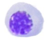
Rubricyte
Also known as polychromatophilic normoblast or polychromatophilic erythroblast
Cell size is 10-15 micrometers
Nucleus
Dark blue, smaller, coarse and clumped
N:C ratio is moderate: 4:1 to 2:1
Nucleoli is no longer visible
Cytoplasm contains various shades of pink and blue; moderate amount with no granules
Reference range: 5-30% of cells in normal bone marrow

Lymphocyte (left) vs rubricyte or polychromatophilic erythroblast (right)


Metarubricyte
Also known as orthochromatic normoblast, orthochromatic erythroblast
Cell size is 8-12 micrometers
Nucleus
Round, usually eccentrically located
N:C ratio is 1:1-1:2
Chromatin: small, pyknotic, condensed, no parachromatin (white areas in the nucleus), completely black appearance, no nucleoli present
Nucleus is extruded during the later stages of the metarubricyte
Cytoplasm is predominantly pink with a slight amount of blue; moderate in amount with no granules
Reference range is 5-10% in bone marrow


Diffusely Basophilic Erythrocyte
Also known as polychromatophilic erythrocyte or reticulocyte
Reported as polychromasia on a CBC (graded as a 0 to 3+)
Cell size is 7-10 micrometers
No nucleus
Cytoplasm is bluish color due to the presence of RNA
New methylene blue or brilliant cresyl blue stains the RNA and organelles to confirm these cells as reticulocytes
Reference range in peripheral blood for reticulocytes is 2.5%-6.0% in newborns and 0.5%-2.0% in adults

Erythrocyte
After the reticulocyte stage, the mature erythrocyte is formed that has the following characteristics:
Functions to transport oxygen to the tissues via hemoglobin
Survives in circulation for 120 days
Metabolizes glucose through anaerobic glycolysis
Has an average diameter of 6 to 8 micrometers
Lacks the ability to make hemoglobin
Lacks a nucleus and functional organelles

What cell is this?
Rubriblasts

What cell is this?
Prorubricyte
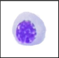
What cell is this?
Rubricyte

What cell is this?
Metarubricyte

What cell is this?
Diffusely Basophilic Erythrocyte
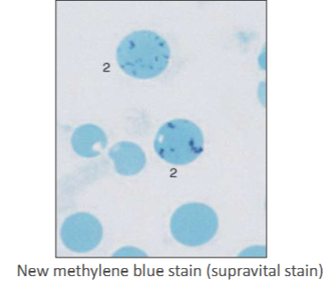
What cell is this?
Reticulocyte
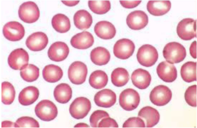
What cell is this?
Erythrocyte
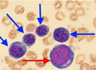
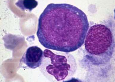
What cells are these?
Red arrow: Rubriblast
Blue arrow: Rubricytes
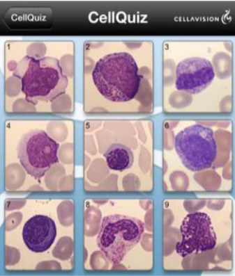
Select the nRBC
5

Select the nRBC
7
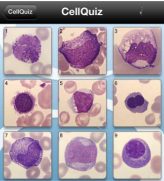
Select the nRBC
6
RBC Structure and Function
Red blood cell survival and function is dependent upon
1. RBC membrane
2. RBC metabolic pathways
3. Hemoglobin (Hgb) structure and function

RBC Membrane
Consists of three areas
1. Outer hydrophilic layer
- Glycolipid
- Glycoprotein
- Protein
2. Central hydrophobic layer
- Protein
- Cholesterol
- Phospholipid
3. Inner hydrophilic layer
- Protein
Membrane Characteristics of Erythrocytes
RBC membrane characteristics
Highly elastic: The shape of the erythrocyte constantly changes as it moves through the circulation and performs extremely complex maneuvers
The cell membrane is deformable and tolerant against mechanical stress and various pH and salt concentrations
Cell shape changes reversibly without fragmentation depending on ATP level in the cell and intracellular calcium ion concentration
The cellular membrane is composed of a protein-lattice lipid bilayer to which the membrane skeleton is attached by peripheral proteins; this network of proteins is responsible for maintaining the strength, shape, stability, and flexibility of the red blood cell to withstand the shear forces in circulation

RBC Membrane
Red blood cell membrane proteins
Integral (transmembrane): Proteins that extend from the outer surface and traverse the entire membrane to the inner cytoplasmic side of the RBC types shown in table 5.3
Peripheral: Proteins located in the cytoplasmic surface of the membrane, which is located beneath the lipid bilayer and forms the RBC cytoskeleton; involves proteins that are:
Membrane associated
Glycosyl phosphatidylinositol (GPI) anchored
Cytoskeletal proteins
RBC Membrane - Peripheral Proteins
Cytoskeleton is composed of peripheral proteins that control cell shape, attachment to other cells, and maintain organization of specialized membrane domains
Types of peripheral proteins
Spectrin (alpha and beta)
Ankyrin (band 2.1) links the membrane skeleton to the bilayer
Actin (band 5)
Band 4.1 and 4.2, adducin, and p55
Together these components form a complex meshwork tethered to the RBC membrane
RBC Membrane - Glycophorin
Principle RBC glycoprotein (20% of membrane protein)
Exposed on the outer RBC membrane
Accounts for most of the membrane sialic acid, which gives RBCs their negative charge causing the RBCs to repel each other as they move through circulation
RBC Membrane - Spectrin
Important factor in RBC membrane integrity
Flexible rod-like molecule composed of 2 helical polypeptide chains (1 alpha and 1 beta)
Binds with other peripheral proteins such as actin, ankyrin, adducin, and others to form a skeletal network of microfilaments on the inner surface of the RBC membrane
Microfilaments strengthen the membrane, control the biconcave shape and deformity, and provide stability to the lipid bilayer interface
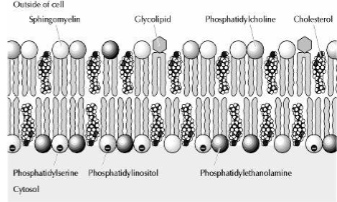
RBC Membrane - Lipids
Red blood cell membrane lipids
Three main types
Phospholipids
Glycolipids
Cholesterol
Red Blood Cell Membrane Lipids - Phospholipids
Choline Phospholipids: Located on the outside of the lipid bi-layer; types include:
Phosphatidyl choline
Sphingomyelin
Amino Phospholipids: Located exclusively on the inside or cytoplasmic side of the RBC membrane; types include:
Phosphatidylethanolamine
Phosphatidylserine
Phosphatidylinositol
Red Blood Cell Membrane Lipids - Glycoproteins
Most are located in the outer half of the lipid bi-layer; they interact with glycoproteins to form many RBC antigens
Red Blood Cell Membrane Lipids - Cholesterol
Located on both sides of the lipid bi-layer inserted between the choline and amino phospholipids
Comprises 25% of the RBC membrane lipid
Continual exchange with plasma cholesterol
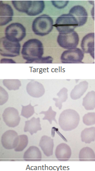
Accumulation of cholesterol will result in the formation of target cells and may cause membrane damage
Acanthocytes have also been associated with an excess accumulation of membrane cholesterol in certain disorders
Liver disease
Inherited lipid disorders such as abetalipoproteinemia and lecithin-cholesterol acyltransferase (LCAT) deficiency
RBC Deformability or Flexibility
RBC’s must be flexible as they flow throughout the circulatory system
Some biochemical changes can alter RBC flexibility and shape change such as:
Low concentration of fatty acids and phospholipids
A variety of amphoteric agents
Elevated intracellular calcium concentration
Decrease ATP levels
RBCs may be sequestered and removed by the spleen when they become aged, damaged, less flexible, or fragmented
RBC Permeability
Permeable to water and anions such as chloride and bicarbonates
Relatively impermeable to cations such as sodium and potassium
K+ is primarily found inside the red cell (25:1 intracellular to extracellular ratio)
Na+ is primarily found outside the red cell (1:12 intracellular to extracellular ratio)
Controls the volume of RBC and water homeostasis
Prevents osmotic hemolysis
Proteins located in the cytoplasmic surface of the membrane located beneath the lipid bilayer and forms the RBC skeleton are known as
Peripheral proteins
The peripheral protein that forms a flexible rod-like dimer responsible for the majority of the RBC membrane integrity is known as
Spectrin
Metabolic Activities of Erythrocytes
The enzymes synthesized during early cell development have to be sufficient to provide the energy needed for several processes
Maintaining hemoglobin iron in an active ferrous (Fe 2+) state
Driving the cation pump needed to maintain intracellular sodium ion (Na+) and potassium ion (K+) concentrations despite the presence of a concentration gradient
Maintaining the sulfhydryl groups of globins, enzymes, and membranes in an active reduced state
Preserving the membrane integrity
If metabolic pathways are blocked or inadequate, the life span of the erythrocyte is reduced and hemolysis results.
Defects in metabolism can include the following:
Failure to provide sufficient reduced glutathione, which protects other elements in the cell from oxidation.
Insufficient energy-providing coenzymes such as reduced nicotinamide-adenine dinucleotide (NADH), nicotinamide-adenine phosphate dehydrogenase (NADPH), and ATP
The two most common erythrocytic enzyme deficiencies, which involves the Embden-Meyerhof glycolytic pathway, are deficiencies of:
Glucose-6-Phosphate dehydrogenase (G6PD): responsible for converting glucose-6-phosphate (G6P) to 6-phosphogluconate (6PG)
Pyruvate kinase (PK): responsible for converting pyruvate (pyruvic acid) to lactic acid
The mature erythrocyte has no nucleus or other organelles but is capable of existing in the blood circulation for an average of 120 days.
An erythrocyte has a limited ability to me