Bio 222 Week 9: Plants + Light - Auxin
1/48
There's no tags or description
Looks like no tags are added yet.
Name | Mastery | Learn | Test | Matching | Spaced | Call with Kai |
|---|
No analytics yet
Send a link to your students to track their progress
49 Terms
When blood sugar levels are low glucagon is secreted into the blood by the alpha cells of pancreas . This leads to an increase in sugar mobilization into the blood. Mobilization is primarily the job of the liver (organ). When blood sugar levels become too high, insulin is secreted by the Beta cells of pancreas . This lowers blood sugar levels in a variety of ways, but uptake from the blood occurs by a hormone mediated increase in numbers of channels in cell membranes of most tissues.
What happens to the rate of NADH production in mitochondria in a pancreatic beta-cell as blood sugar levels increase?
NADH production increases
As blood sugar increases above fasting both insulin levels in the blood and cellular glucose uptake levels increase. The hormone induced increase in sugar uptake in fat and muscle cells is due to an increase in transporters exocytosed to the plasma membrane. An increase in muscle cell sugar uptake can also be caused by muscle contractions.
The enzyme that adds a phosphate to glucose to begin glycolysis in pancreatic beta cells is called glucokinase . This enzyme has a lower affinity for glucose than do the enzymes in most of the cells of the body. This allows the phosphorylation of sugar in beta cells to remain proporional to blood sugar levels throughout the physiological range. In muscle cells this reaction is carried out by hexokinase, which operates at Vmax throughout the normal physiological range of blood sugar levels.
In pancreatic beta cells aerobic respiration rate is regulated by glucokinase activity. And because that is true these cells generate more ATP when blood sugar levels rise. This in turn leads to the inhibition of potassium channels, which leads to cell depolarization.
The Km of pancreatic beta cell sugar importers is relatively high in comparison to the importers found in muscle and fat cells
True or False:
In a wild type plant, there is more iron in the grain than in the leaves, on a per gram basis.
False
True or False:
There is significantly more iron in brown rice than in white rice.
True
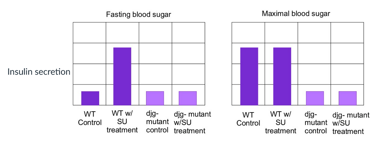
Insulin secretion of wild type and djg- (loss of function) mutant Beta cells with and without SU treatment
Given the data provided in the graphs, and the added information that djg- and wild type cells do not differ in their responses to fructose added to the media, it is possible* that SU is an inhibitor of ligand gated potassium channel activity and DJG is a depolarization gated calcium channel.
*Assume these are channels we discussed in class.
Hexokinase has a lower Km for glucose tin comparison to glucokinase. If you replaced the version of these isozymes found in beta cells with the version found in most cells of the body in quantities that didn't change the Vmax you begin to suffer from low blood sugar.
Uptake of glucose into muscle cells is a function of circulating insulin in a resting individual but uptake will be increased by muscle contractions .
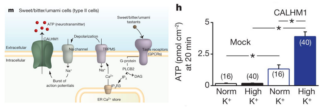
Taste bud cells deliver ATP as a signaling molecule to neurons to indicate when the specific tastant it is their job to monitor has been detected. Panel m provides the signal transduction pathway for this process, which involves a cascade of membrane potential relevant events that eventually opens the channel that allows taste cell ATP delivery to neurons. This is a rare case of signal being exported not from vesicles, but through
Panel h shows the results of experiments in which the ATP secretion of cells with (the two bars on right) and without (two bars on left) the ATP channel CALHM1 that are bathed in normal and in high K+ solutions. (Stars represent significant differences between bars 1&3, 2&4 and 3&4. #s in parenthesis are just number of cells tested)
Here we revisit Homework one. The results of this experiment demonstrate that depolarization of these cells suffices to activate the ATP channel CALHM1. You know from Bi221 that ATP has a negative charge so the driving force on it is is negative . Although the signal transduction pathway in these cells involves opening a series of cell membrane spanning sodium channels, the researchers added potassium to the fluid surrounding the cells because the leak channels that allow this ion to move through the membrane have the highest conductance. It's also true that if you have 150/10 as a ratio, adding 20 to the numerator has a much smaller effect than adding 20 to the denominator.
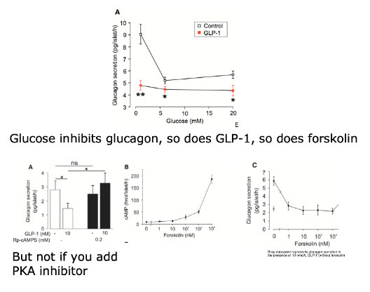
How does GLP1 influence glucagon secretion?
Decreases it via cAMP → PKA —|
When does the liver typically carry out gluconeogenesis?
When it detects hormones produced by pancreatic alpha cells
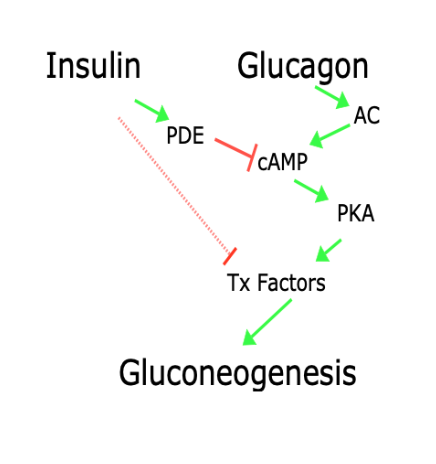
Insulin and glucagon have opposing influences on gluconeogenesis
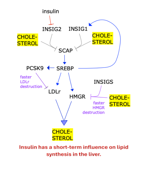
Insulin has a short-term influence on lipid synthesis in the liver
(Indirectly activates SREBP?)
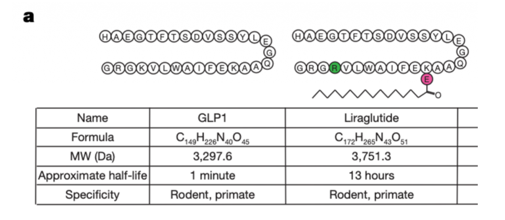
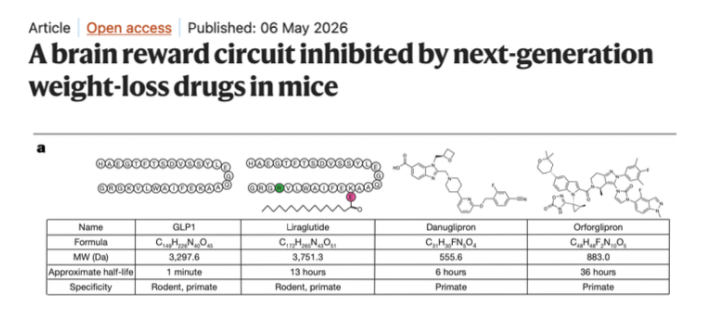
GLP1 analog drugs such as Liraglutide bind to the same receptors to create the same response, but they live for much longer in the body because they have a fatty acid that blocks the enzyme from digesting/breaking down GLP1.
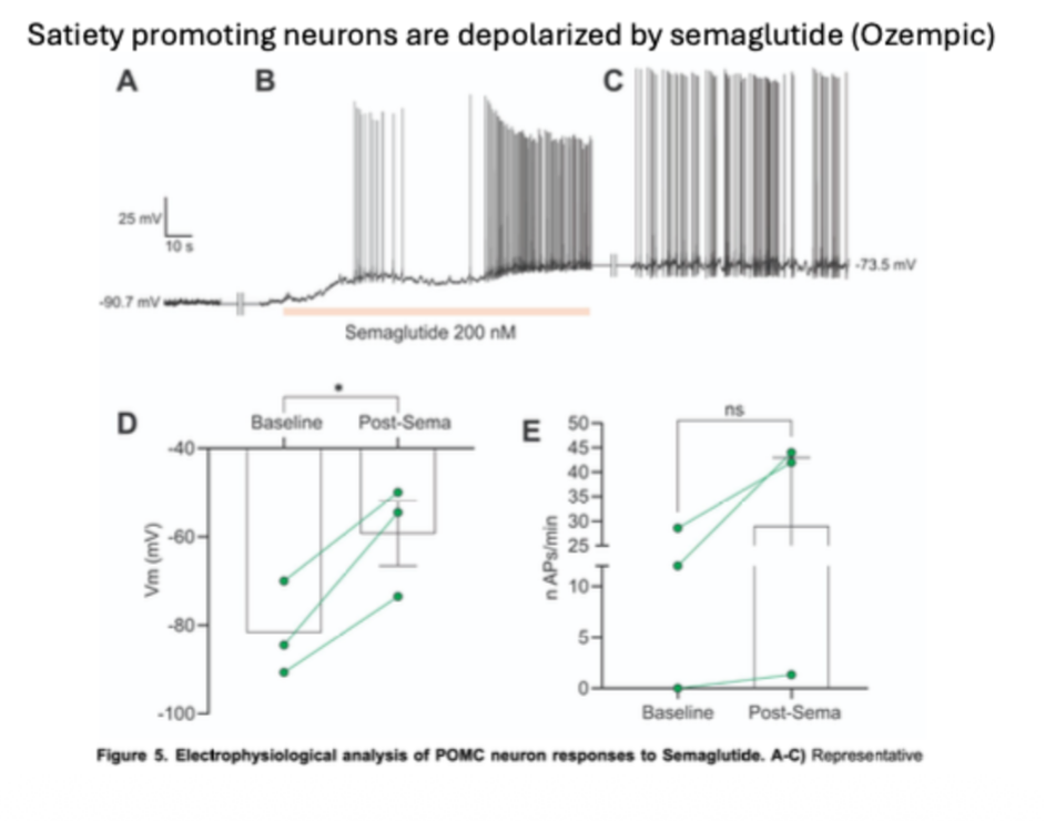
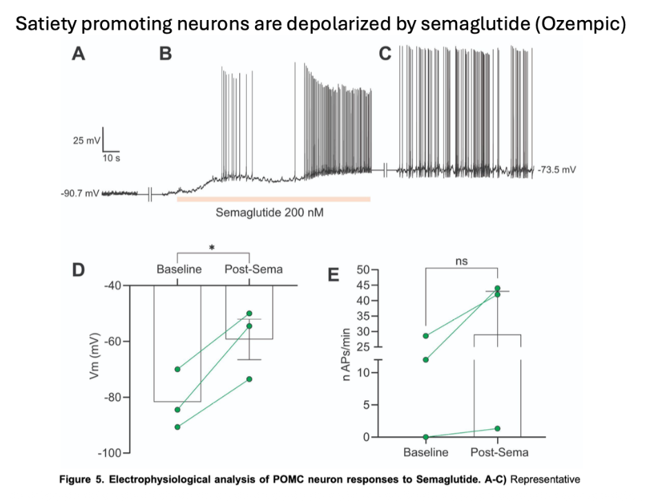
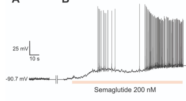
Satiety promoting neurons are depolarized by semaglutide (Ozempic).
Ozempic is a GLP1 receptor agonist. Ozempic differs in structure to ozempic so that…
The enzymes that eliminate GLP1 break ozempic down more slowly.
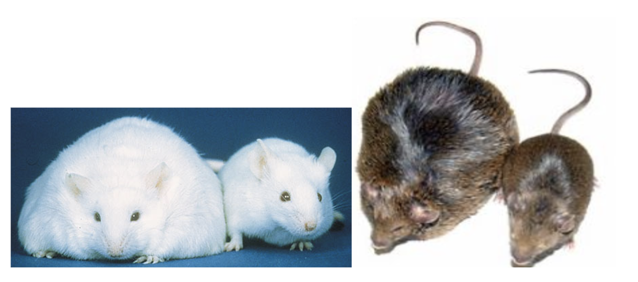
A hormone known as Leptin made by white adipose tissue (white fat cells) informs cells with Leptin receptors of how much fat is present/available in their body.
Fat cells seem to behave differently after a large enough gain of weight to be considered obesity.
Leptin receptor gene loss of function (db-/db-) and wild type, and leptin gene mutant (ob-/ob-) and wild type mice. The leptin gene was named OB for the obese phenotype of those missing it.
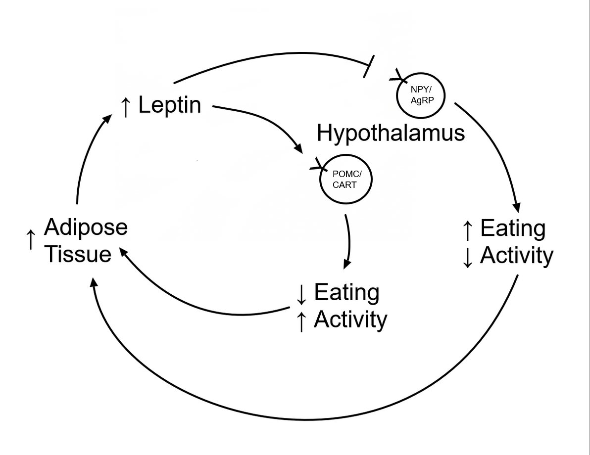
Leptin Pathway!
A much more confusing leptin pathway provided by Mark…
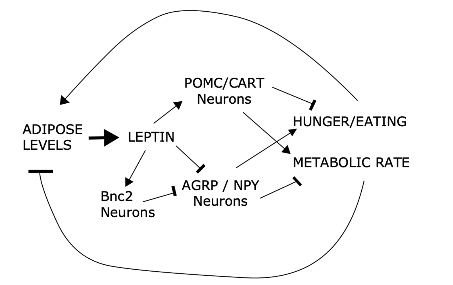
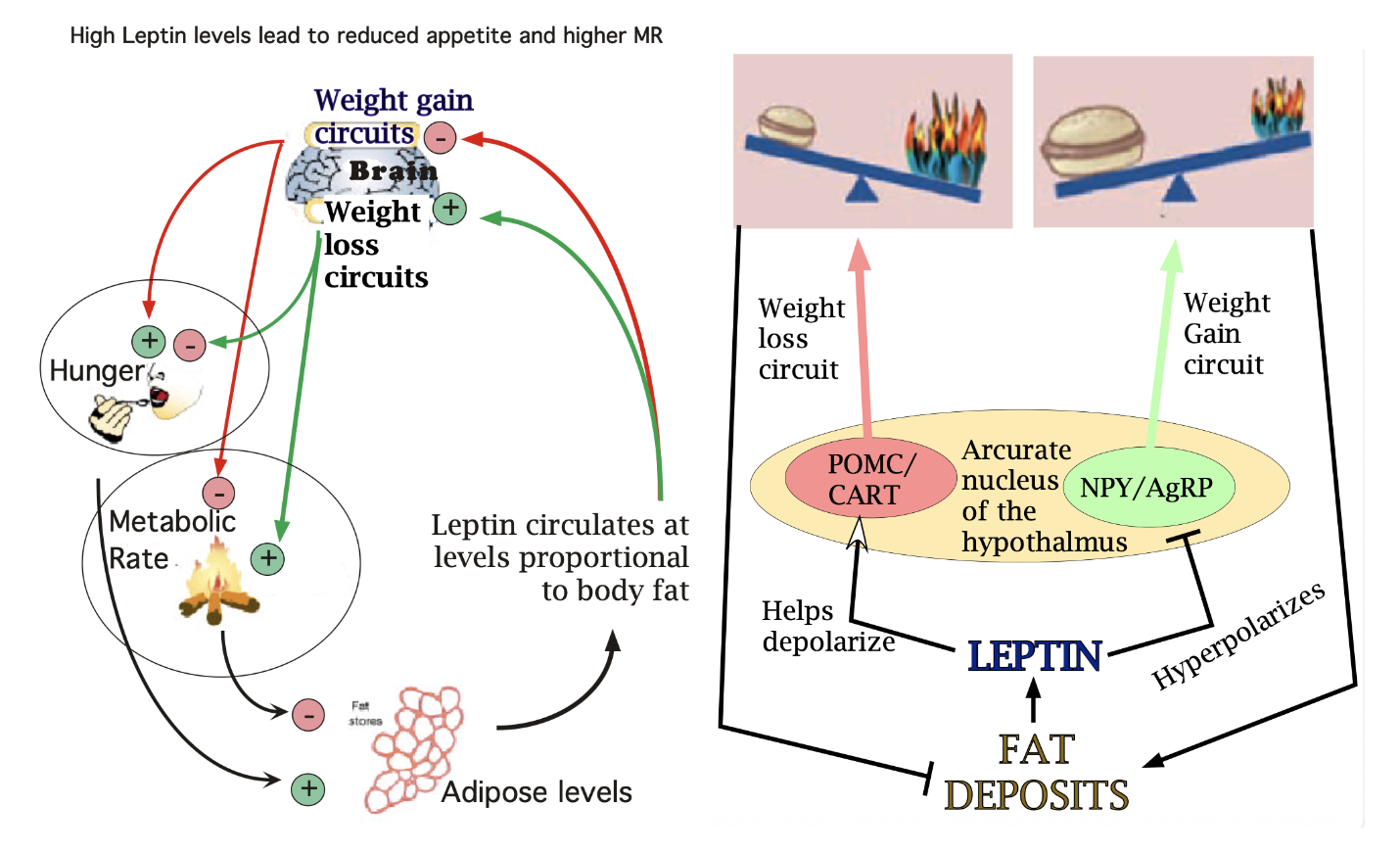
Weight Gain/Loss Circuit
Ablation of leptin receptors on either BN2 or AGRP neurons causes weight gain. Both cell types live in the arcuate nucleus of the hypothalamus.
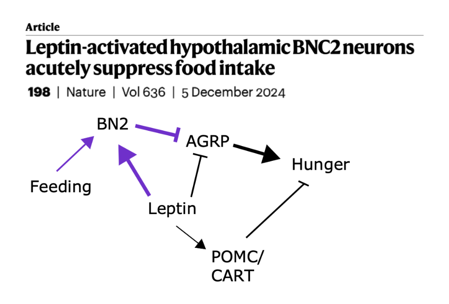
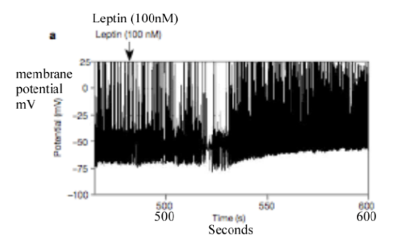
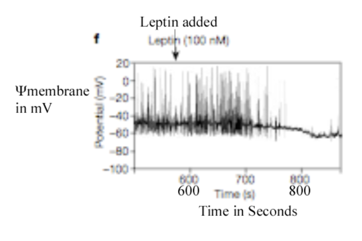
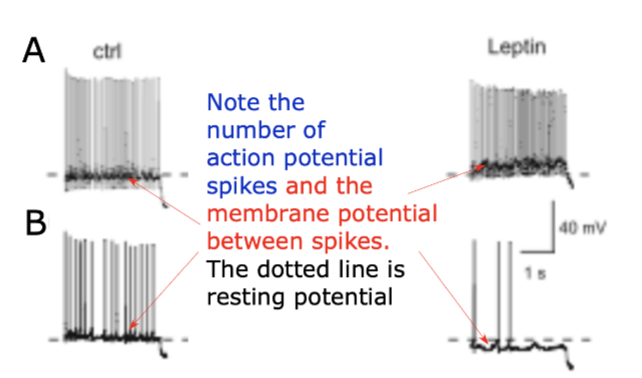
Leptin seems to depolarize this neuron (POMC/CART) after a brief delay.
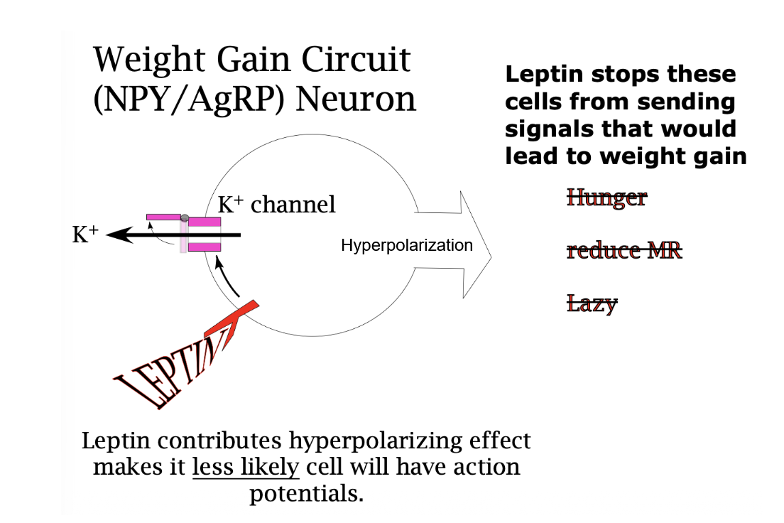
Leptin seems to hyperpolarize this neuron (NGY/AgRP) after a brief delay
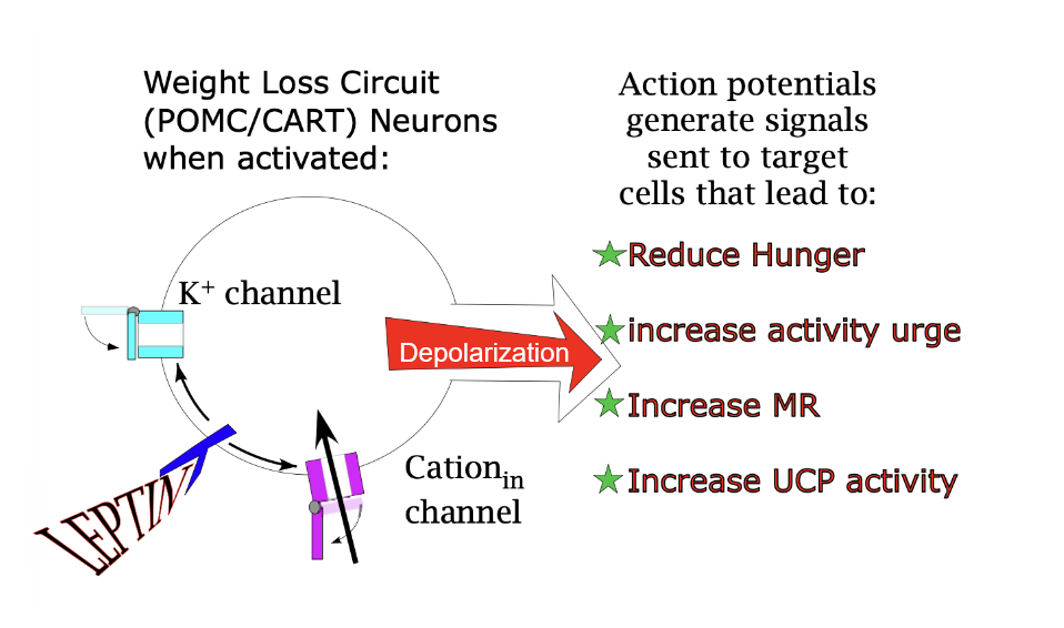
Weight Loss Circuit (POMC/CART) Neuron
Actions potentials caused by depolarization generate signals sent to target cells that lead to…
↓ Eating/Hunger, ↑ Activity, ↑ MR, and ↑ UCP —| ↑ Fat
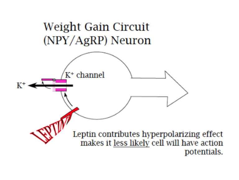
Wait Gain Circuit (NPY/AgRP) Neuron
A lack of action potentials caused by hyperpolarization means that less signals will be sent to target cells, leading to…
↑ Eating, ↓ Activity, and ↓ MR —> ↑ Fat
Because the driving force on Na+ is much stronger (thousands of times more of it waiting outside to come in), it is sodium that brings change to a threshold in this (and many) settings that use “cation in” channels.
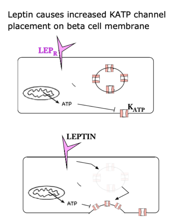
Leptin is a hyperpolarizing influence on beta cells as it causes KATP channels to exocytose (?) and allow more potassium to exit the cell.
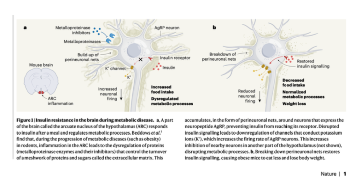
This figure describes how obesity related inflammation in the part of the brain in which body weight homeostasis neurons are housed leads to the inability of INSULIN to reach the insulin receptors on AgRP neurons!
Notice the influence this has on K+ channel activity
Leptin concentration dictates how much of the lower level depolarization occurs here. Less Leptin means a more negative membrane potential. So, the more Leptin in the body the less “food signal” is required to bring these cells to threshold. This means that when the system is working, it takes less food to make you feel full if you have plenty of fat stored.
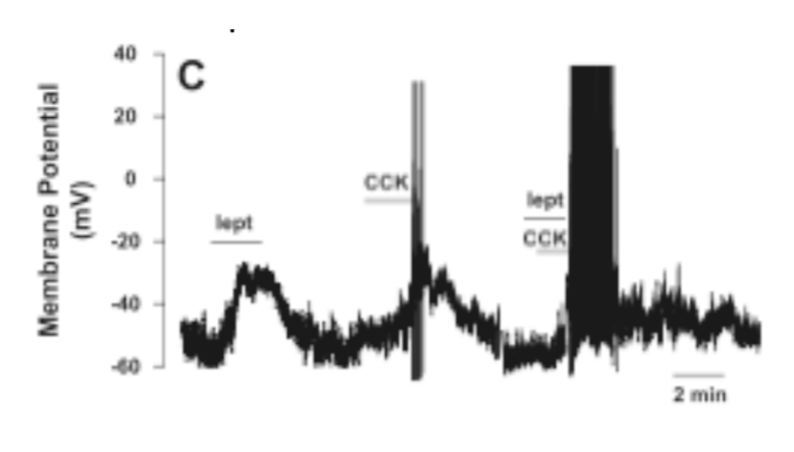
Satiety promoting neurons depolarize to sub-threshold levels in response to leptin, which allows smaller levels of signals that indicate the uptake of nutrients to generate action potentials in these cells. Notice how much stronger the cck + leptin output of this neuron is than the output when there is just cck. CCK is an indicator that food has been consumed.
Satiety promoting neurons are depolarized in response to semaglutide (Ozempic)
This slide is repeated from the previous lecture. It shows the influence of a GLP1 analog on POMC neurons involved in satiety signaling. Notice that GLP1 receptor activation is having a veery similar influence on these neurons as does leptin!
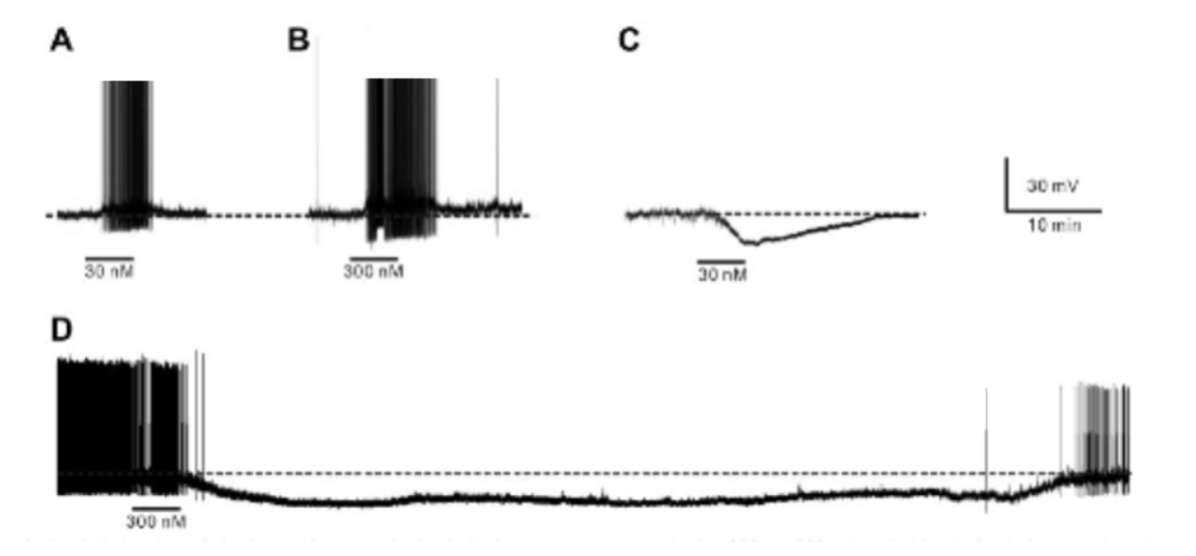
Which tracings represent weight loss circuit neurons? (Depolarization/more action potentials)
A and B (C and D would represent weight gain circuit neurons)
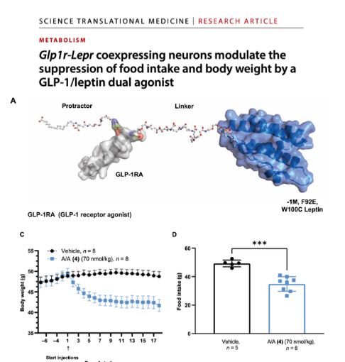
This study identified a set of hypothalamus neurons with receptors for both leptin and GLP1.
Neurons in the brain region NTS that cause meal termination but don’t generate nausea lead to less food intake
Neurons in the brain region AP that cause nausea cause meal termination and nausea, and lead to less food intake
Both respond to GLP1
GLP1 inhibition of EITHER set leads to less food intake, and seemingly in a non-additive way—so the same amount of weight loss occurs with either of these brain regions silenced.
Food reward circuit is double treated by an even newer drug.
This diagram shows how GLP-1 receptors that bind a treatment which combines GLP-1 and a molecule that inhibits activation by glutamate (a neurotransmitter) activation of neurons involved in the reward circuitry that responds to nutrient uptake. This inhibition is accomplished by stopping Na+ import.
Remember that I told you that Ozempic has been shown to reduce drug seeking as well as food seeking behaviors. This diagram demonstrates that GLP-1 receptors exist on neurons in the reward circuitry.
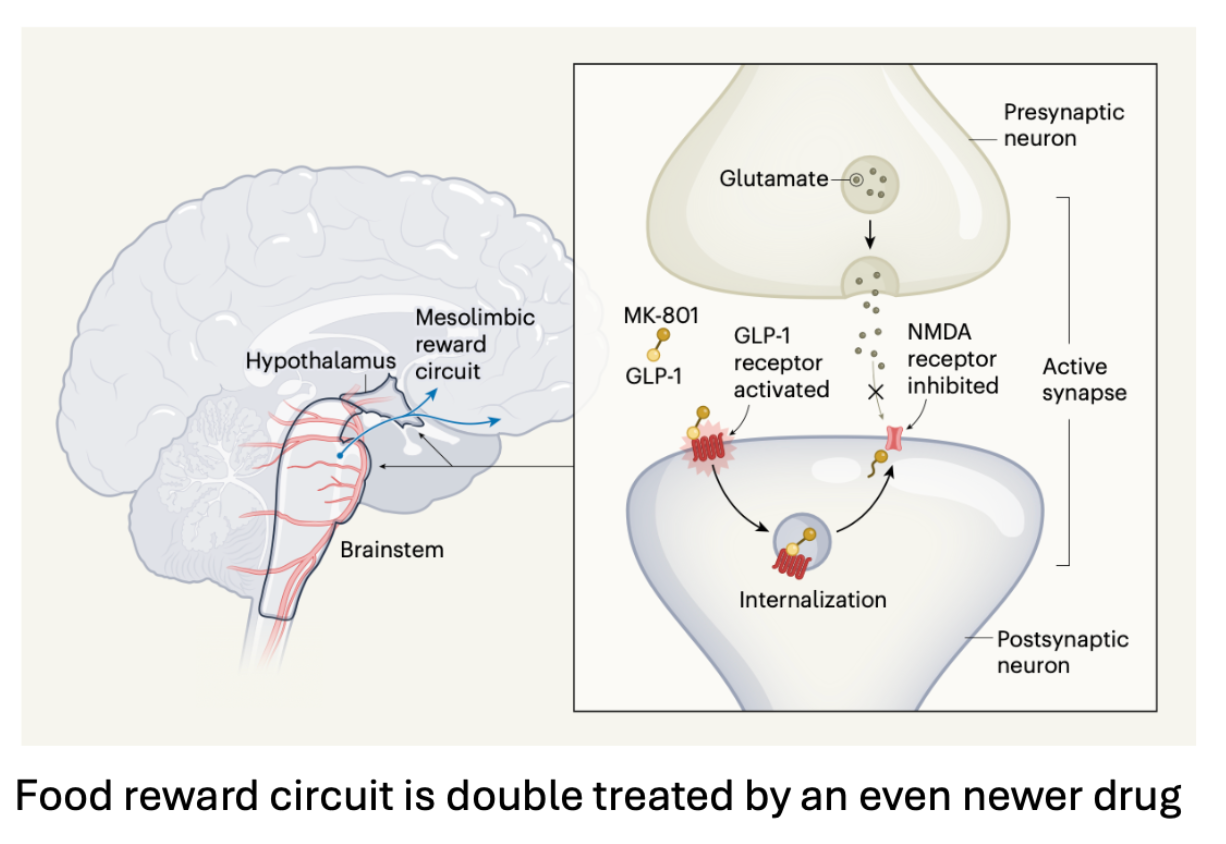
Smaller GLP1 analogs have been developed. They work in humans, but not rodents. Because this is true you can add humanized GLP1 receptors to specific brain regions and measure very specific mechanisms of drug activity!
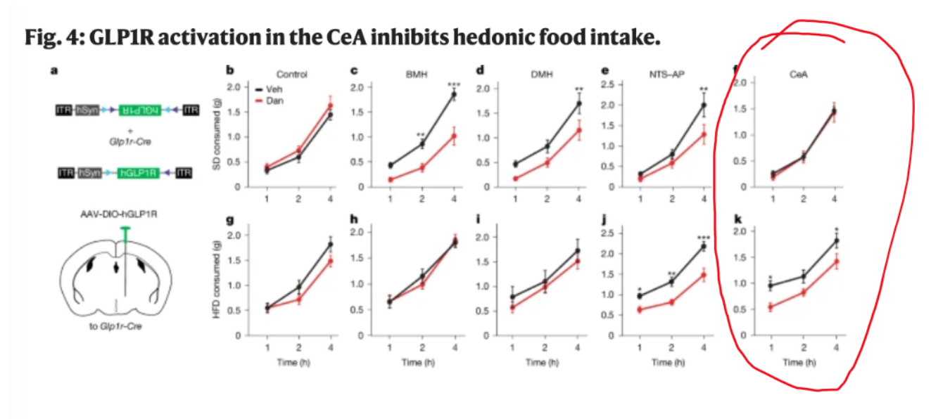
GLP1 receptor activation in the central amygdala (reward center) inhibits hedonic (pleasure seeking) food intake!
Essentially, it stops the drive to eat high fat diet, which when fed ad libitum (without restriction) to untreated rodents will lead to obesity.
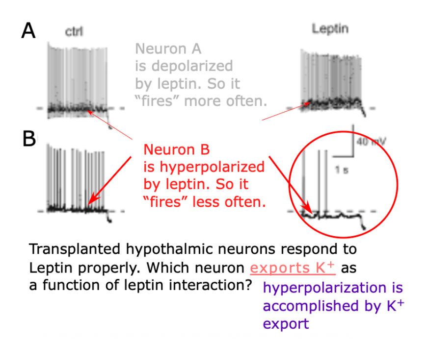
Transplanted hypothamic neurons respond to Leptin properly. Which neuron exports K+ as a function of leptin interaction?
Neuron B is hyperpolarized by leptin as potassium leaves, so it “fires” less often.
If weight gain circuits fail to bind leptin they will have increased activity, which will lead to…
weight gain (?)
Upregulating transferrin receptor expression in AgRP neurons leads to leptin and insulin insensitivity in those cells.
Increased transferrin receptor activity most directly leads to…
Increased cellular iron uptake.
Upregulating transferrin receptor expression in AgRP neurons leads to leptin and insulin insensitivity in those cells.
So upregulating transferrin receptor expression in these cells would…
Increase their activity.
Upregulating transferrin receptor expression in AgRP neurons leads to leptin and insulin insensitivity in those cells.
Excess iron uptake in these cells would lead to __________ animal UCP1 activity.
Excess iron uptake in these cells would lead to decreased animal UCP1 activity.
Upregulating transferrin receptor expression in AgRP neurons leads to leptin and insulin insensitivity in those cells.
When functional insulin signaling to these cells leads to movement of an ion through a channel that has a…
Positive driving force and negative equilibrium potential.
Leptin binding by weight gain circuit cells opens K+ channels in those cell membranes, leading to membrane hyperpolarization.
Leptin binding by weight loss circuit cells closes K+ channels and opens Na+ channels in those cell membranes, leading to membrane depolarization.
Leptin is made by adipose cells.
All the channels would be ligand gated, by the way.
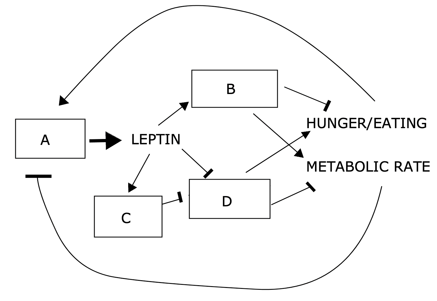
animals with B cells lacking leptin receptors would gain weight
animals with D cells lacking leptin receptors would gain weight
animals with C cells lacking leptin receptors would gain weight
B cells are POMC/CART neurons
C cells are BNC2 neurons
D cells are NPY/AgRP neurons
A cells are WAT (white adipose tissue)
Inactivation of D cells would increase metabolic thermogenesis
Given the results of ozempic treatment on humans it is likely that the cell whose membrane potential measurements are shown above is a weight loss circuit neuron . If the mechanism of action leading to the results shown involves regulation of a potassium channel, semaglutide signal transduction must lead to the closure of that channel. If the mechanism of action leading to the results shown involves regulation of a sodium channel semaglutide signal transduction must lead to the opening of that channel.
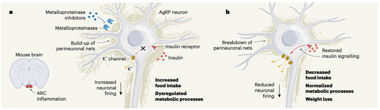
This figure from a recent Nature paper illustrates a role that insulin plays in regulating AGRP neurons, although the purpose of the figure is to illustrate how weight gain leads to inflammation responses that block insulin signaling. I am not sure if the words in bold for both panels are intended to tell us the cause or the effect of the processes illustrated. Makes sense either way. So let's break it down...
Obesity increases the perineural netting around the cells of the ARC in an inflammation- related process. This prevents insulin to reach receptors on the cell surface.
Insulin/insulin receptor signal transduction in the illustrated cells leads to increased potassium efflux from the cell, and consequently to cell hyperpolarization which in turn means the cell becomes less active.
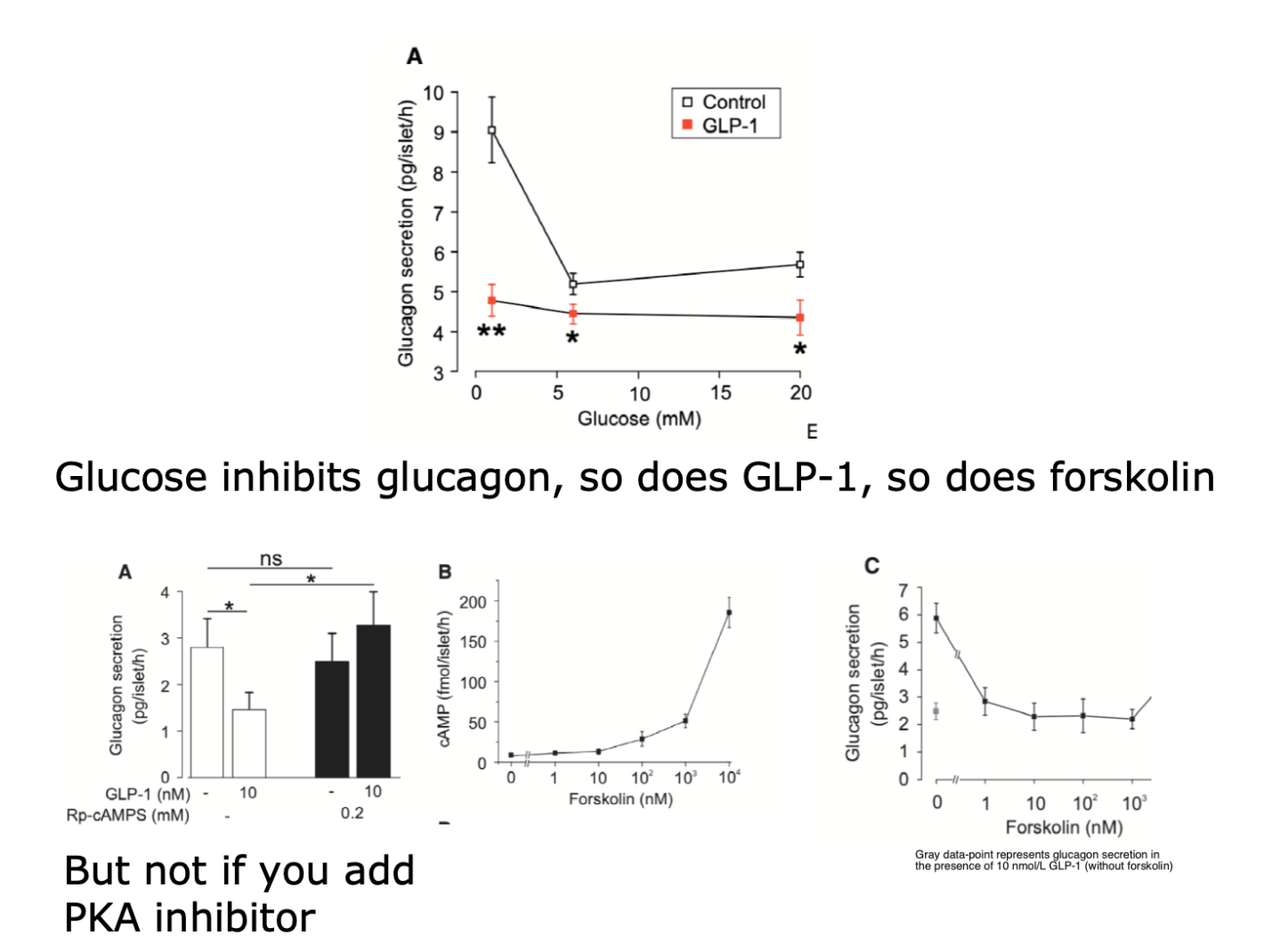
How does GLP1 influence glucagon secretion?
Activates cAMP which activates PKA which inhibits secretion
LEPTIN —| INSULIN
Adding more KATP channels to the pancreatic beta cell membrane would blunt the response to small increases in blood sugar. This is how Leptin influences these cells.
INSULIN —> LEPTIN
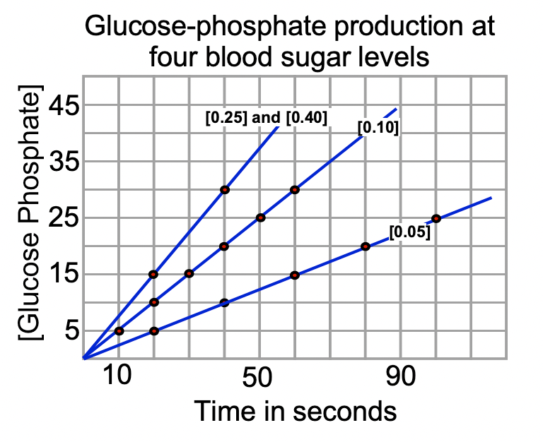
Glucose-phosphate production by an enzyme extracted from cells taken from the much-feared Lilyputin in media with four different glucose levels is provided above. The fasting blood sugar levels of this animal fall between 0.04 and 0.06 sugar units.
The enzyme catalyzing this reaction must be and the cells from which the enzyme was extracted could have been pancreatic beta cells
The substrate of this enzyme is glucose.
If you added insulin to the media you would expect the enzyme rate to stay the same.
Glucose-phosphate production by an enzyme extracted from cells taken from the much-feared Lilyputin in media with four different glucose levels is provided above. The fasting blood sugar levels of this animal fall between 0.40 and 0.60 sugar units.
The enzyme catalyzing this reaction must be and the cells from which the enzyme was extracted could have been muscle cells.
The Km of this enzyme to three decimal places is 0.075.


Glucose-phosphate production by an enzyme from cells taken from the much-feared Lilyputin in media with four different glucose levels is provided above. The fasting blood sugar levels of this animal fall between 0.04 and 0.06.
What rate of glucose phosphate production you would expect from this enzyme under these conditions if provided a substrate concentration of 0.043?
0.215 units GP/Second
In pancreatic beta cells the membrane spanning sugar importing carrier/channel has higher Km for glucose than those found in muscle cells. Increases in the rate at which this step occurs lead to decreased ACTIVITY of a channel that allows potassium to exit the cell. Changes in this channel activity alter the likelihood that calcium will enter the cell through another channel. The coincidence detector in beta cells that allows coordination of response to multiple forms of carbohydrate is a channel that dictates whether sodium will enter the cell. ultimately it is calcium flow through the cell membrane that dictates whether or not insulin will exit the cell via exocytosis.
The cells of the liver are the primary target of the hormone glucagon, which is made in the pancreas. These cells respond by increasing their rate of gluconeogenesis , and decreasing their rate of glycogen synthesis.

What is the insulin secretion level of the double mutant at fasting blood sugar?
Above 10% but below 40%
What is the insulin secretion level of the double mutant at elevated blood sugar?
60% maximal
How much insulin secretion would you expect in wild type b-cells in the presence of elevated blood sugar but no fructose present?
60% maximal
It is evident that the allosteric regulator that the GCK-HH mutation fails to bind is an inhibitor. Patients with this allele would have symptoms of low blood sugar.

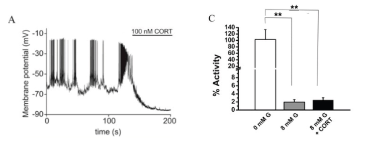
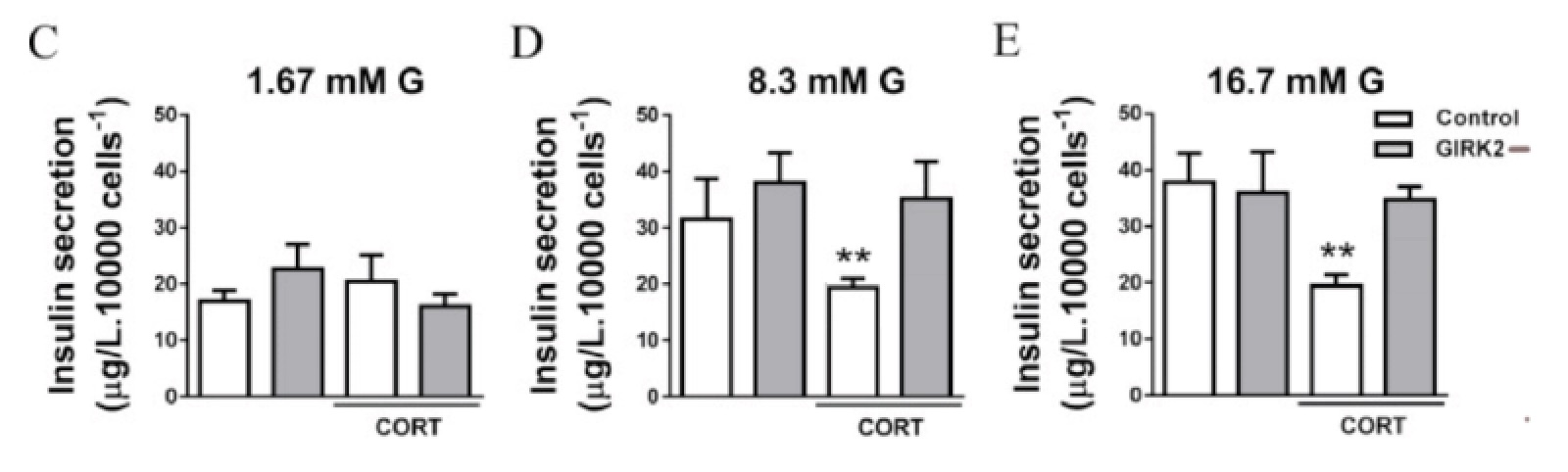
Panel A tells us that the hormone CORT changes the membrane potential of pancreatic beta cells in a medium containing high levels (8mM) of glucose. Panel C provides the activity of the KATP channels in beta cells in low and high sugar media and in high sugar plus CORT. Stars indicate that the only significantly difference is between the 0 mM glucose activity data and the high sugar treatments (the two short bars are not different). In panel A CORT delivery timing is indicated by the line above the graph.
This data shows us that CORT hyperpolarizes beta cells after a short delay and that it does this without altering KATP function.
Panels C-E in this next figure show us CORT activity in both control and GIRK2 loss of function beta cells.
This data supports the hypothesis that CORT activates GIRK which in turn inhibits insulin secretion. (CORT —> GIRK —| Insulin Secretion)
Given all the data provided on this page, what ion is moving through the beta cell membrane as a result of CORT activity? Potassium
Human systemic iron homeostasis regulated by the hormone hepcidin. This hormone is a protein. An increase in the activity of this hormone in the blood leads directly to a reduction in the activity of ferroportin. This leads to a decrease in the number of Fe+++ bound to transferrin that is circulating in the blood.
What is driving iron through ferroportin?
Concentration
The following is an excerpt from a report in the November 2019 issue of the journal Haematologica.
“Conversely, [loss of function] mutations in the TMPRSS6 gene lead to overexpression of hepcidin. In this case, patients suffer from the condition IRIDA. These individuals suffer from a form of IRIDA that typically does not improve with oral iron treatment
... novel thiazolidinone derivatives appear to act on hepcidin expression by decreasing Tmprss6 activity.”
Based on this report:
IRIDA seems to be a condition that includes low blood iron.
TMPRSS6 seems to be an inhibitory transcription factor.
Patients with IRIDA don't improve with oral iron treatment because this condition deactivates ferroportin.
Additional phosphodiesterase activity would not change insulin secretion in beta cells treated with GLP-1 during fasting blood sugar levels.
Additional phosphodiesterase activity would increase glucagon secretion in alpha cells treated with GLP-1 during fasting blood sugar levels.
Additional phosphodiesterase activity would not change insulin secretion in beta cells treated with GLP-1 during fasting blood sugar levels with added fructose.
Additional phosphodiesterase activity would decrease insulin secretion in beta cells treated with GLP-1 during elevated blood sugar levels but with no fructose present
The three structures that are classified as plant organs are root leaf stem.
The order of organs generated in a phytomer is leaf - bud - stem.