All of AQA Uniy 3
1/106
There's no tags or description
Looks like no tags are added yet.
Name | Mastery | Learn | Test | Matching | Spaced | Call with Kai |
|---|
No analytics yet
Send a link to your students to track their progress
107 Terms
What is transpiration?
The loss of water vapor through the stomata of leaves by evaporation
Factors that affect the rate of transpiration
1. Light intensity
- causes more stomata to open
- therefore larger SA for evaporation
2. Temperature
- more heat = more kinetic energy
- so molecules move faster
- therefore more evaporation
3. Humidity
- more water vapour in the air will make the water potential outside the leaf more positive
- therefore reduces the water potential gradient
4. Wind
- more wind will blow away humid air containing water vapour
- therefore marinating water potential gradient
Explain how the structure of xylem tissue is adapted to its function
1. Long tubes w no end walls
- continuous water columns
2. No organelles to obstruct flow
- allows easier water flow
3. Has lignin
- support
- waterproof
4. Pits in walls
- allows lateral movement
Describe the cohesion-tension theory of water transport in the xylem.
Evaporation of water molecules through the stomata by transpiration, which lowers the WP of leaf cells
Water molecules "stick" together by H-bonds, forming continuous water column
these H-bonds maintains column
Water pulled up the xylem, creating tension
Adhesion of water molecules to walls of xylem
Describe how a high pressure is produced in the leaves.
sucrose actively transported in the phloem
WP becomes lower as sugar enters phloem
Water enters phloem by osmosis
Increased volume of water causes increased pressure
What is adhesion in water?
the sticking of water molecules to the xylem wall
- causes capillarity
Explain 2 ways in which companion cells are adapted for the transport of sugars between cells
1. Mitochondria are present to release energy for active transport
2. Ribosomes make proteins which are needed for carrier proteins/enzymes
State and explain the ways in which sieve cells are adapted for mass transport
1. No/few organelles
2. Little cytoplasm
3. Hollow
4. Large vacuole
5. Thick walls
explanation for all:
easier flow
stronger resistance to pressure
Describe the mass flow hypothesis for the mechanism of translocation in plants
translocation is how organic materials are transported around the plant
In source, sugars are actively transported in the phloem by companion cell
this lowers the WP of sieve tube & water enters by osmosis.
Increase in pressure causes mass movement towards the sink
Sugars used in root for respiration for storage
Give evidence for the mass flow hypothesis of translocation.
Cutting the stem of a plant results in phloem sap being released
therefore there must be hydrostatic pressure in the sieve tubes.
There is a higher sucrose concentration in the leaves than the roots.
Give evidence against the mass flow hypothesis of translocation.
The structure of sieve tubes seems to hinder mass flow
Not all solutes move at the same speed, as you would expect in mass flow.
How can tracing experiments be used to investigate transport in plants?
Plants are grown in the presence of radioactive CO2, which will be incorporated into the plant's sugars
- Can determine which tissue carries the radioactively labelled sucrose by:
take thin horizontal sections of plant tissue
Place against photographic film in dark for several hours and carry out autoradiography
we can see that the areas exposed to radiation correspond to where the phloem is.
How can ringing experiments be used to investigate transport in plants?
The bark and phloem of a tree are removed in a ring, leaving behind the xylem.
Eventually the tissues above the missing ring swells due to accumulation of sucrose and the tissue below begins to die.
- Therefore sucrose must be transported in the phloem.
A student wanted to determine the rate of water loss per mm2 of surface area of the leaves of the shoot.
Outline a method she could have used to find this rate. You should assume that all water loss from the shoot is from the leaves.
draw around each leaf on graph paper and count the squares on both sides of the leaf
divide the rate of water loss by the total surface area of the leaf
The rate of water movement through a shoot in a potometer may not be the same as the rate of water movement through the shoot of a whole plant.
Suggest one reason why.
plants have roots
What is digestion?
hydrolysis of large insoluble substances to smaller soluble substances
- that can be absorbed through the bloodstream
roles of enzymes in the complete breakdown of starch
Amylase: hydrolyses starch into maltose
by hydrolysing the glycosidic bonds
Maltase: hydrolyses maltose to glucose
by hydrolysing the glycosidic bonds
the process of starch digestion
glucose moves into epithelial cell with sodium via carrier protein
sodium removed from epithelial cell by AT and moves into the blood, maintaining a low concentration of sodium
glucose moves into blood by facilitated diffusion
In a person with a blocked pancreatic duct, starch digestion is affected.
Explain how
blocked pancreatic ducts means that less amylase can enter the small intestine
so less starch is digested
Healthy people have amylase in their blood. Explain why this doesn’t cause any harmful effects,
amylase is specific to starch
also theres no starch in human blood
Explain how the epithelial cels that line the small intestine are adapted for the absorption of glucose.
microvilli which provide large SA
many mitochondria which produce ATP
carrier proteins for active transport
carrier proteins for facilitated diffusion
co-transport fo sodium (ions) and glucose
membrane-bound enzymes that digest disaccharides, producing glucose
structure of proteins
proteins are polymer of amino acids joined by peptide bonds, formed by condensation reaction
primary structure: order of AA
secondary structure: folding of polypeptide chain due to hydrogen bond
forms alpha helix and beta pleated sheets
tertiary: unique 3D shape formed by the folding of polypeptide chain
due to hydrogen bonding, ionic bonding and disulfide bridges
quaternary structure: 2 or more polypeptide chain
Explain why releasing protein-digesting enzymes into the blood can be harmful the body
these enzymes could digest hormones/antibodies in the blood
Co-transport of sodium and glucose
1. Sodium ions actively transported from ileum to blood
- maintains concentration gradient for sodium
2. glucose enters by facilitated diffusion with sodium ions
Explain why the combined action of endopeptidases and exopeptidases are more efficient that exopeptidases on their own
1. Endopeptidases hydrolyse internal peptide bonds and exopeptidases hydrolyse amino acids at the ends
2. so increase in SA
Role of enzymes in digestion of proteins
1. Hydrolysis of peptide bonds
2. Exopeptidases act at the end of the polypeptide chain
hydrolyse amino acids at the ends of the chain
3. Endopeptidases act in the middle of polypeptide chain and hydrolyse internal peptide bonds
4. Dipeptidases acts b/w 2 amino acids
Trypsin is a protease.
Suggest the advantage of producing trypsin in an inactive form inside cells in the pacreas
doesnt digest proteins inside the cells
so pancreatic cells are not destroyed
What are lipids digested by?
Lipase and the action of bile salts
Where is lipase produced and how does lipase digest lipids?
Pancreas and small intestine
Hydrolyses the ester bond in triglycerides to form the monoglycerides and fatty acids
Where are bile salts produced and what do they do?
liver
Emulsify lipids to form tiny droplets called micelles
What are micelles?
water soluble vesicles formed of fatty acids, glycerol and bile salts
describe the action of membrane-bound dipeptidases and explain their importance
hydrolyses peptides to release amino acids
amino acids can cross cell membranes, whereas dipeptides cannot cross cell membrane
Explain the advantages of lipid droplet and micelle formation
Droplets increase surface areas for lipase / enzyme action
So faster hydrolysis/digestion of lipids
Micelles carry fatty acids and glycerol through membrane to epithelial cell
Golgi apparatus in lipid transport
1. Modifies lipids
2. Combines triglycerides with proteins
3. Packages them for exocytosis
Digestion and absorption of lipid molecules
1. Micelles contain bile salts and fatty acids
- this makes fatty acids more soluble in water
2. Micelles carry fatty acids to lining of epithelium
3. Fatty acids absorbed by diffusion
When in cell:
1. Triglycerides reform
2. Vesicles move to cell membrane
cooperative binding
first oxygen binds to haemoglobin, changing tertiary structure
this creates/uncovers another binding site
change in shape (of haemoglobin) allows more oxygen to bind easily
How does partial pressure of oxygen affect oxygen-haemoglobin binding?
1.As partial pressure of oxygen increases, the affinity of haemoglobin for oxygen also increases
-so oxygen binds tightly to haemoglobin.
2.When partial pressure is low:
-oxygen is released from haemoglobin.
Bohr effect OxyHb graph
When a high carbon Dioxide concentration causes the oxyhemoglobin curve to shift to the right
The affinity for oxygen decreases b/c the acidic carbon dioxide changes the shape of haemoglobin slightly
Bohr effect
As partial pressure of carbon dioxide increases, the conditions become acidic due to the increase in H+ ions, causing haemoglobin to change shape.
The affinity of haemoglobin for oxygen therefore decreases
so oxygen is released from haemoglobin.and so more oxygen can be delivered to cells for respiration
How does saturation of haemoglobin with oxygen affect oxygen-haemoglobin binding?
It is hard for the first oxygen molecule to bind but once it does:
first oxygen molecule binds to haemoglobin, changing the tertiary structure
this reveals/uncovers another binding site
it changes the shape of haemoglobin to make it easier for the second and third oxygen molecules to bind
known as positive cooperativity.
It is then slightly harder for the fourth oxygen molecule to bind
because there is a low chance of finding a binding site.
Explain why oxygen binds to haemoglobin in the lungs.
Partial pressure of oxygen is high.
Low concentration of carbon dioxide in the lungs, so affinity of Hb to oxygen is high.
Positive cooperativity
Explain why oxygen is released from haemoglobin in respiring tissues.
Partial pressure of oxygen is low
High concentration of carbon dioxide in respiring tissues, so affinity of Hb to oxygen decreases.
if oxyhaemoglobin dissociation curve goes towards the left
haemoglobin has higher affinity for oxygen
so it releases less oxygen/ uploads more oxygen
it becomes saturated at lower partial pressure
if oxyhaemoglobin dissociation curve shifts to the right
haemoglobin has a lower affinity for oxygen
so it unloads/dissociates more oxygen, more readily into cells for respiration
therefore greater (rate of ) respiration
at a particular partial pressure, more oxygen released
Foetal haemoglobin
has higher affinity for oxygen (than adult haemoglobin), even at the same partial pressure
loads oxygen from mothers haemoglobin/blood
so more oxygen moves from the mother to the fetus
advantage of replacing fetal haemoglobin with adult haemoglobin
adult haemoglobin has a lower affinity for oxygen
so more oxygen is released and delivered to respiring cells
easier unloading of oxygen for aerobic respiration
how oxygen is loaded, transported and unloaded in the blood
haemoglobin has a high affinity for oxygen
at high partial pressure: oxygen is uptaken into the lungs
at low partial pressure: oxygen is released into respiring cells
this is due to higher CO2 conc. (bc respiration)
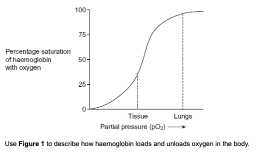
Figure 1 shows the oxygen dissociation curve for human haemoglobin.
loading of oxygen at high partial pressure
in the lungs, haemoglobin has a high affinity for oxygen
haemoglobin unloads oxygen at low partial pressure
Explain how the shape of a red blood cell allows it to take up a large amount of oxygen in a short time.
large SA:V ratio
for diffusion
thin
so oxygen can release all haemoglobin
Explain how oxygen in a red blood cell is made available for respiration in active tissues.
low pH due to increased CO2
increased dissociation of oxygen from haemoglobin
oxygen diffuses from rbc to tissues
What is meant by the term partial pressure?
the measure of concentration of a gas
When do the AV valves open and close?
Open: when pressure is higher in atria compared to ventricles
Close: when pressure is higher in ventricles than atria
When do semilunar valves open and close?
Open: when the pressure is higher in the ventricle compared to the arteries
Close: when the pressure is higher in the arteries compared to the ventricles
role of septum
separates oxygenated and deoxygenated blood
maintains high conc. of O2 in oxygenated blood
to maintain conc. gradient to therefore enable diffusion at respiring cells
some babies are born with a hole between the right and left ventricles. suggest why these babies are unable to get enough oxygen to their tissues
mixing of oxygenated and deoxygenated blood
- therefore lower volume of oxygenated blood leaves the left ventricle and flows into the aorta
role of tendons
prevents inversion of valves due to high pressure
2 ways in which blood moves in one direction as it passes through the heart
blood moves down a pressure gradient
blood moves from high to low pressure
2. valves stop backflow of blood
structure of arteries
thick walls: enabling it to carry blood at high pressures
elastic tissue: smooths out blood flow and maintains pressure
muscles in wall: to control blood flow
smooth endothelium: to reduce friction
contrast the structure of arterioles and arteries
Arterioles have thicker muscle layer than arteries, which contract to control the flow of blood
Arterioles elastic layer is relatively thinner than the elastic layers in arteries, because blood pressure is lower
why is an arteriole described as an organ?
made up of different tissues/made up of more than one tissue
how do the muscle fibres in arterioles reduce blood flow to the capillaries?
the muscle contracts
so the arterioles narrows/constricts
vasoconstriction
structure of aorta
elastic tissue: to allow stretching/recoil to maintain pressure
elastic tissue stretches when ventricles contract
muscles: contraction/vasoconstriction
thick walls: to withstand pressure
smooth endothelium: to reduce friction
SLV: to prevent back flow of blood
suggest why the rise and fall in blood pressure in the aorta is greater than in the small arteries
aorta is directly linked to the heart, therefore the pressure is higher in the aorta than in the small arteries
aorta has elastic tissue, so it can stretch/recoil
describe the difference in the thickness of aorta wall during each time in cardiac cycle
1. during systole: aorta wall stretches bc ventricles contract
2. during diastole: aorta walls recoil bc ventricles relax
this helps maintain smooth blood flow
explain how the highest blood pressure is produced in the left ventricle
left ventricle has thicker, muscular walls
therefore stronger contractions
structure of veins
thin walls due to lower pressure, therefore requiring valves to ensure blood doesn’t flow backwards
less muscular and elastic tissue bc they don’t have to control blood flow
smooth endothelium to reduce friction
explain the difference in thickness between pulmonary artery and pulmonary vein
pulmonary artery contains muscle and elastic fibres
b/c pulmonary arteries handle high pressures and smooth out blood flow
structure of capillaries
permeable capillary membrane
thin, one-cell thick walls: short diffusion distance/pathway
small diameter: short diffusion distance
flattened endothelial cells: short diffusion distance
numerous and highly branched: provides large SA
narrow lumen: reduces blood flow rate to give more time for diffusion
advantage of capillaries being narrow
short distance b/w blood and the outside of the capillary
therefore fast diffusion
why is the blood flow in capillaries slow?
so that theres more time for diffusion
what factor limits the internal diameter of the lumen of a capillary?
width/diameter of blood cell
how can the widening of blood vessels reduce blood pressure
widening of blood vessels causes larger lumen, which reduces the blood pressure in blood vessels
therefore less friction/resistance in the blood vessels
suggest why pulse can be used to measure heart rate
pulse is caused by the pressure from one contraction/beat of the heart
role of heart in formation of tissue fluid
1. contraction of ventricles produces high hydrostatic pressure
2. this forces water and some dissolved substances out of the capillaries
how tissue fluid is formed and returned to circulatory system
high blood/hydrostatic pressure at arterial end of capillary which forces water out
large proteins remain
water potential in capillary becomes lower/negative due to these proteins
water potential now lower than hydrostatic pressure
so water enters venous end of capillary by osmosis
water moves out at the arteriole end and back in at the venule end
lymph system collects any excess tissue fluid which returns to blood/circulatory system
how does high blood pressure lead to an accumulation of tissue fluid
1. high blood pressure = high hydrostatic pressure
2. increases outward pressure from arterial end of capillary
3. more fluid forced out of capillary due to high pressure
4. so more tissue fluid formed
5. less return of fluid into capillary (due to high pressure)
Explain why a lack of protein in the blood causes a build up of tissue fluid.
water potential in the capillary is higher
- therefore less water removed by osmosis
suggest how an increase in volume of blood entering the heart reduces angina
1. large amount of blood leaves the heart
2. therefore more blood/oxygen flow to the heart muscle via coronary arteries
explain how blood in a vein in the leg is returned to the heart
1. muscles surrounding the vein contracts and press on the walls of the vein, squeezing blood along the veins
2. valves prevent backflow
3. contraction of heart pumpls blood through arteries into vein (systole)
4. the recoil of heart muscle after contraction/ during diastole draws the blood from the veins into the atria
5. veins have wide lumens therefore theres little resistance
how to calculate something with eyepiece graticule
measure using eyepiece graticule
calibrate eyepiece graticule against stage micrometer
take a number of measurements to calculate a mean
Contrast the trachea of a mammal and the trachea of an insect
1. Mammals have just one trachea whereas insects have multiple trachea
2. Trachea of mammals have a larger diameter than trachea of insects
3. Mammal trachea made up of cartilage whereas insect trachea made up of chitin
4. Mammal trachea is longer than insect trachea
5. Mammal trachea branch into bronchi whereas insect trachea branch into tracheoles
Describe the difference in the composition of gases in inhaled and exhaled air.
Explain how these differences are caused.
inhaled air contains more oxygen that exhaled air
inhaled air contains less carbon dioxide than exhaled air
inhaled air contains less water vapour
water vapour diffuses from moist surface
respiration results in higher blood carbon dioxide and lower blood oxygen
oxygen enters blood and carbon dioxide leaves blood in alveoli by diffusion
describe the gross structure of the human gas exchange and how we breathe in and out
trachea, bronchi, bronchioles, alveoli
1. when you breathe in: the diaphragm contracts and moves down and the external intercostal muscles contract
contraction of diaphragm muscles flattens diaphragm
causes an increase in volume and pressure decrease in thoracic cavity (to below atmospheric), resulting in air moving in down pressure gradient
2. when you breathe out: the diaphragm relaxes and internal intercostal muscles contract
diaphragm moves up and becomes dome shaped
contraction of intercostal muscles raises ribcage
causes a decrease in volume and a pressure increase in thoracic cavity to above atmospheric
pressure in lungs is higher than pressure outside
resulting in air moving out
apart from reduced elasticity, explain how changes to the lung tissue reduce the efficiency of gas exchange
alveolar walls thicken
so longer diffusion pathway
scarred tissue
reduces SA for gas exchange
role of diaphragm in breathing in
diaphragm contracts and moves down
increases volume in thorax and lowers pressure in thorax
air moves in down pressure gradient
Reduced pressure allows air to enter
role of diaphragm in breathing out
diaphragm moves up and becomes dome-shaped
reduces volume of thorax and increases pressure in thorax
so pressure in thorax is higher than the outside
how paralysis of diaphragm leads to breathing difficulties
diaphragm will not contract and move down
thoracic cavity/lung volume is not increased, so cannot breathe in
movement of ribs when a person breathes in
up and out
explain whether breathing out is active or passive
active because it involves contraction of muscles
three ways in which an insects tracheal system is adapted for efficient gas exchange
tracheoles have thin walls
so short diffusion distance to cells
large number of tracheoles
so short diffusion distance to cells
large number of tracheoles
so large surface area
Tracheae provide tubes full of air
so fast diffusion into insect tissues
Fluid in the end of the tracheoles that moves out during exercise
so larger surface area
the structure through which gases enter and leave the body of an insect
spiracle
name the small tubes that carry gases directly to and from the cells of an insect
tracheole
explain the movement of oxygen into the gas exchange system of an insect when it is at rest
oxygen used in respiration
oxygen moves down a diffusion gradient
The damesfly larva is a carnivore that actively hunts prey. It has gills to obtain oxygen from water.
Explain how the presence of gills adapts the damesfly to its way of life.
Damselfly larvae has higher metabolic / respiratory rate
so it uses more oxygen
explain 5 ways in which the structure of fish gills is adapted for efficient gas exchange
Gills have many lamellae / filaments so large surface area
Thin epithelium surface so short diffusion pathway
Countercurrent maintains concentration gradient along gill
equilibrium not reached
Circulation replaces blood saturated with oxygen
Ventilation replaces water as oxygen is removed
counter-current mechanism
water and blow flow in opposite directions
blood always passing water with a higher oxygen concentration
diffusion gradient maintained throughout length of gill/lamella
describe how oxygen in the air reaches capillaries surrounding alveoli in the lungs.
details of breathing are not required.
the oxygen moves through the trachea, bronchi and bronchioles
down a pressure and diffusion gradient
across alveolar epithelium and capillary epithelium
how oxygen in the air in the alveoli enters the blood in the capillaries
the oxygen moves by diffusion across alveolar epithelium
Explain why a large number of small alveoli is more efficient in gas exchange than a smaller number of larger alveoli
small alveoli has larger SA
so more diffusion
Forced expiration volume (FEV1) is the volume of air a person can breathe out in 1 second.
One of the severe disabilities that results from emphysema is that walking upstairs becomes difficult.
Explain how a low FEV1 value could cause this disability.
less CO2 removed
less oxygen uptaken
less respiration