IOS1_RESPIRATORY_ANATOMY
1/53
There's no tags or description
Looks like no tags are added yet.
Name | Mastery | Learn | Test | Matching | Spaced |
|---|
No study sessions yet.
54 Terms
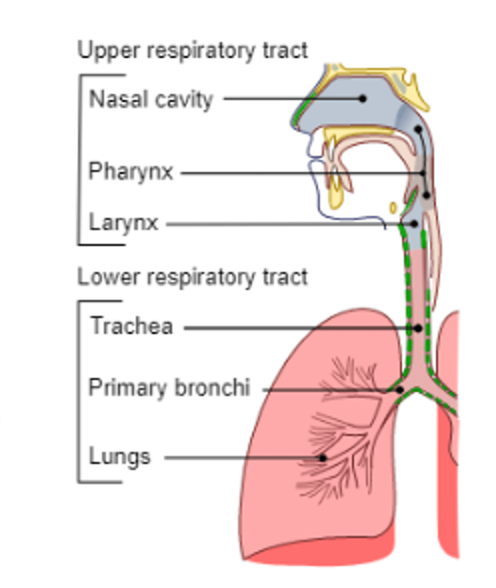
What are the parts of the upper respiratory tract?
nose, mouth
nasal cavity
pharynx
larynx
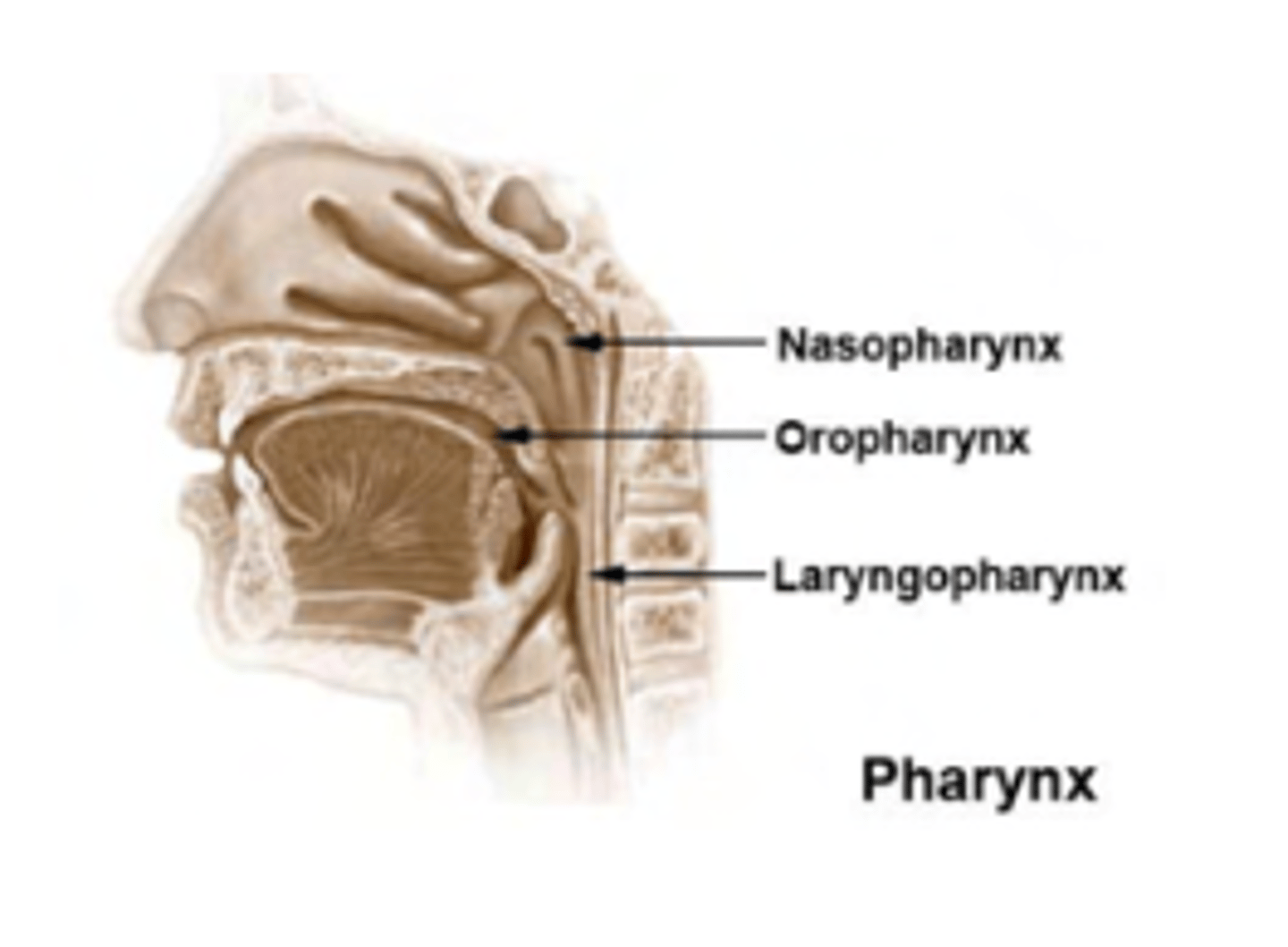
What are the divisions of the pharynx?
. Nasopharynx (adenoids and openings to Eustachian tubes)
. Oropharynx (pathway for food and air)
. Laryngopharynx (connects esophagus and larynx)
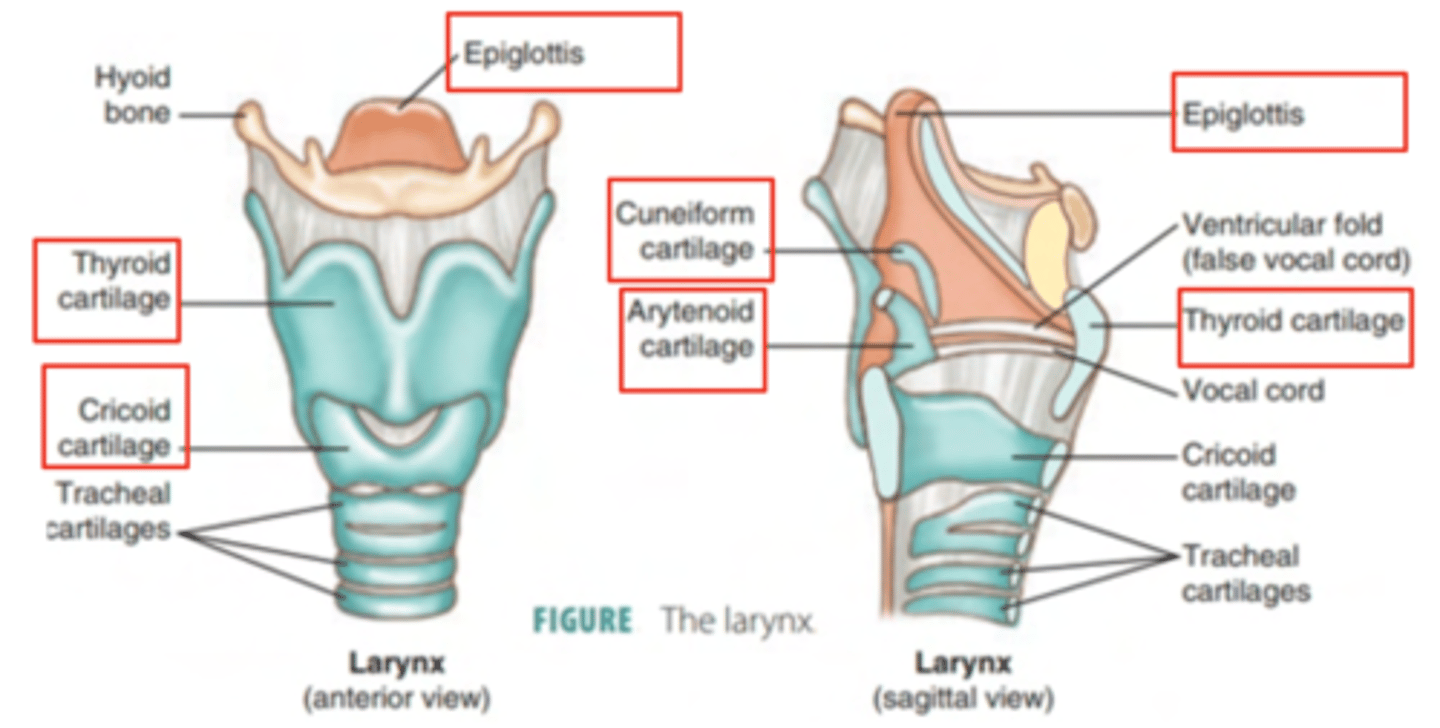
What are the structures of the laryngopharinx?
Epiglottis: Acts as a valve, preventing food entry into the trachea.
Thyroid cartilage: Largest cartilage
Cricoid cartilage:
Arytenoid
Cuneiform
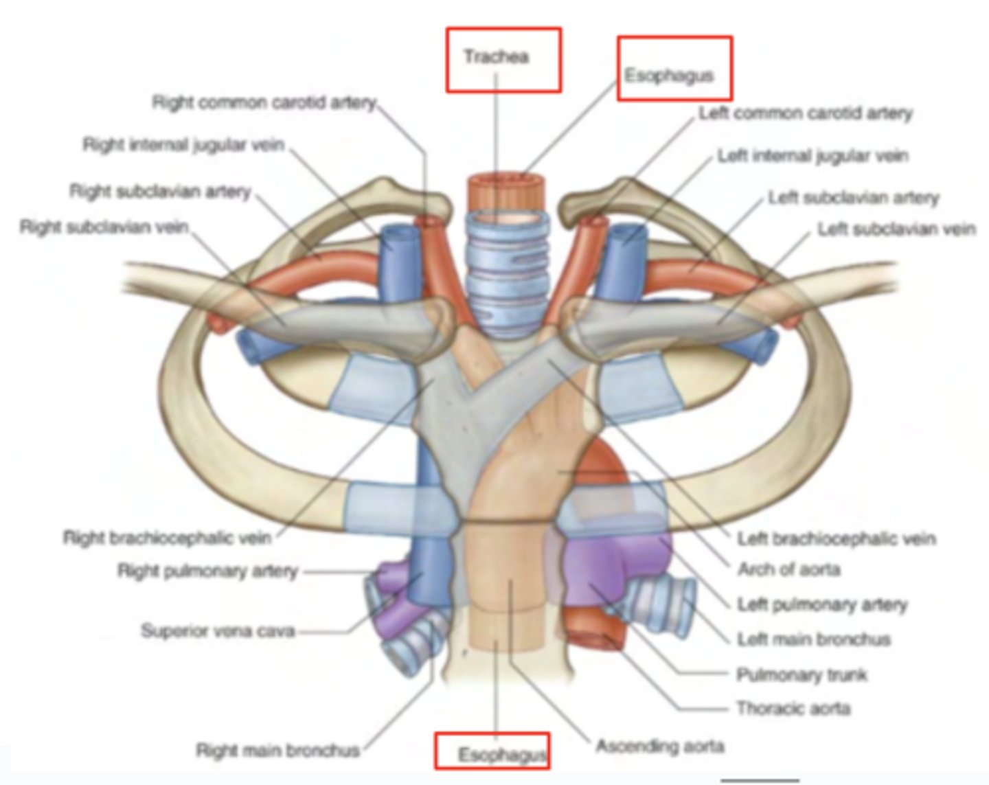
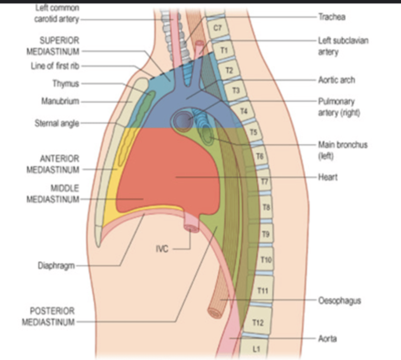
Where is located the trachea?
superior medianistum
anterior to esophagus
behind the manubrium of the sternum
FROM LARYNX TO T4-T5
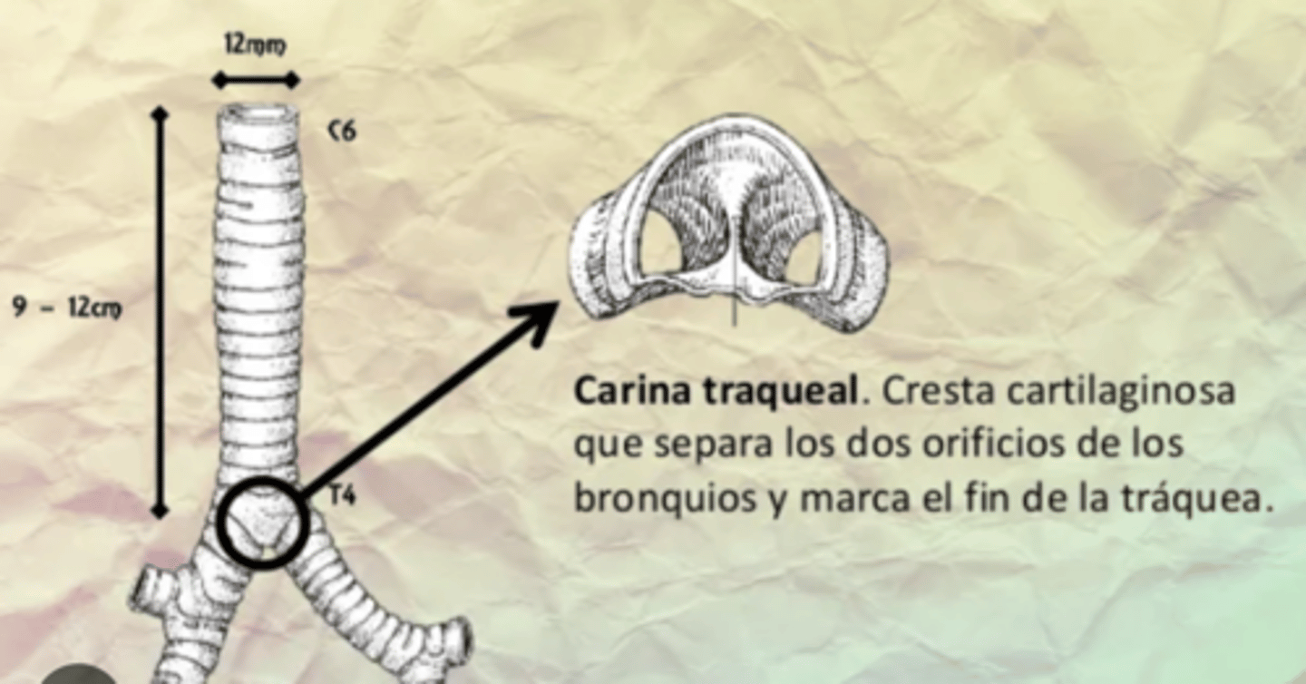
What's the structure of the trachea?
Supported by C-shaped hyaline cartilage rings
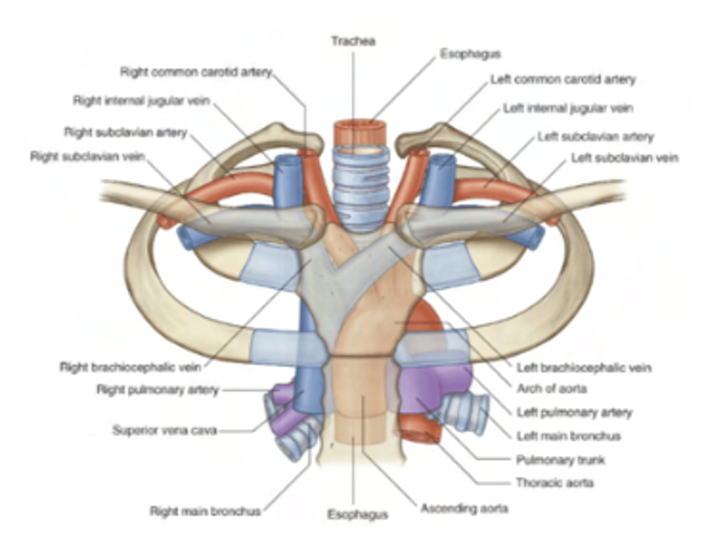
What's the name of the bifurcation of the trachea?
carina
it happens at T4-T5 posteriorly/sternal angle anteriorly
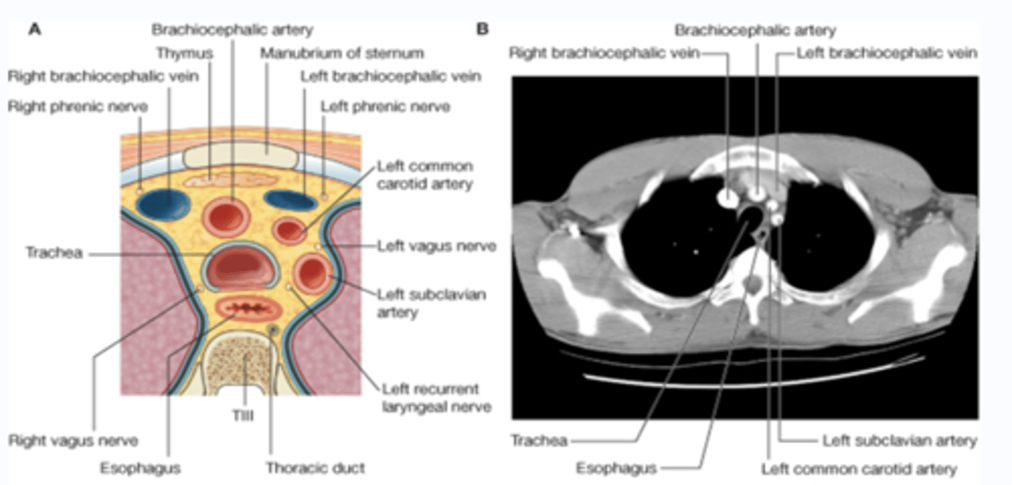
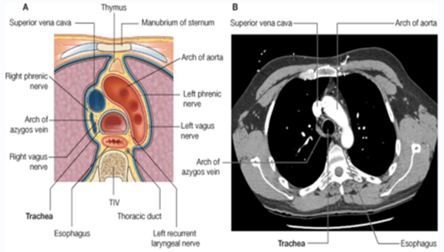
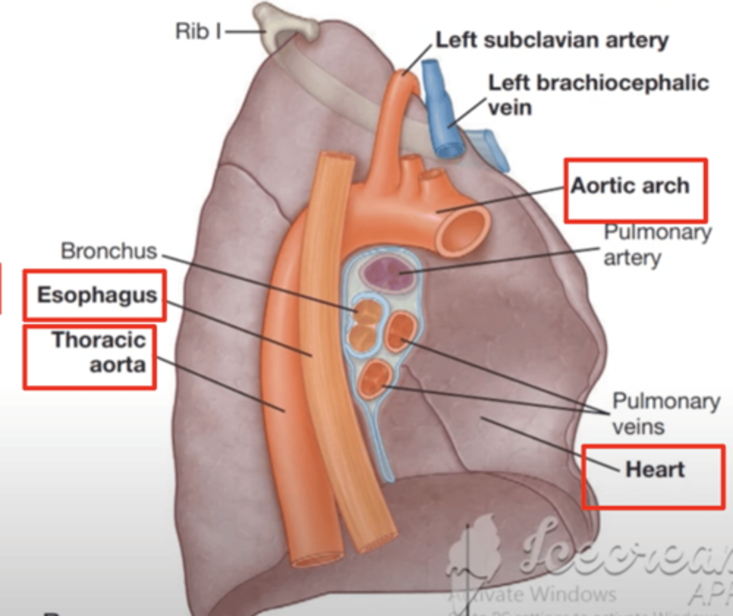
What important elements do we have at T4-T5?
. Bifurcation of the trachea
. Arch of the aorta
. Supervior Vena Cava enters RA
. Divison of superior and inferior mediastinum
Axial T3 view
Axial T4 view
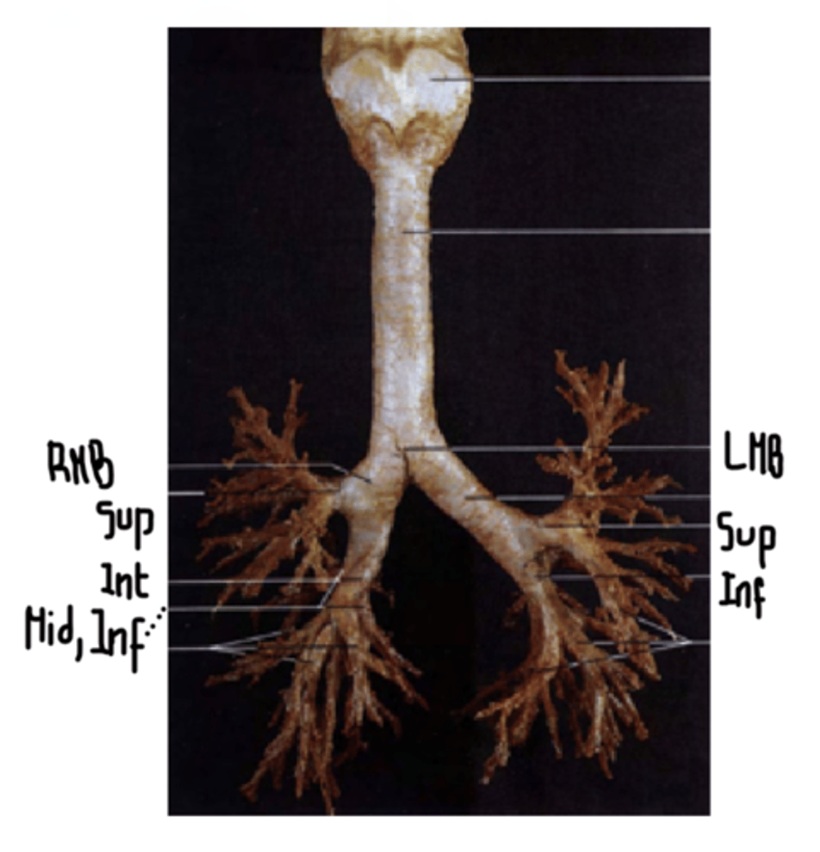
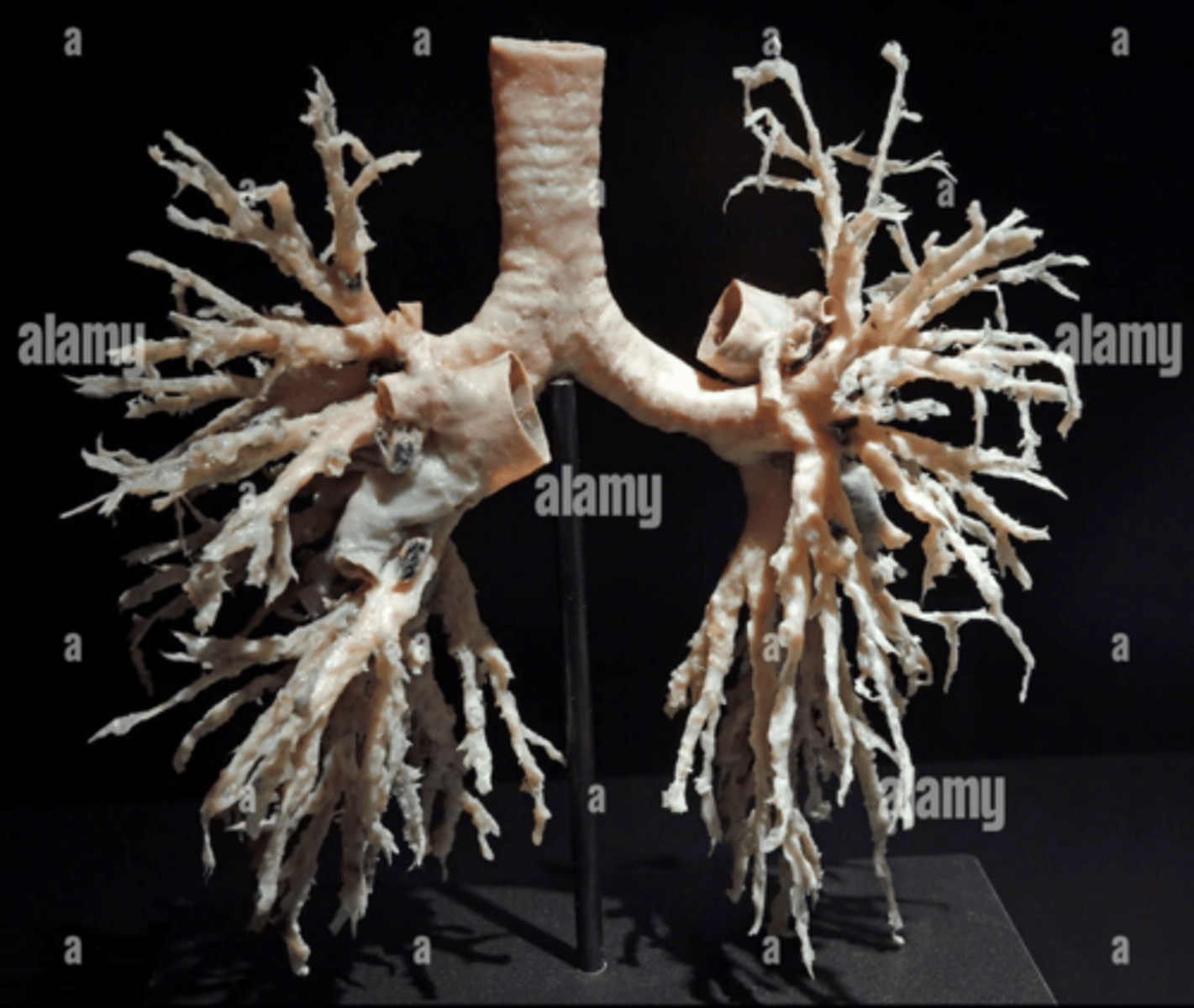
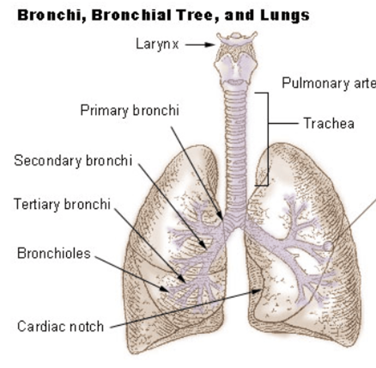
How is dividing the bronchial tree?
. 2 main bronchi
. Left bronchus
. Right bronchus
. Secondary lobar bronchi
.2 on the left
.3 on the right
. Segmental lobar bronchi
. 8 segmental on the left
. 10 segmental on the right
What is the structure of the bronchial tree?
horse-shoe shaped cartilage plates
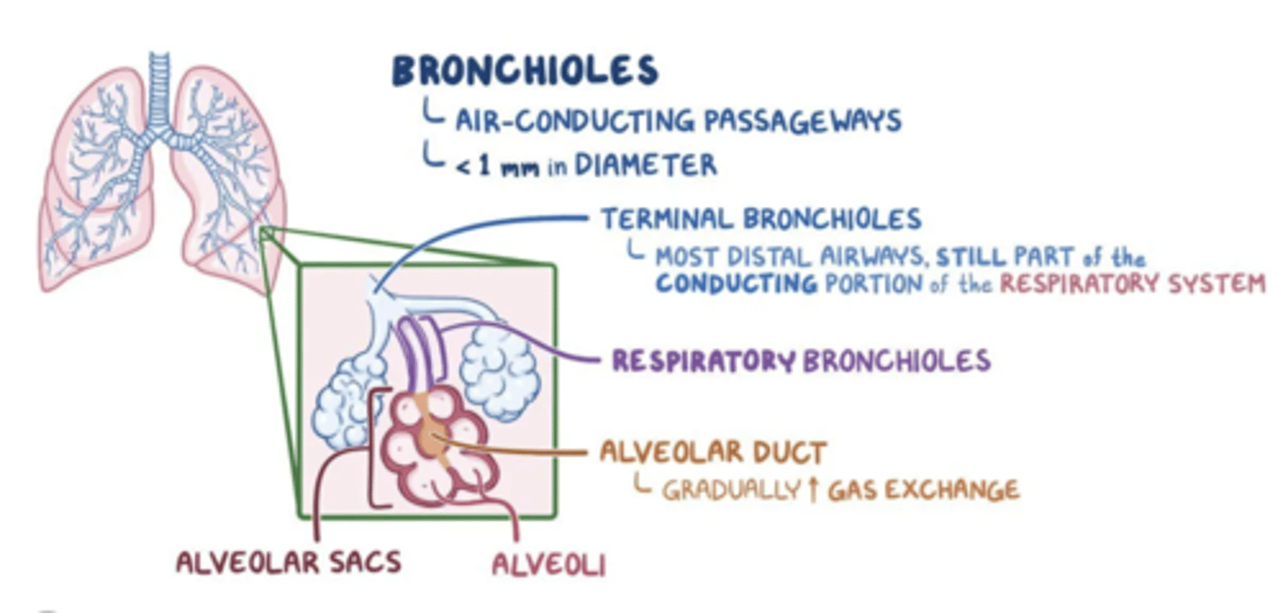
What is the structure and type of bronchioles?
. Segmental bronchi lose their cartilage (from 6th generation) and further divide into branchs
Two types:
. terminal bronchioles
. respiratory bronchioles
What are differences between left and right bronchi?
. Right wider, shorter
and enters more vertically
ASPIRATED FOREIGN OBJECTS TEND TO OBSTRUCT RIGHT BRONCHUS
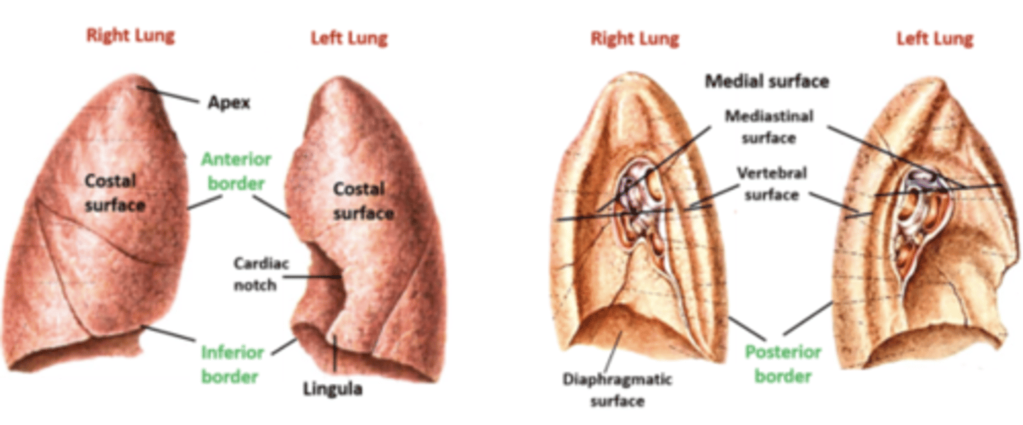
What are the anatomical difference between lungs?
Left is smaller due to heart.
Right is bigger but diaprhagmatic surface is more concave and dome-shaped as it sits on top of the liver
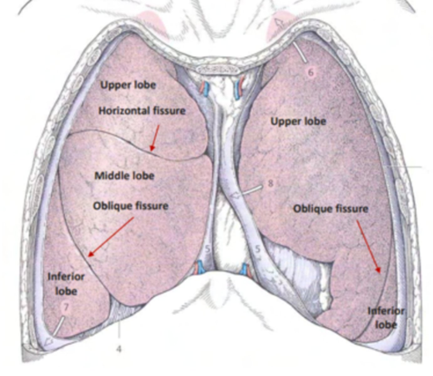
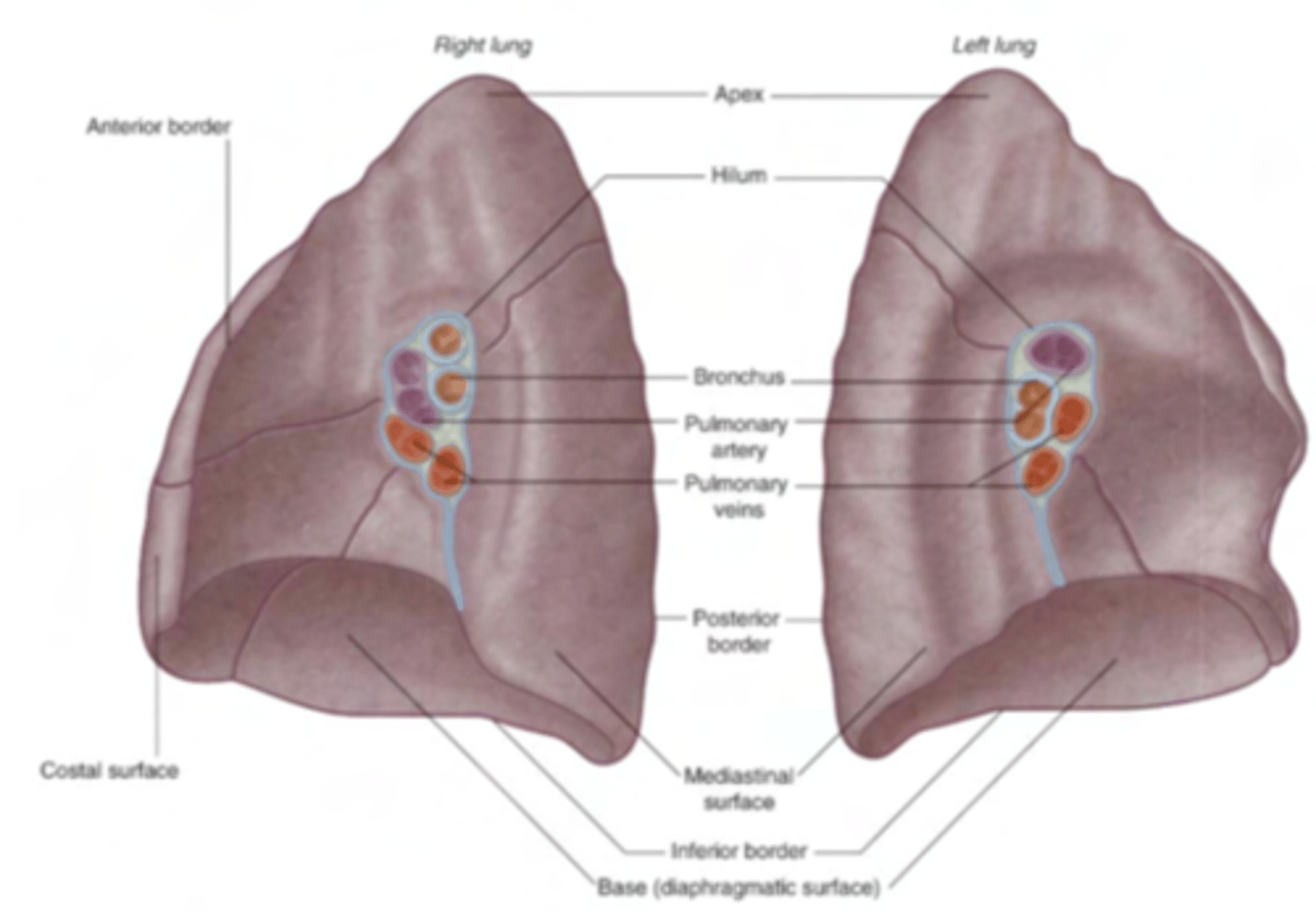
What is the structure of the lungs?
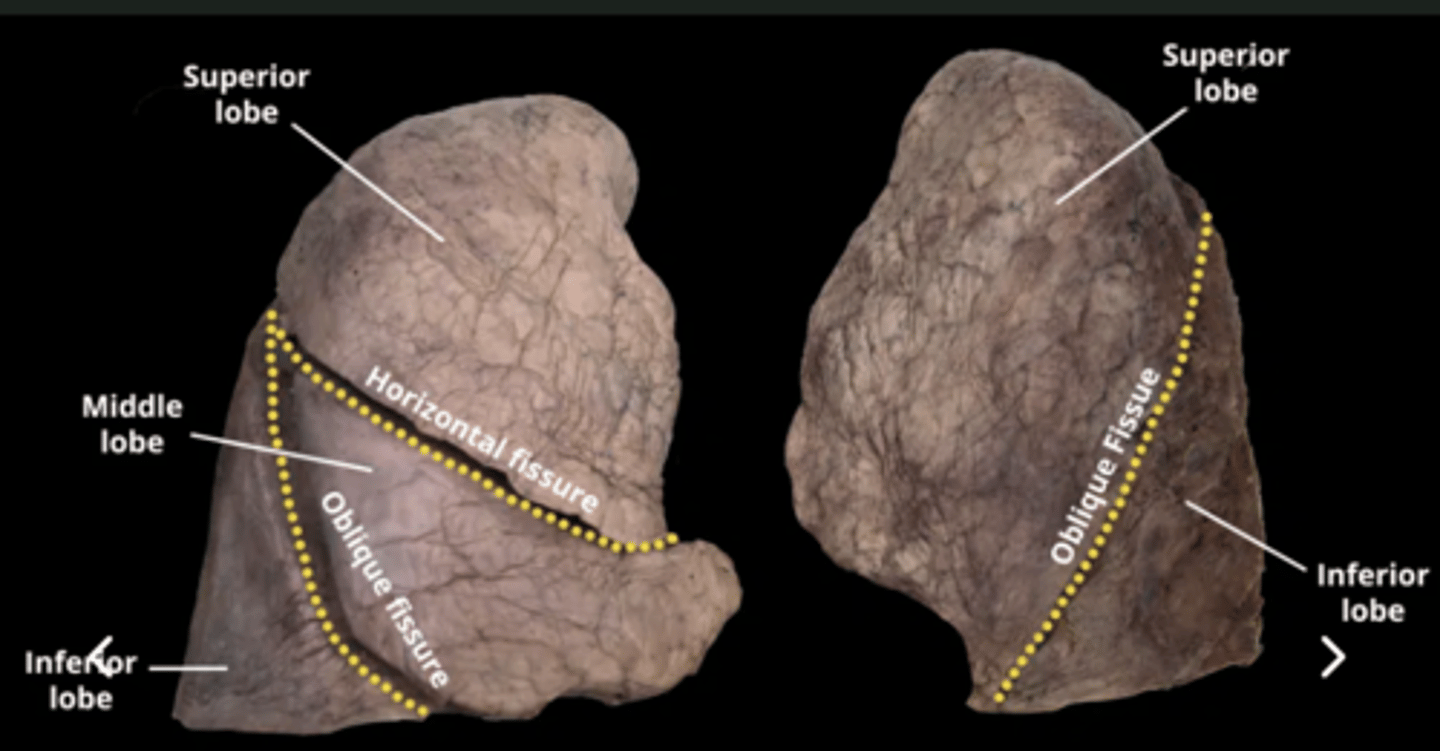
. Right lung : 3 lobes, separated by right oblique fissure and horizontal fissure
. Left lung: 2 lobes, one unique, left oblique fissure

What are the lung surfaces?
. Mediastinal -> contact with mediastium
. Costal -> contact with the ribcage
. Diaprhagmatic -> dome-shaped contact with the diaphragm
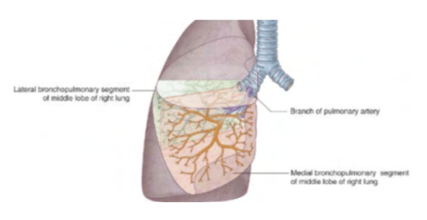
What is a lung or bronchopulmonary segment?
. smallest functional independent region
Each have their own bronchus and their own artery
Veins are intersegmental
It can be dissected and removed (important in cancer surgery)
What are the lobes of the right lung?
. Superior
. Middle
. Inferior
What are the segments of the right superior lobe?
. Apical
. Anterior
. Posterior
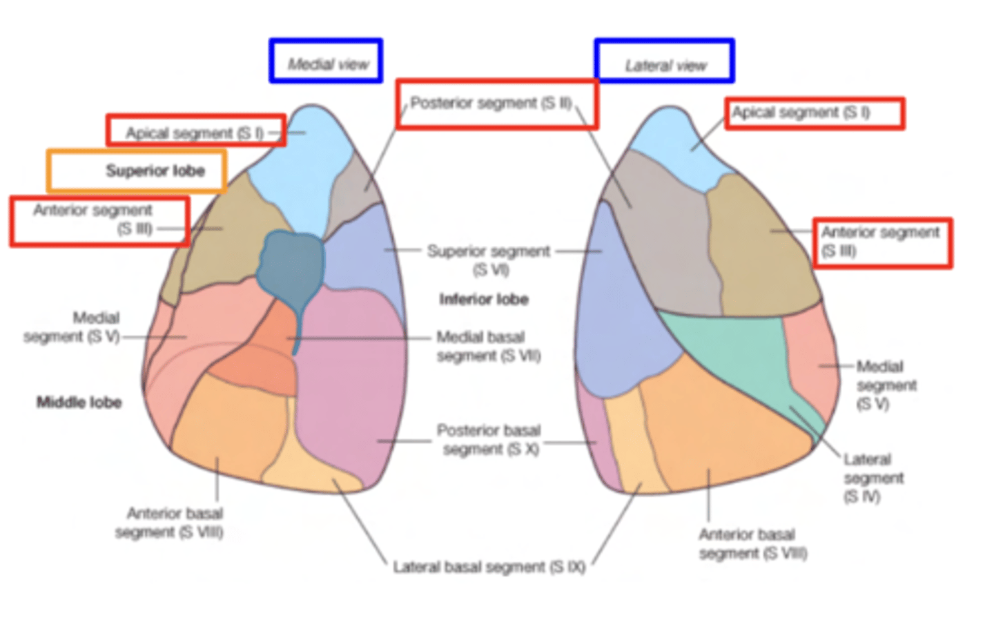
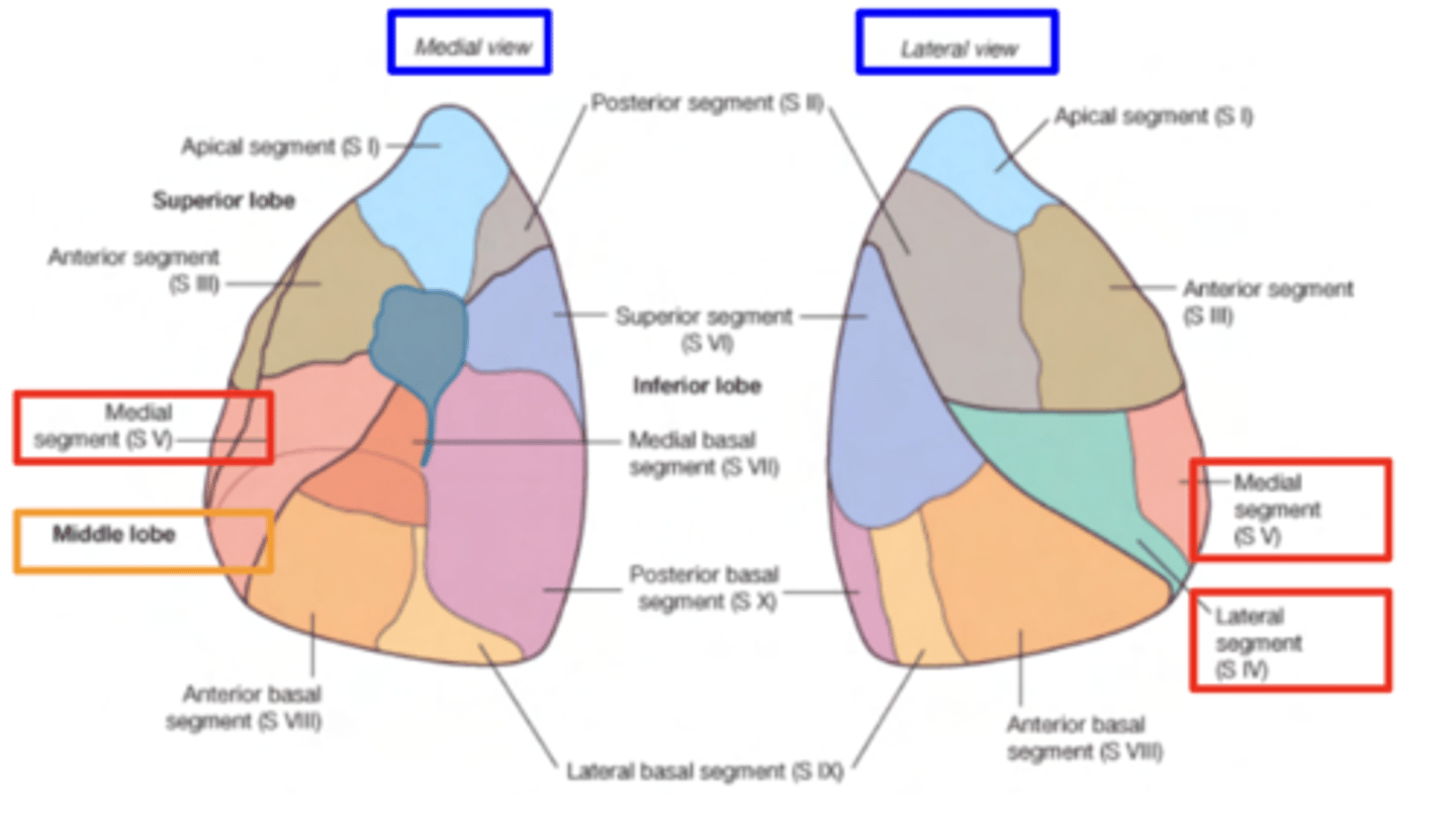
What are the segments of the middle right lobe?
. Medial
. Lateral
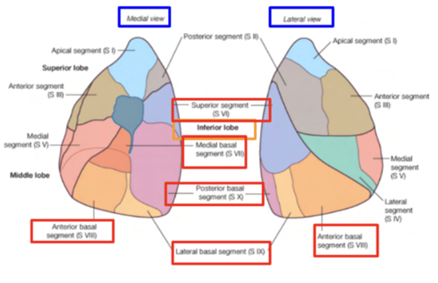
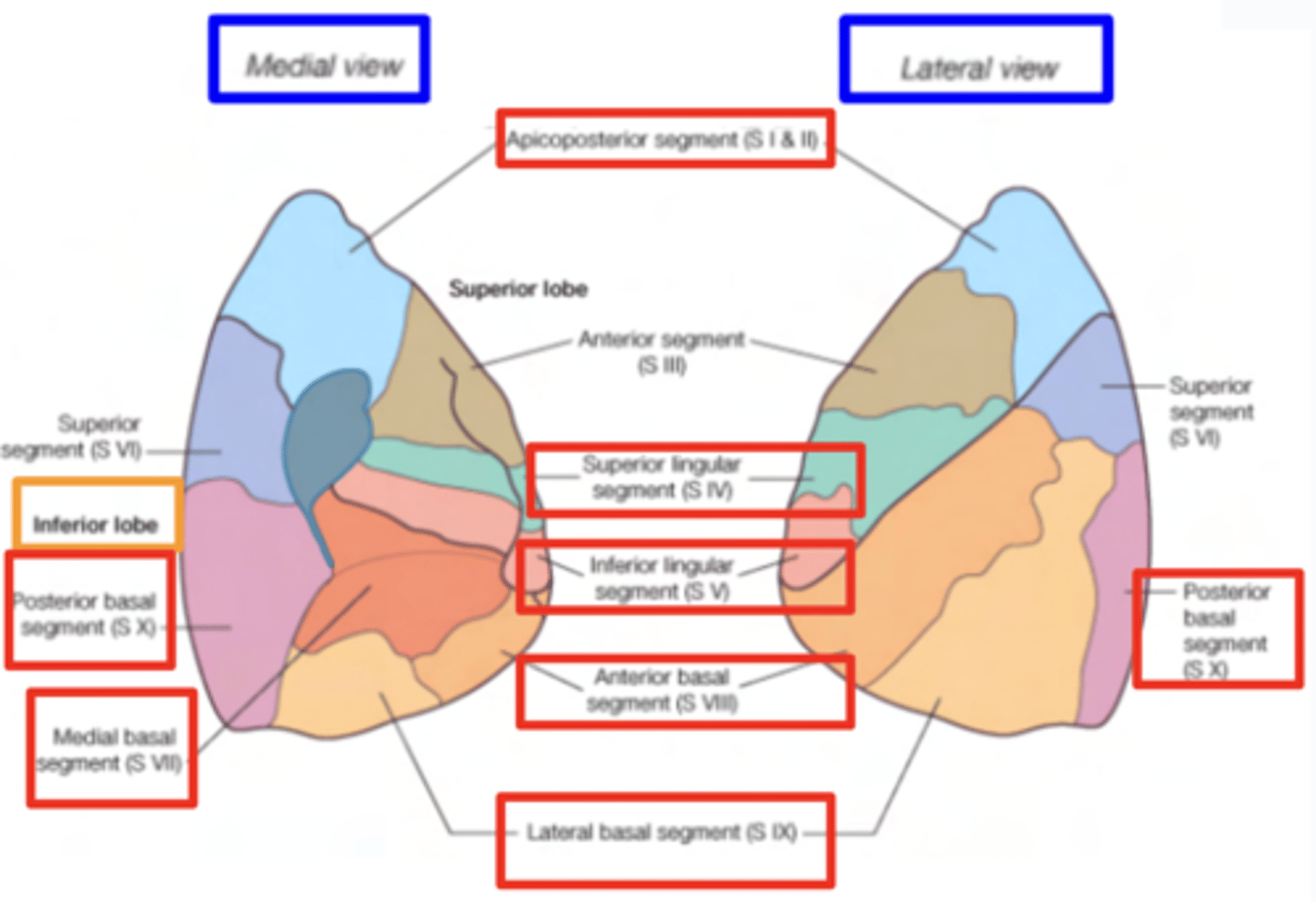
What are the segments of the inferior right lobe?
. Superior
. Basal anterior
. Basal lateral
. Basal posterior
. Basal medial (only seen on the mediastinal face)
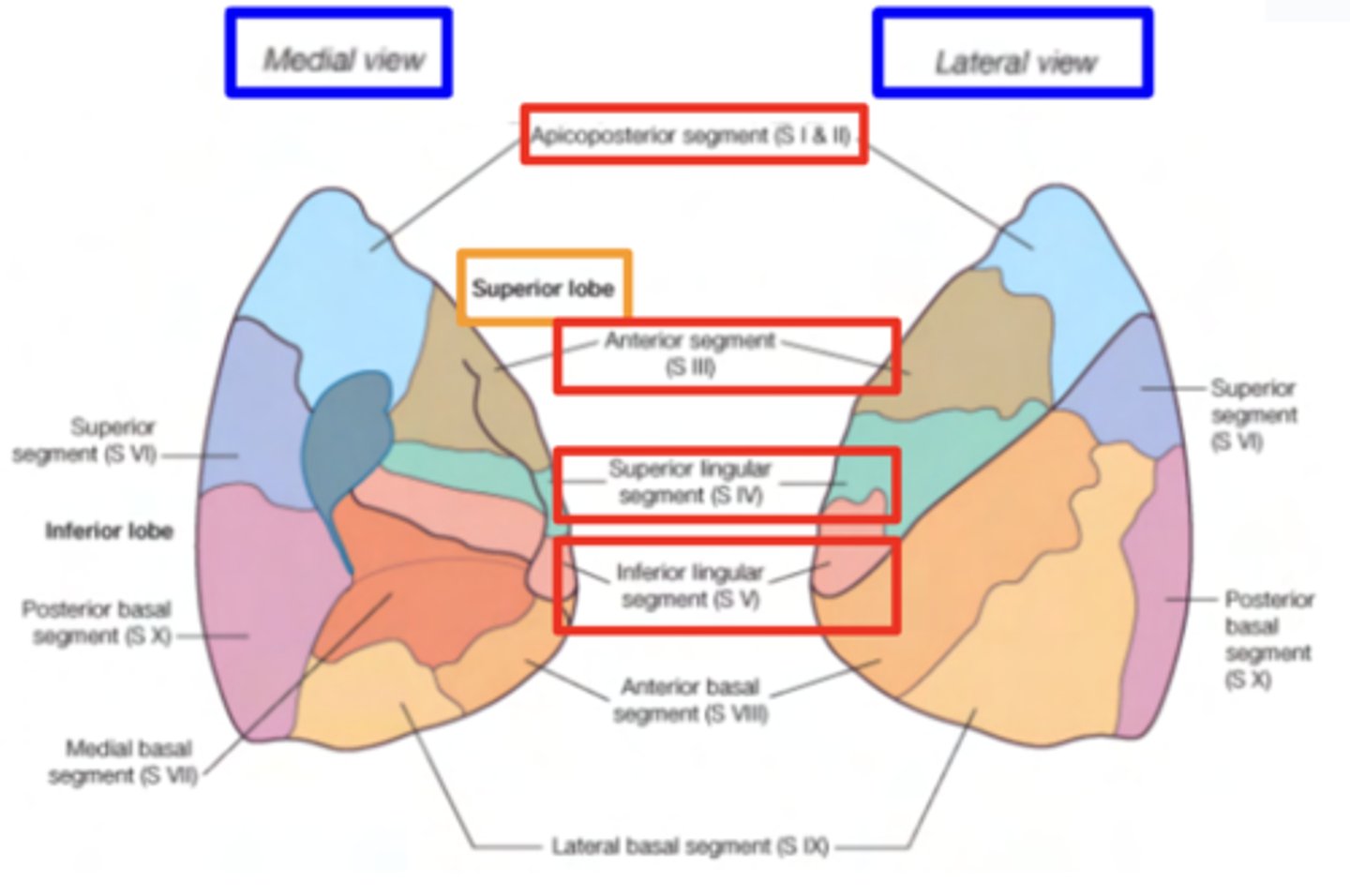
What are the segments of the left superior lobe?
. Apical
. Posterior (normally fused as apicoposterior segment)
. Anterior segment
. Lingula (superior and inferior)
What are the segments of the left inferior lobe?
. Superior
. Basal anterior
. Basal lateral
. Basal posterior
. Basal medial (only seen on the mediastinal surface)
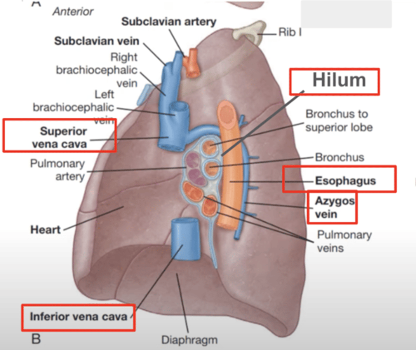
What are the impressions of the right lung?
Superior cava vein
Inferior cava vein
A little curved groove can be seen, corresponding to the arch
of the azygos vein
Esophageal impression
HILUM a hollow or narrow opening in an organ through which nerves, blood vessels, and tubes enter and leave:
What are the impressions of the left lung?
. Arch of the aorta
. Descending aorta (more posterior)
. Esophageal impression (in front of the DA)
. Cardiac impression (most important!)
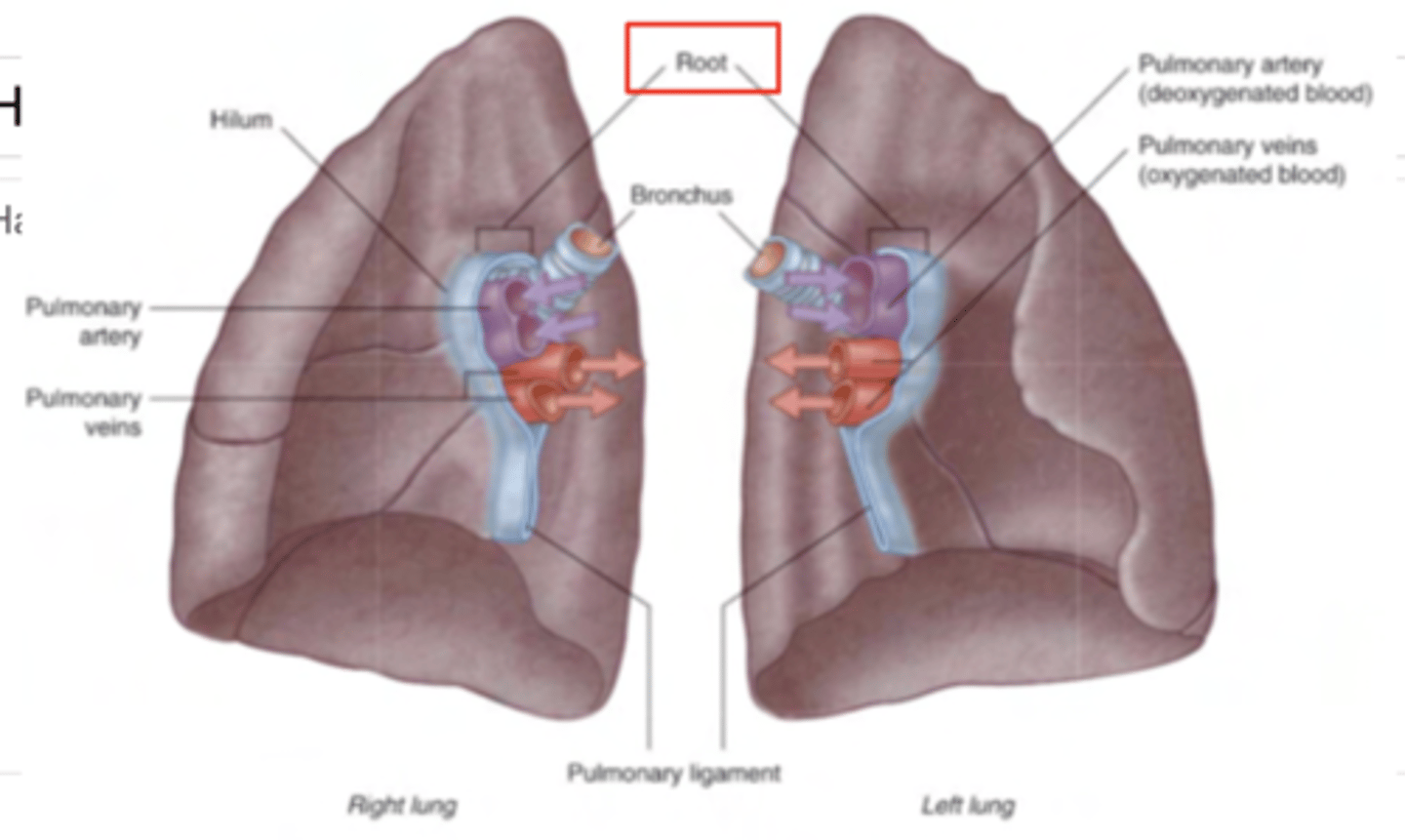
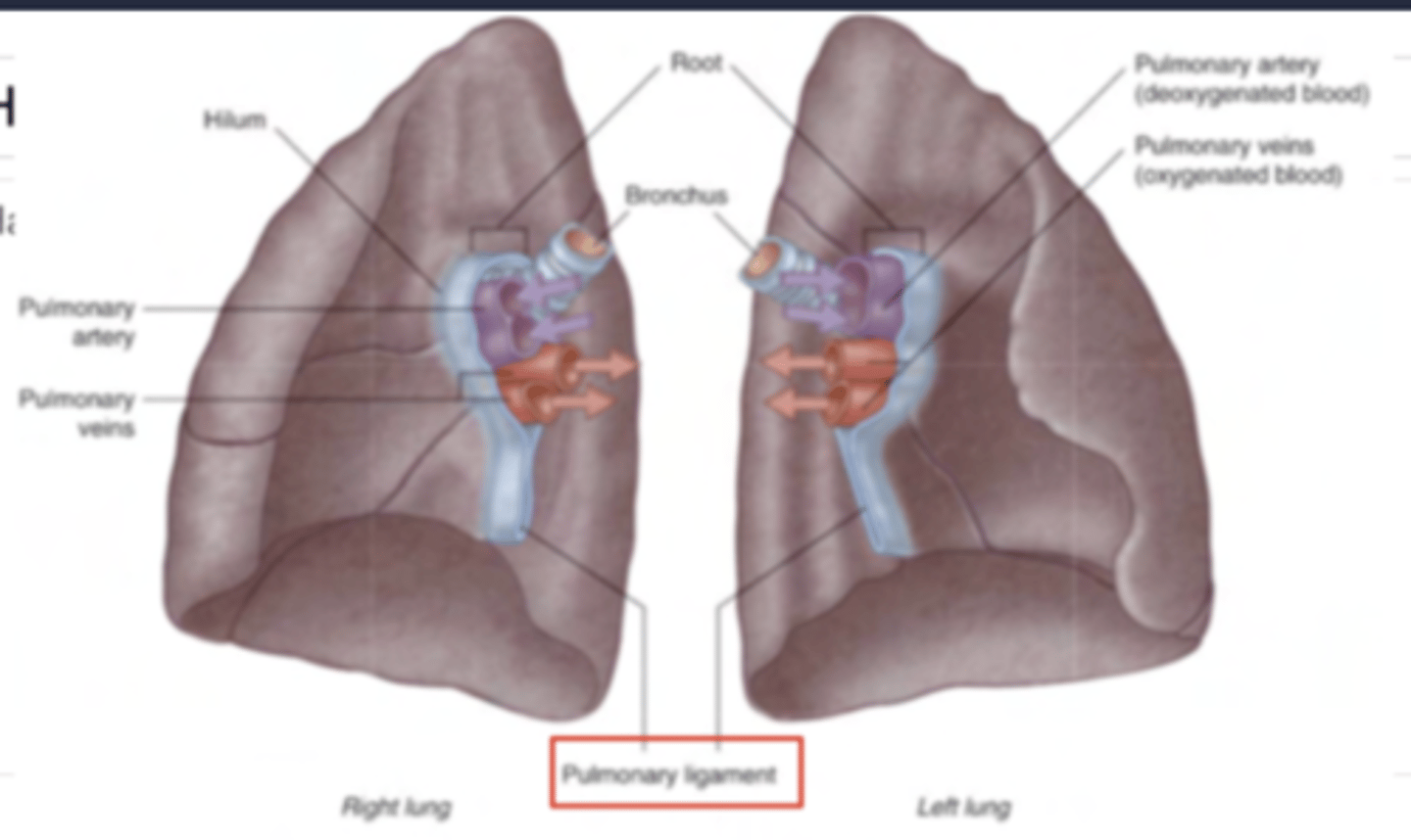
Where are entering the vessels and bronchi in the hilum?
. bronchi (sometimes already divided, sometimes not,
depending on the cut): they’ll be posterior
. Arteries: superior and anterior
. Veins: inferior
What is the pulmonary root?
pedicle which is covered by parietal pleura and
contains the structures which penetrate into the hilum
What is the pulmonary ligament?
A double fold of parietal pleura coming down from the pulmonary root
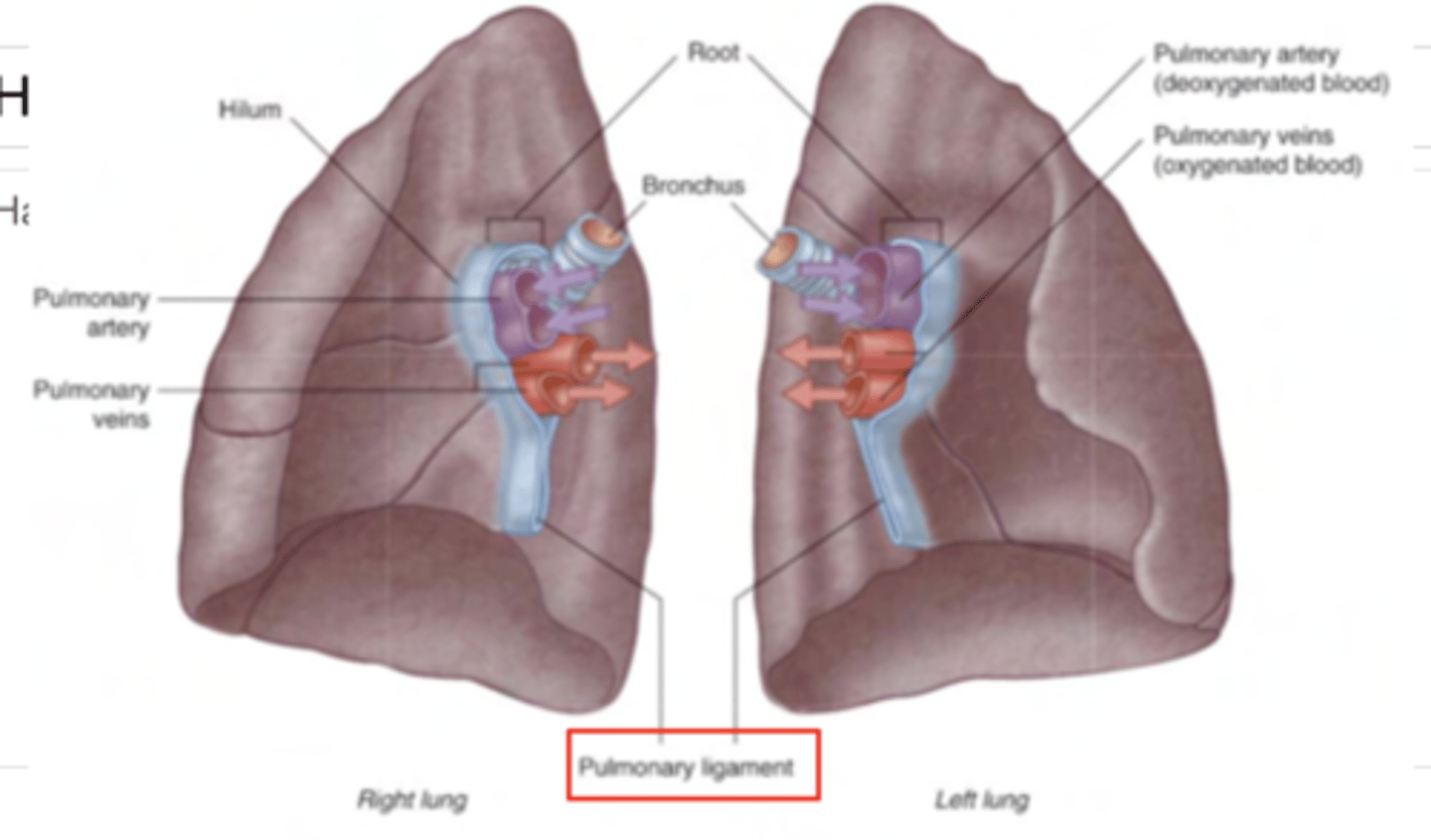
What is the function of the pulmonary ligament?
To maintain each structure in their correct position within the root of the lung.
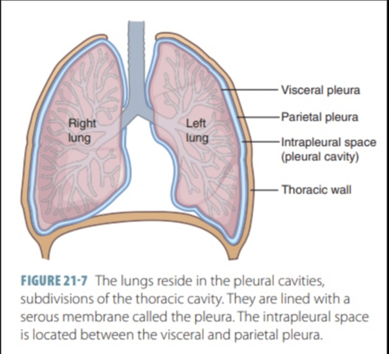
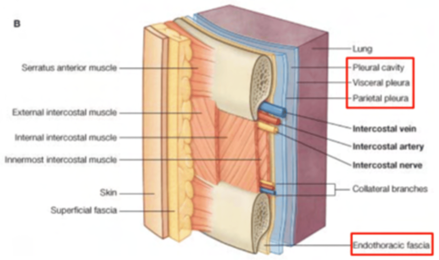
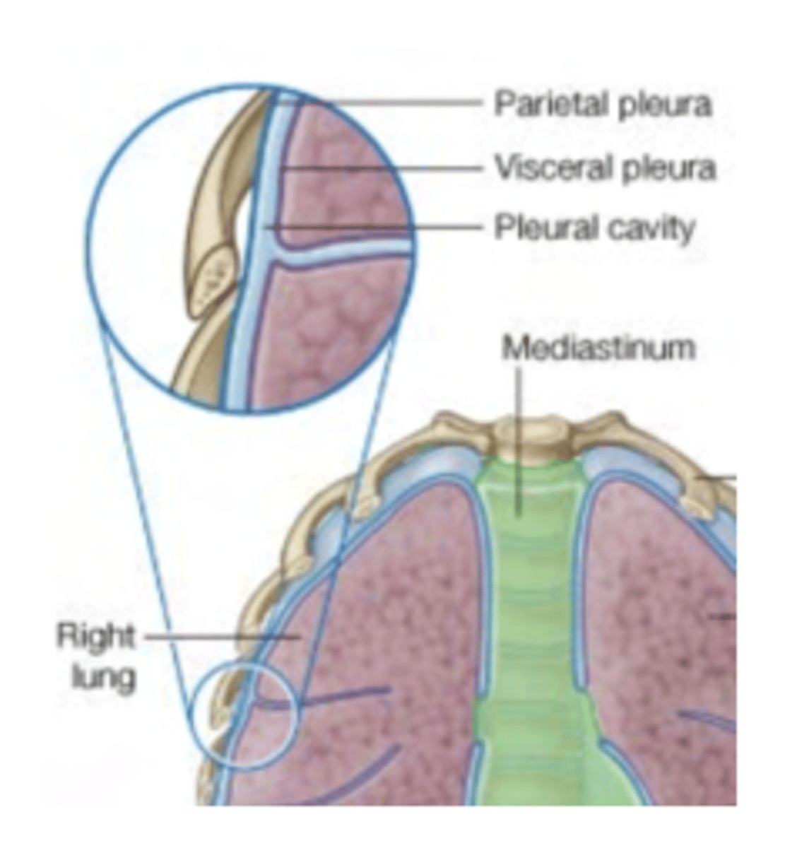
What are the pleural cavities?
are thin serous membranes surrounding the lungs
The pleural cavities, which are relatively small virtual cavities, are delimited by 2 layers of pleura
- Visceral pleura, in contact with the lungs
- Parietal pleura, in contact with the thoracic wall and spaces
that enclose and surround the lung (such as the mediastinum)
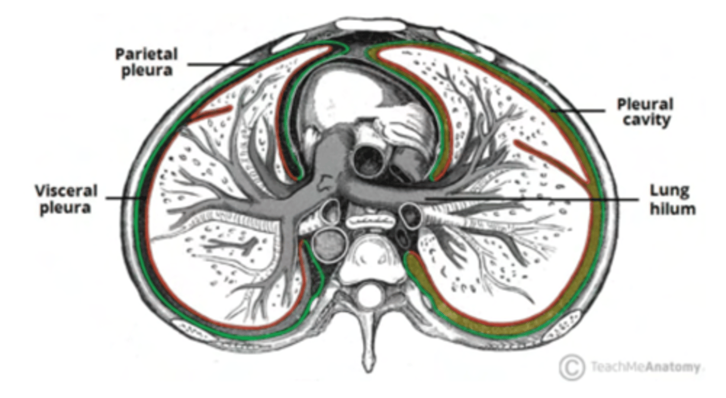
Describe the visceral pleura?
Directly adheres to the surface of the lungs, including into the fissures.
It is tightly attached and moves with the lung during respiration.

Describe the parietal pleura?
Lines the inner surface of the thoracic wall, diaphragm, and mediastinum.
It is continuous with the visceral pleura at the lung's root (hilum).

Explain the pleural cavity
The pleural cavity is the potential (virtual) space between the visceral and parietal pleura.
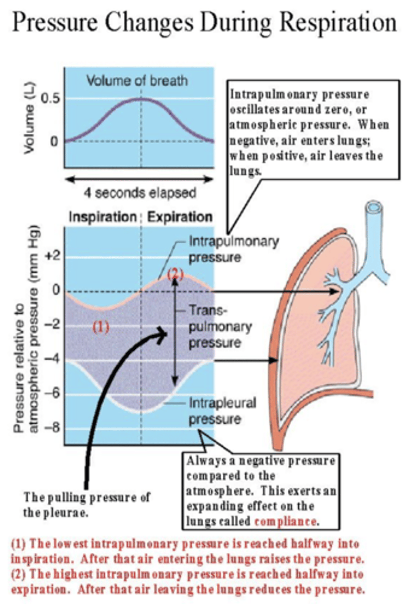
What is the importance of negative pressure within the pleural cavity?
Keeps the lungs expanded against the thoracic wall.
Prevents lung collapse by maintaining a suction effect.
What are the functions of the pleural fluid?
Lubrication: Reduces friction between the pleura during respiratory movements.
Cohesion: Keeps the two pleural layers in close contact, facilitating lung expansion during inspiration.
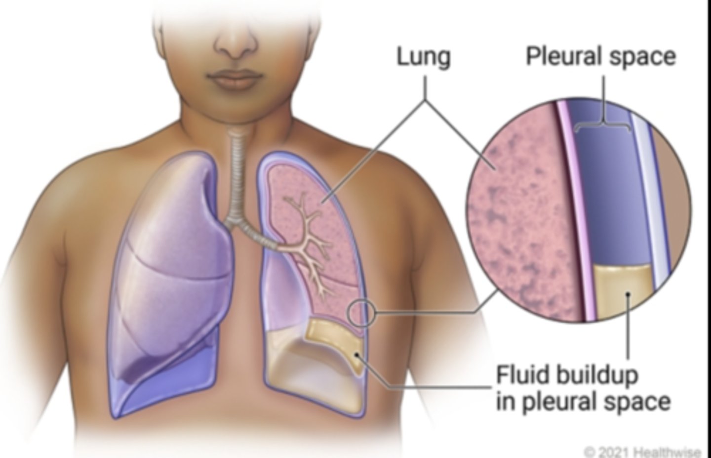
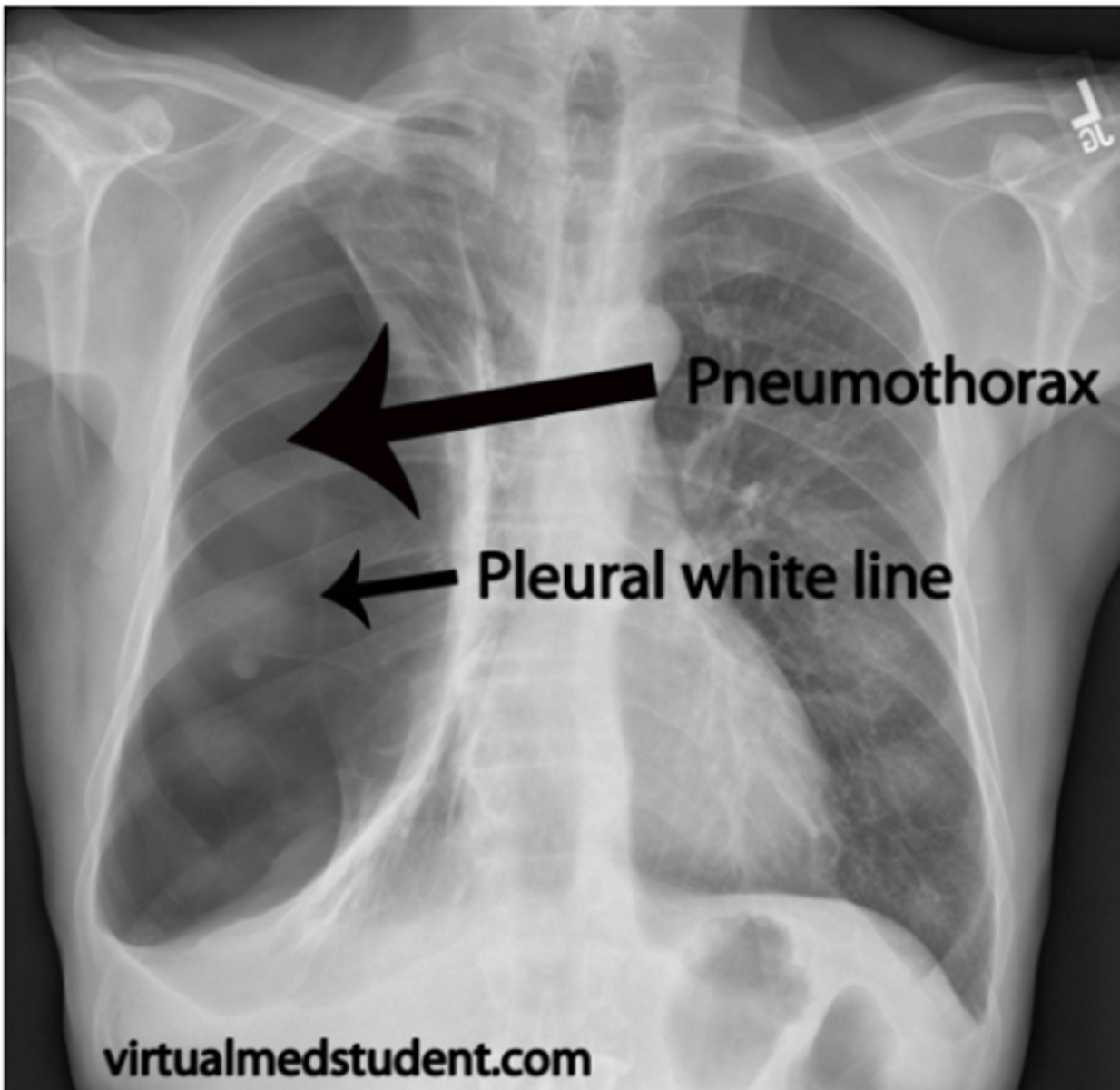
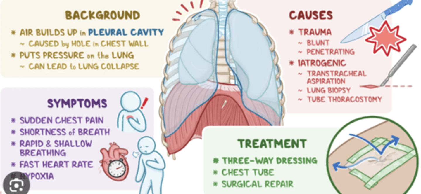
What is a pneumothorax?
If air enters the pleural cavity (e.g., due to trauma or lung rupture), the negative pressure is lost, creating a real cavity instead of a virtual one.
This condition leads to:Lung collapse.
What are the symptoms of a pneumothorax?
Difficulty breathing, chest pain, and in severe cases, respiratory distress.
What is the endothoracic fascia?
A layer of connective tissue located between the parietal pleura and the structures of the thoracic wall, such as:Intercostal musclesRibs
It provides a protective layer and serves as a cushion, ensuring that the parietal pleura is not directly in contact with the thoracic wall.
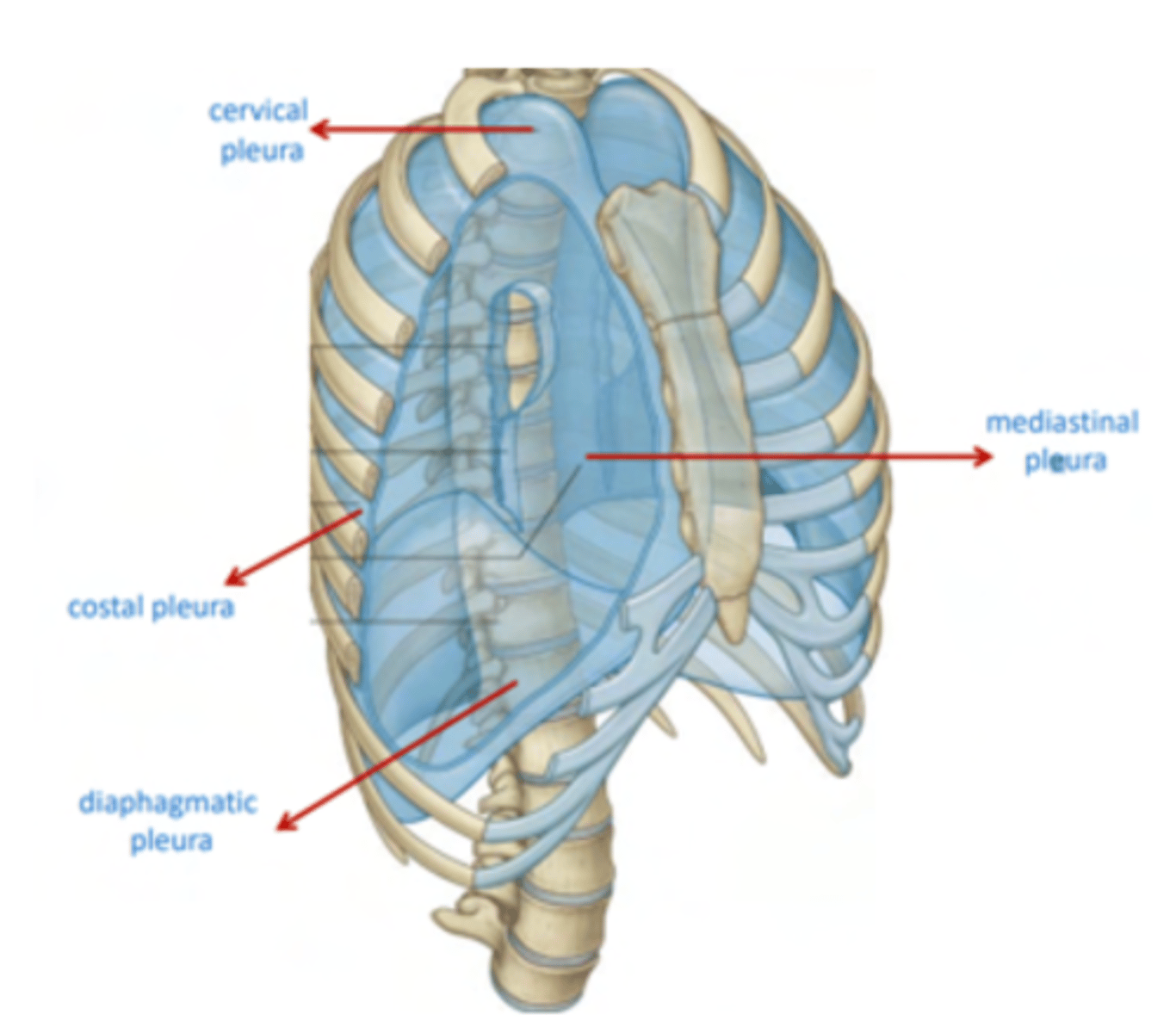
What are the parts of the parietal pleura?
classified depending on the part it’s in contact with
.Cervical pleura / cupula pleura. A little thicker, associated with some ligaments
. Costal pleura
. Mediastinal pleura
. Diaphragmatic pleura
Visceral pleura structure
Associated with the lungs. It ENTERS into each of the lobes through their fissures, as a double layer of visceral pleura, almost reaching the hilum
What are the reflections of the pleura?
reflects (folds back) into the parietal pleura at the level of the hilum, where the lung root (pedicle) is located.
The parietal pleura extends beyond the lungs, creating the pleural cavity.
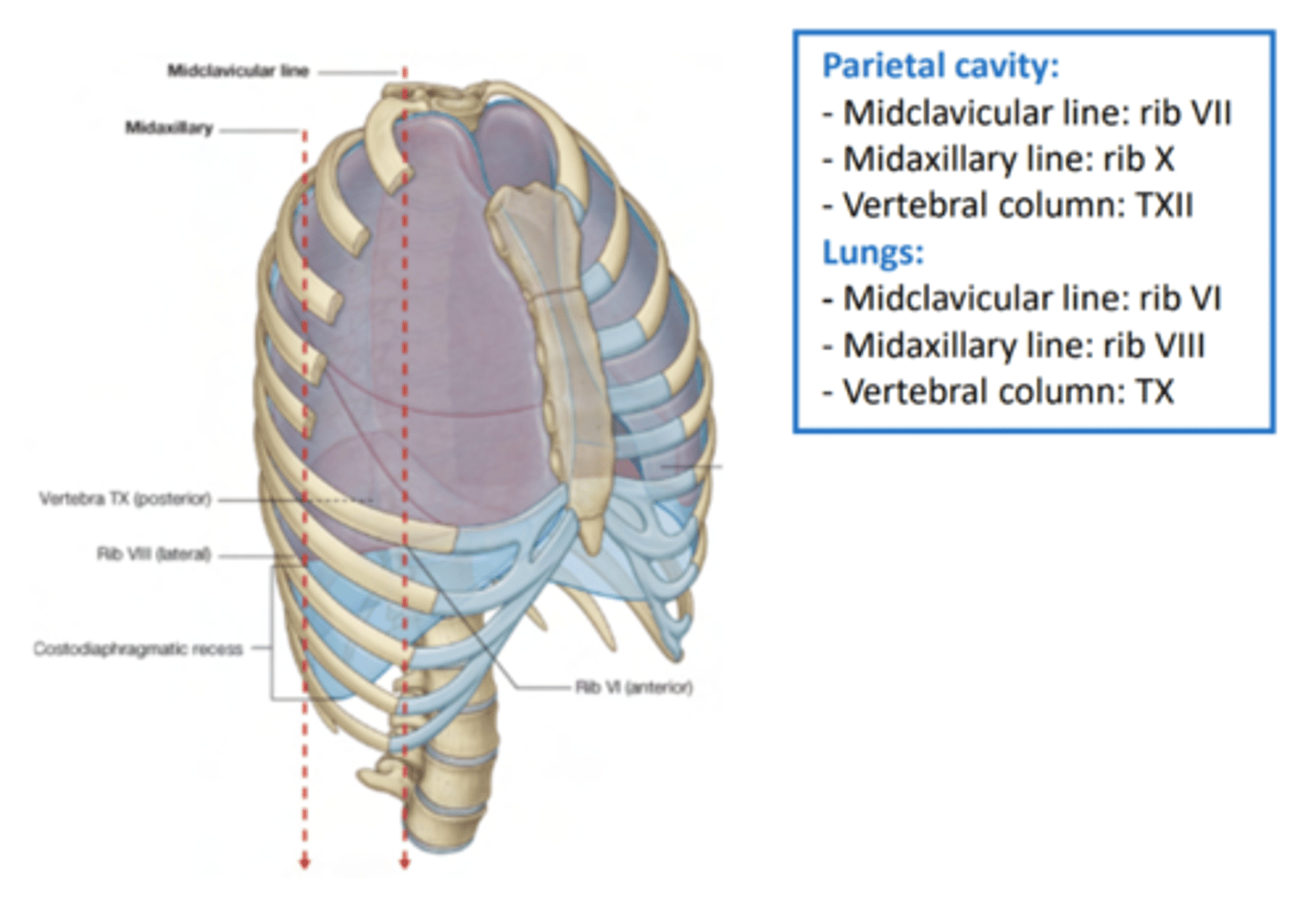
What is the coverage of the lungs?
- At the midclavicular line: end at rib VI
- At the midaxillary line: end at rib VIII
- At the vertebral column: end at TX

What is the coverage of the parietal cavity?
- At the midclavicular line: ends at rib VII
- At the midaxillary line: ends at rib X
- At the vertebral column: ends at TXII
LUNGS DON'T COMPLETELY FILL THE PLEURAL CAVITIES

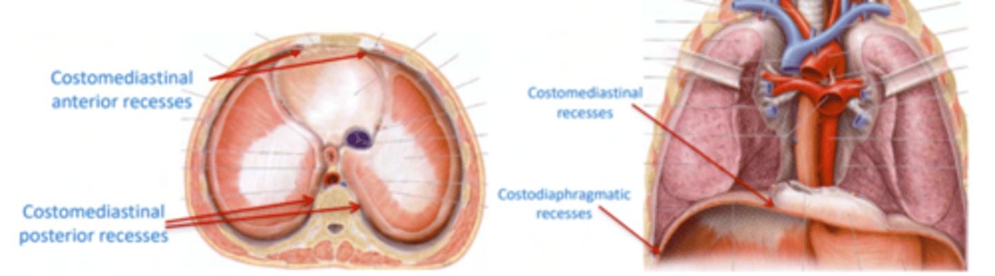
What are pleural recesses?
regions in which the pleura isn’t surrounding any lung tissue.
Why is pleural recessess relevant?
They are spaces where the lungs expand into during inspiration.
Pathological fluid can be collected in those spaces, without directly affecting the lungs but with some repercussions during forced inspiration.
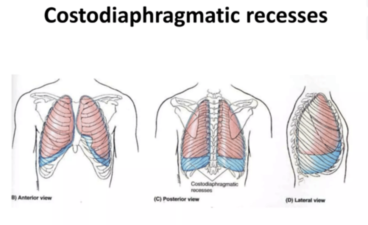
What are the two pleural recesses?
- Costodiaphragmatic recess. Most clinically important
- Costomediastinal recesses
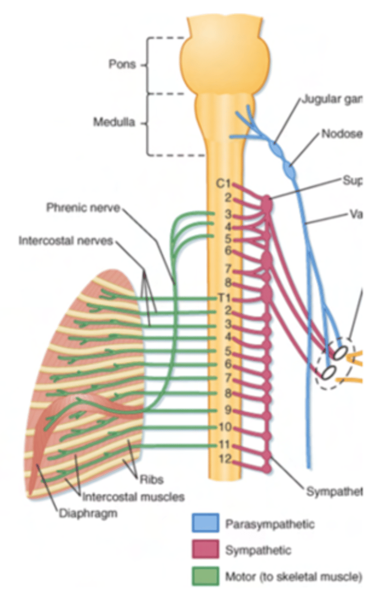
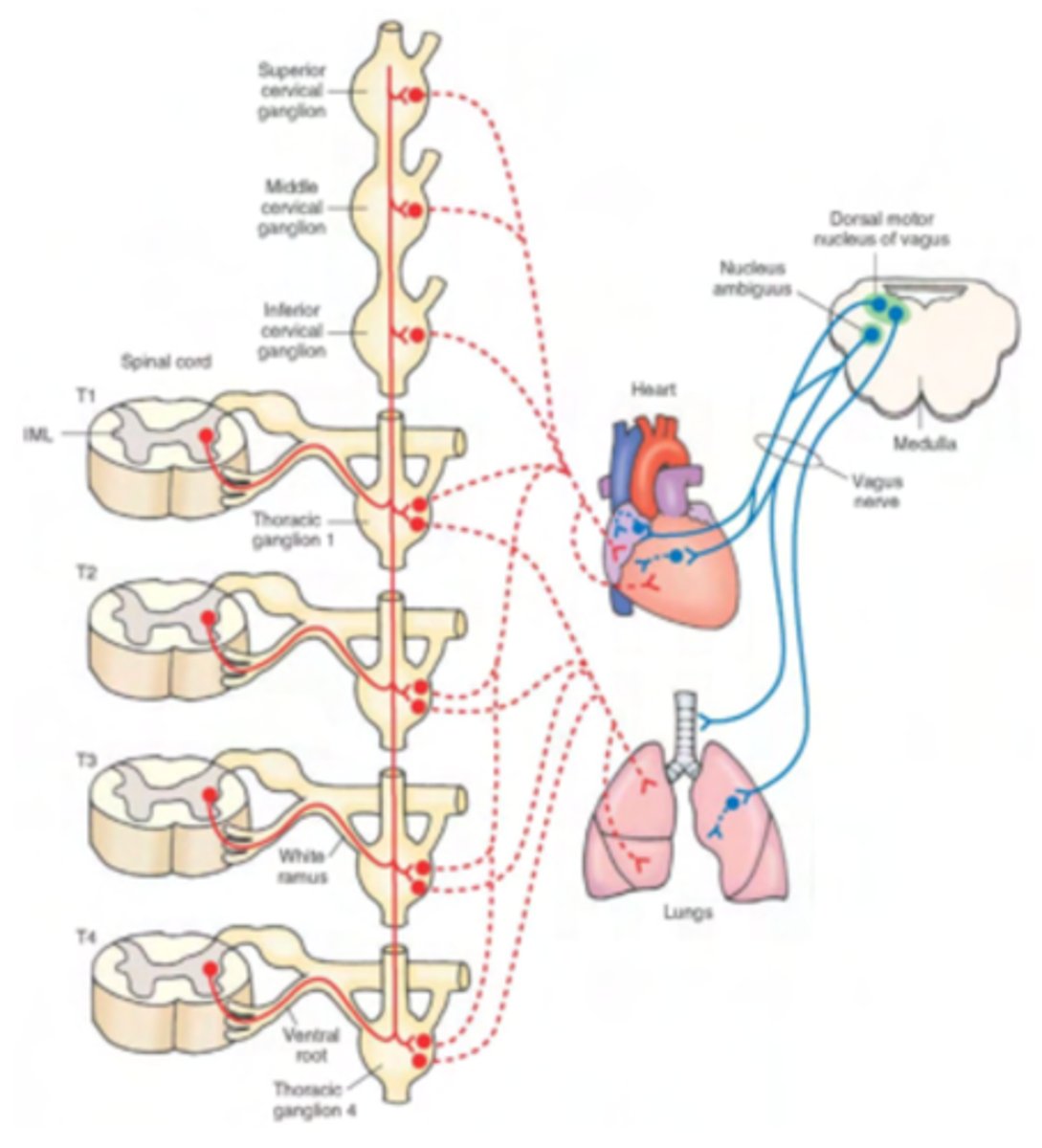
How is done the innervation of the lungs?
A pulmonary plexus formed by
. Sympathetic branches
. Parasympathetic branches
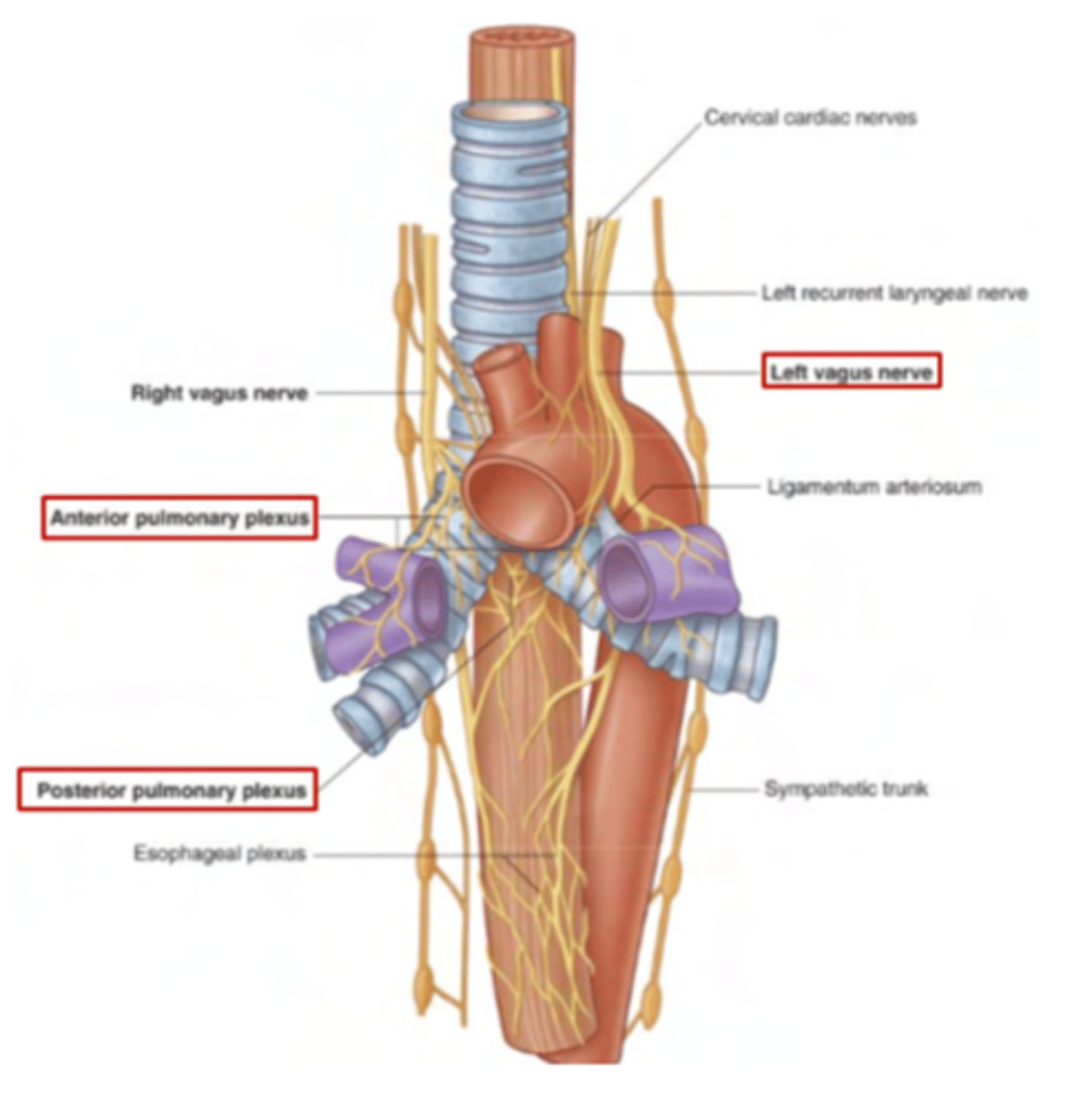
How are the sympathetic branches?
postganglionic fibers coming out of the sympathetic trunk. They originate from the thoracic portion T1-T4 of the spinal cord and synapse at the trunk, exiting as postganglionic fibers.
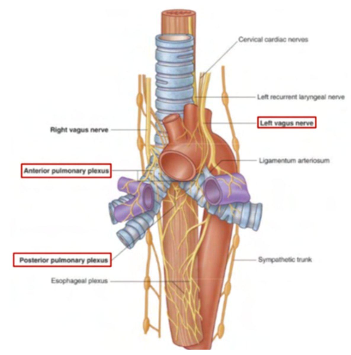
How are the parasympathetic fibers?
From the vagus nerve. are preganglionic fibers as they’ll synapse in PS microganglia located close to the organ of interest.
Come down through the vagus nerve, which gives fibers for the innervation of the lungs before continuing down to innervate further structures (heart).

Where is the pulmonary plexus bifurcated?
Found at the level of the bifurcation of the trachea.
We find both anterior (over the trachea, along with the deep
cardiac plexus) and posterior (over the esophagus) pulmonary plexuses.
- Sympathetic effect: bronchodilation
- Parasympathetic effect: bronchoconstriction
What are the respiratory muscles involved in respiration?
Innervation is provided by somatic fibers from the CNS.
Diaphragm: will be provided with somatic motor fibers by the phrenic nerve (C3-C4-C5)
Intercostal muscles: provided with somatic motor fibers by the
intercostal nerves (which are the anterior/ventral branches of spinal nerves at thoracic level)
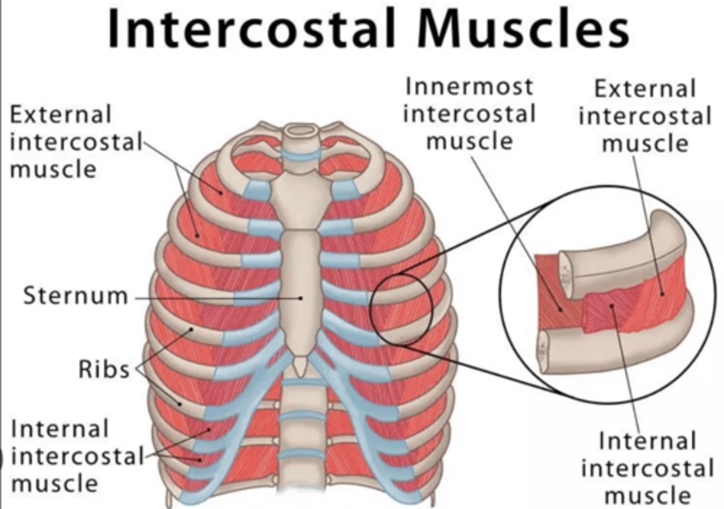
What are the muscles involved in respiration?
. diaphragm
. intercostal muscles
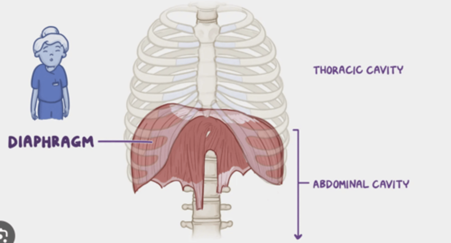
What is the nerve that innervates the diaphragm?
somatic motor fibers by the prhenic nerve (C3-C4-C5)
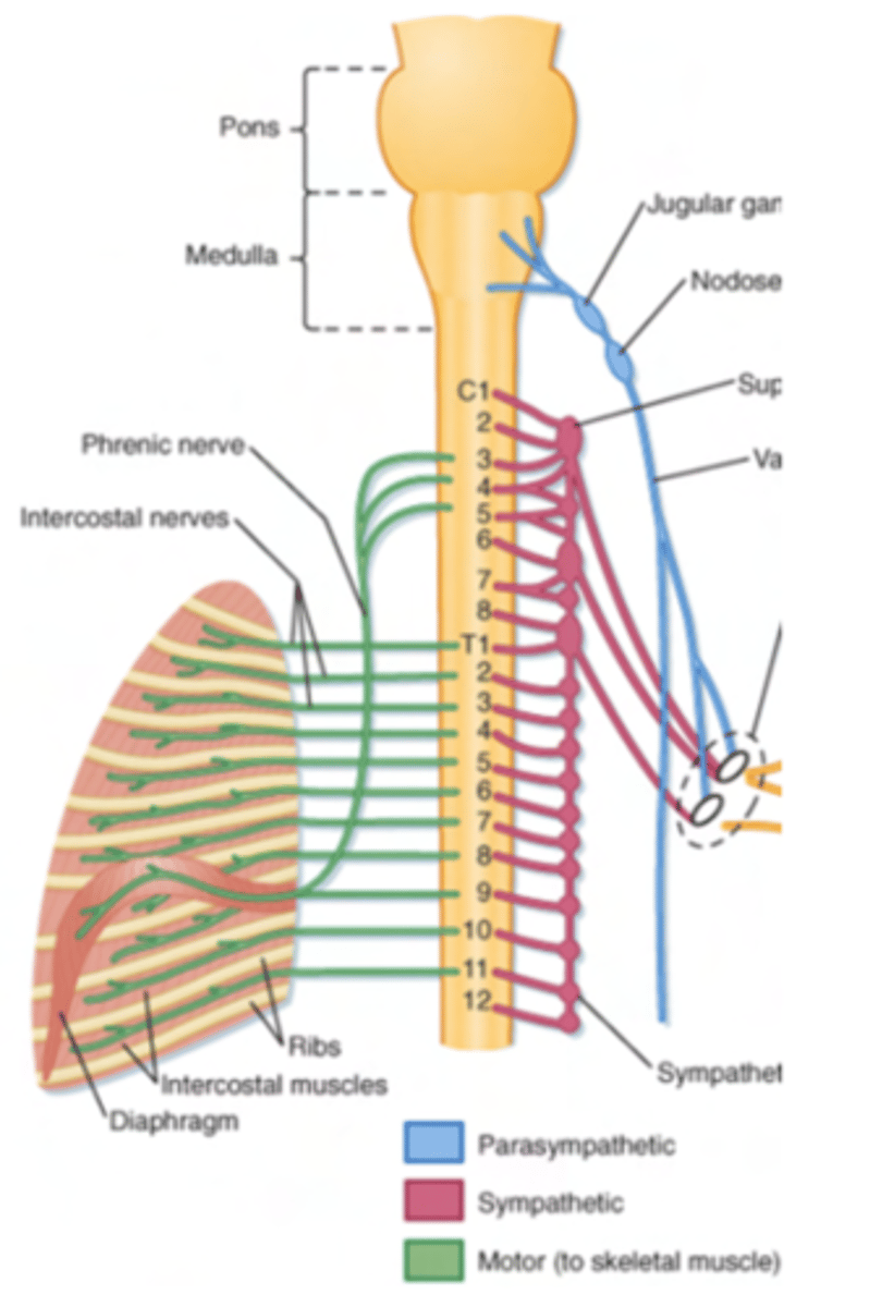
What is the nerve that innervates the intercostal muscles?
intercostal nerves (which are the anterior/ventral branches of spinal nerves at thoracic level)