10 - Gram Stain Limitations
 Call Kai
 Learn
Learn Practice Test
 Spaced Repetition
 Match
Match Flashcards
 Knowt Play
1/150

There's no tags or description
Looks like no tags are added yet.
Name | Mastery | Learn | Test | Matching | Spaced | Call with Kai | Chat |
|---|
No analytics yet
Send a link to your students to track their progress
151 Terms
b. They are very thin
[Spirochete]
Reason Gram stain cannot be used for Spirochetes
a. They are too thick to stain
b. They are very thin
c. They are intracellular
d. They lack a cell wall
a. Leptospira interrogans
[Spirochete]
The organism identified as:
Ice fangs
Shepherd's crook question mark shaped
a. Leptospira interrogans
b. Treponema pallidum
c. Borrelia burgdorferi
d. Campylobacter jejuni
c. Directly through contaminated fluids
[Spirochete]
Route of transmission of Leptospira enterrogans
a. Droplet
b. Fecal-oral
c. Directly through contaminated fluids
c. Wading on flooded water
[Spirochete]
Example of exposure leading to Leptospira enterrogans infection
a. Eating reheated rice
b. Cat bite or dog bite
c. Wading on flooded water
d. Inhaling contaminated air
a. Leptospira interrogans
[Spirochete]
The organism that can be transmitted even without a wound
a. Leptospira interrogans
b. Treponema pallidum
c. Borrelia burgdorferi
d. Campylobacter jejuni
c. Leptospirosis
[Spirochete]
Disease caused by Leptospira enterrogans
a. Brucellosis
b. Tularemia
c. Leptospirosis
d. Cat-scratch disease

c. Leptospiremic phase
[Spirochete]
Leptospirosis phase where organism is found in blood
a. Leptuspiuric phase
b. Immune phase
c. Leptospiremic phase
d. Convalescent phase

d. Leptuspiuric phase
[Spirochete]
Leptospirosis phase where organism is found in urine
a. Leptospiremic phase
b. Convalescent phase
c. Immune phase
d. Leptuspiuric phase
Fever
Calf tenderness
Conjunctival suffusion
No jaundice
[Spirochete]
Clinical manifestations of leptospirosis [3]
d. Weil syndrome
[Spirochete]
Complication to watch out for in Leptospira enterrogans infection
a. Hemolytic uremic syndrome
b. Guillain-Barre syndrome
c. Bubonic plague
d. Weil syndrome
a. Weil syndrome
[Spirochete]
The condition characterized by jaundice, lung hemorrhage, and acute kidney injury
a. Weil syndrome
b. Typhoid fever
c. Brucellosis
d. Bubonic plague
Jaundice
Lung hemorrhage
Acute Kidney Injury (AKI)
[Spirochete]
The findings associated with Weil syndrome
a. Jaundice, lung hemorrhage, and Acute Kidney Injury (AKI)
b. Fever, calf tenderness, and conjunctival suffusion
c. Rose spots, fever, and abdominal pain
d. Buboes, septicemia, and death
Lepto-MAT
Culture (blood, CSF, Urine)
[Spirochete]
Diagnostic tests for Leptospira enterrogans
a. Widal test, Elek test
b. Lepto-MAT, Culture (blood, CSF, Urine)
c. Skirrow's media, Urea breath test
d. Schick test, Dick test
c. Doxycycline
[Spirochete]
Treatment for mild, early leptospirosis
a. Penicillin G
b. Erythromycin
c. Doxycycline
d. Ceftriaxone
d. Penicillin G
[Spirochete]
Treatment for moderate to severe leptospirosis
a. Doxycycline
b. Erythromycin
c. Ceftriaxone
d. Penicillin G
c. Doxycycline
[Spirochete]
The prophylaxis for leptospirosis that is contraindicated in pregnant women
a. Penicillin G
b. Erythromycin
c. Doxycycline
d. Ceftriaxone
c. Azithromycin
[Spirochete]
The alternative prophylaxis for leptospirosis that is safe in pregnant women
a. Penicillin G
b. Doxycycline
c. Azithromycin
d. Ceftriaxone
b. Treponema pallidum
[Spirochete]
The organism identified by: an axial filament and corkscrew motion
a. Leptospira interrogans
b. Treponema pallidum
c. Borrelia burgdorferi
d. Campylobacter jejuni
b. Congenital syphilis
[Spirochete]
Route of vertical transmission of Treponema pallidum leading to this condition
a. Syphilis
b. Congenital syphilis
c. Leptospirosis
d. Bacterial vaginosis
Hutchinson's teeth
Mucus patches
Saddle nose
[Spirochete]
Clinical manifestations of congenital syphilis caused by Treponema pallidum [3]
d. Syphilis
[Spirochete]
Route of sexual transmission of Treponema pallidum leading to this condition
a. Leptospirosis
b. Brucellosis
c. Chancroid
d. Syphilis
c. Chancre
[Spirochete]
Clinical manifestation of primary syphilis caused by Treponema pallidum
a. Macules, papules
b. Gumma formation
c. Chancre
d. Condylomata lata
Hard chancre
Painless chancre
Self-limiting chancre
[Spirochete]
The lesion in primary syphilis
a. Hard, painless, self-limiting chancre
b. Painful, soft ulcer
c. Beefy red velvety ulcer
d. Painless, shallow ulcer
Macules
Papules
Erythematous palm and sol
Condylomata lata
[Spirochete]
Clinical manifestations of secondary syphilis caused by Treponema pallidum
a. Chancre, hard, painless, self-limiting
b. Gumma formation, neurosyphilis, Argyll-Robertson pupil
c. Symptoms
d. Macules, papules, erythematous palm and sol, condylomata lata
d. 1 month after chancre
[Spirochete]
Condylomata lata in secondary syphilis appears this many months after chancre and is self-limiting
a. 3 months after chancre
b. 6 months after chancre
c. 2 months after chancre
d. 1 month after chancre
c. No Symptoms
[Spirochete]
Clinical characteristic of latent syphilis caused by Treponema pallidum
a. Chancre
b. Macules, papules
c. No Symptoms
d. Gumma formation
Gumma formation
Neurosyphilis
Argyll-Robertson pupil (prostitute's eyes)
[Spirochete]
Clinical manifestations of tertiary syphilis caused by Treponema pallidum
a. No symptoms
b. Chancre, hard, painless
c. Macules, papules, condylomata lata
d. Gumma formation, neurosyphilis, Argyll-Robertson pupil "prostitute's eyes"
a. Darkfield microscopy
[Spirochete]
The diagnostic test for syphilis
a. Darkfield microscopy
b. Gram stain
c. Lepto-MAT
d. Skirrow's media
c. RPR and VDRL
[Spirochete]
Non-treponemal screening tests for syphilis
a. FTA-Abs and MHA
b. Darkfield microscopy and culture
c. RPR and VDRL
d. Lepto-MAT and Widal test
d. FTA-Abs and MHA
[Spirochete]
Treponemal confirmatory tests for syphilis
a. RPR and VDRL
b. Darkfield microscopy and culture
c. Lepto-MAT and Widal test
d. FTA-Abs and MHA
d. Penicillin G
[Spirochete]
Treatment for Treponema pallidum
a. Doxycycline
b. Erythromycin
c. Ceftriaxone
d. Penicillin G
a. Borrelia burgdorferi
[Spirochete]
The organism that is the largest medically important bacteria
a. Borrelia burgdorferi
b. Treponema pallidum
c. Leptospira interrogans
d. Campylobacter jejuni
c. Ticks (Ixodes spp.)
[Spirochete]
Vector of Borrelia burgdorferi
a. Mosquito
b. Flea
c. Ticks
d. Mite
a. Borrelia burgdorferi
[Spirochete]
The causative agent of Lyme disease
a. Borrelia burgdorferi
b. Treponema pallidum
c. Leptospira interrogans
d. Brucella spp.
d. Lyme disease
[Spirochete]
Borrelia burgdorferi is the causative agent of this disease
a. Tularemia
b. Brucellosis
c. Cat-scratch disease
d. Lyme disease
c. Zoonotic (deer, forest animals)
[Spirochete]
Lyme disease caused by Borrelia burgdorferi is classified as this type of infection
a. Nosocomial
b. Anthroponosis
c. Zoonotic
d. Vector-borne only
d. Erythema chronicum migrans
[Spirochete]
Pathognomonic clinical manifestation of Lyme disease caused by Borrelia burgdorferi
a. Buboes
b. Condylomata lata
c. Saddle nose
d. Erythema chronicum migrans
d. Doxycycline
[Spirochete]
Treatment for early Lyme disease caused by Borrelia burgdorferi
a. Penicillin G
b. Erythromycin
c. Ceftriaxone
d. Doxycycline
c. Penicillin G
[Spirochete]
Treatment for late Lyme disease caused by Borrelia burgdorferi
a. Doxycycline
b. Erythromycin
c. Penicillin G
d. Ceftriaxone
a. Mycobacterium spp.
[Mycobacterium spp]
The organisms that have a high fatty acid content
a. Mycobacterium spp.
b. Streptococcus spp.
c. Staphylococcus spp.
d. Escherichia spp.
a. Mycobacterium avium
[Mycobacterium spp]
The organism associated with MAC (Mycobacterium avium complex) in HIV/AIDS
a. Mycobacterium avium
b. Mycobacterium bovis
c. Mycobacterium tuberculosis
d. Mycobacterium leprae
b. Mycobacterium bovis
[Mycobacterium spp]
The organism that is a milk-borne pathogen
a. Mycobacterium avium
b. Mycobacterium bovis
c. Mycobacterium tuberculosis
d. Mycobacterium leprae
d. Aerobe
[Mycobacterium spp]
Oxygen requirement of Mycobacterium tuberculosis
a. Anaerobe
b. Microaerophile
c. Facultative anaerobe
d. Aerobe
c. Acid fast bacilli (AFB)
[Mycobacterium spp]
Staining characteristic of Mycobacterium tuberculosis
a. Gram positive
b. Gram negative
c. Acid fast bacilli (AFB)
d. Non-acid fast
b. Serpentine arrangement
[Mycobacterium spp]
Arrangement of Mycobacterium tuberculosis
a. Palisade "chinese character"
b. Serpentine arrangement
c. Box car shaped
d. Lollipop, drumstick, tennis racket
d. Airborne
[Mycobacterium spp]
Route of transmission of Mycobacterium tuberculosis
a. Fecal-oral
b. Direct contact
c. Vector-borne
d. Airborne
Pulmonary tuberculosis
Extra pulmonary tuberculosis
[Mycobacterium spp]
Two classifications of tuberculosis based on location
a. Active and latent tuberculosis
b. Pulmonary and extra pulmonary tuberculosis
c. Primary and secondary tuberculosis
d. Contagious and non-contagious tuberculosis
b. Initial infection, children, less contagious, affects middle part of the lungs
[Mycobacterium spp]
Characteristics of Primary Pulmonary tuberculosis
a. Reaction, adults, more contagious, affects apex of lungs
b. Initial infection, children, less contagious, affects middle part of the lungs
c. Reactivation, elderly, less contagious, affects apex of lungs
d. Initial infection, adults, more contagious, affects middle part of lungs
c. Ghon complex
[Mycobacterium spp]
Primary complex in Primary PTB is also known as
a. Pott's complex
b. Scrofula complex
c. Ghon complex
d. Laryngeal complex
c. Reaction, adults, more contagious, affects apex of the lungs
[Mycobacterium spp]
Characteristics of Post primary/secondary PTB
a. Initial infection, children, less contagious, affects middle part of lungs
b. Reactivation, elderly, less contagious, affects middle part of lungs
c. Reaction, adults, more contagious, affects apex of the lungs
d. Initial infection, adults, less contagious, affects apex of lungs
Cough >2 weeks
Weight loss
Night sweat
Late afternoon fever
📌Presumptive PTB"
[Mycobacterium spp]
Signs and symptoms of PTB; if all present this is the diagnosis
a. Cough >1 week, fever, fatigue, weight loss
b. Cough >2 weeks, weight loss, night sweat, late afternoon fever = "presumptive PTB"
c. Cough >3 weeks, hemoptysis, chest pain, dyspnea
d. Fever, chills, productive cough, pleuritic chest pain
Chest X-ray
Xpert MTB/Rif or Gene Xpert
Sputum microscopy-AFB
Culture (gold standard)
[Mycobacterium spp]
Diagnostic tests for Pulmonary tuberculosis (PTB)
a. Widal test, Elek test, Schick test
b. Lepto-MAT, culture, Skirrow's media
c. Chest X-ray, Xpert MTB/Rif or Gene Xpert, Sputum microscopy-AFB, Culture (gold standard)
d. Darkfield microscopy, RPR, VDRL
d. Culture
[Mycobacterium spp]
Gold standard diagnostic test for PTB
a. Chest X-ray
b. Sputum microscopy-AFB
c. Xpert MTB/Rif or Gene Xpert
d. Culture
a. Mantoux test
[Mycobacterium spp]
The test that uses purified protein derivative administered intradermally
a. Mantoux test
b. Widal test
c. Lepto-MAT
d. VDRL test
c. Purified protein derivative
[Mycobacterium spp]
Mantoux/TB skin test uses this substance injected intradermally
a. Live attenuated bacteria
b. Killed bacteria
c. Purified protein derivative
d. Toxoid
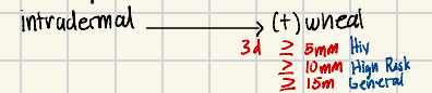
b. wheal
[Mycobacterium spp]
Result of Mantoux/TB skin test injected intradermally
a. redness
b. wheal
c. blister
d. induration
d. 3 days
[Mycobacterium spp]
Mantoux/TB skin test result read after this number of days
a. 1 day
b. 5 days
c. 7 days
d. 3 days
c. HIV positive patients
[Mycobacterium spp]
Mantoux/TB skin test result of ≥5mm is considered positive in this population
a. General population
b. High risk population
c. HIV positive patients
d. Children
d. High risk population
[Mycobacterium spp]
Mantoux/TB skin test result of ≥10mm is considered positive in this population
a. HIV positive patients
b. General population
c. Low risk population
d. High risk population
c. General population
[Mycobacterium spp]
Mantoux/TB skin test result of ≥15mm is considered positive in this population
a. HIV positive patients
b. High risk population
c. General population
d. Low risk population
b. Latent TB infection
[Mycobacterium spp]
A positive Mantoux/TB skin test result indicates this condition
a. Active TB infection
b. Latent TB infection
c. Primary PTB
d. Post primary PTB
a. TPT (TB Preventive Therapy)
[Mycobacterium spp]
The treatment for latent TB infection
a. TPT (TB Preventive Therapy)
b. Directly Observed Therapy
c. Four-drug regimen
d. Steroids
c. Lymph node TB
[Mycobacterium spp]
Most common form of extra pulmonary tuberculosis also known as "scrofula"
a. Laryngeal TB
b. Spinal TB
c. Lymph node TB
d. Miliary TB
d. Neck area
[Mycobacterium spp]
Location of lymph node TB also known as scrofula
a. Axilla area
b. Groin area
c. Chest area
d. Neck area
d. Laryngeal TB (pagmagsalita nakakahawa)
[Mycobacterium spp]
Most infectious form of extra pulmonary tuberculosis
a. Lymph node TB
b. Spinal TB
c. Miliary TB
d. Laryngeal TB
c. Pott's disease
[Mycobacterium spp]
Spinal TB is also known as
a. Scrofula
b. Ghon complex
c. Pott's disease
d. Laryngeal complex
b. Directly observed therapy short course
[Mycobacterium spp]
Full name of TB DOTS
a. Directly observed treatment short course
b. Directly observed therapy short course
c. Direct observation therapy short course
d. Directly observed therapy standard course
b. Isoniazid, Rifampicin, Pyrazinamide, Ethambutol
[Mycobacterium spp]
First line drugs for TB DOTS (HRZE)
a. Hydrazide, Rifampicin, Zinc sulfate, Ethambutol
b. Isoniazide, Rifampicin, Pyrazinamide, Ethambutol
c. Isoniazide, Rifampicin, Zinc sulfate, Erythromycin
d. Hydrazide, Rifampicin, Pyrazinamide, Erythromycin
c. Per orem (PO) — all oral regimen
[Mycobacterium spp]
Route of administration of first line TB DOTS drugs (HRZE)
a. Intravenous (IV)
b. Intramuscular (IM)
c. Per orem (PO)
d. Subcutaneous (SQ)
d. Streptomycin (IM)
[Mycobacterium spp]
This drug is no longer first-line because of its route of administration
a. Isoniazid
b. Rifampicin
c. Pyrazinamide
d. Streptomycin
d. Inhibit mycolic acid synthesis
[Mycobacterium spp]
Mechanism of action of Isoniazid
a. Inhibit arabinosyl transferase
b. Inhibit RNA synthesis
c. Misreading of mRNA
d. Inhibit mycolic acid synthesis
c. Cidal vs rapidly dividing
[Mycobacterium spp]
Effect of Isoniazid on Mycobacterium tuberculosis
a. Static, retard growth
b. Cidal vs slowly dividing
c. Cidal vs rapidly dividing
d. Cidal, weak
c. Enzyme inhibitor
[Mycobacterium spp]
Isoniazid is classified as this type of enzyme
a. Enzyme inducer
b. Enzyme activator
c. Enzyme inhibitor
d. Enzyme substrate
d. Peripheral neuropathy
[Mycobacterium spp]
Watch out for side effect of Isoniazid due to decreased pyridoxin
a. Red-orange discoloration
b. Hepatotoxicity
c. Color blindness
d. Peripheral neuropathy
c. Give Pyridoxine (Vitamin B6)
[Mycobacterium spp]
Treatment for peripheral neuropathy caused by Isoniazid
a. Give Cyanocobalamin (Vitamin B12)
b. Give Thiamine (Vitamin B1)
c. Give Pyridoxine (Vitamin B6)
d. Give Folic Acid (Vitamin B9)
d. Inhibit RNA synthesis
[Mycobacterium spp]
Mechanism of action of Rifampicin
a. Inhibit mycolic acid synthesis
b. Misreading of mRNA
c. Inhibit arabinosyl transferase
d. Inhibit RNA synthesis
d. Cidal vs slowly dividing
[Mycobacterium spp]
Effect of Rifampicin on Mycobacterium tuberculosis
a. Cidal vs rapidly dividing
b. Static, retard growth
c. Cidal, weak
d. Cidal vs slowly dividing
d. Enzyme inducer
[Mycobacterium spp]
Rifampicin is classified as this type of enzyme
a. Enzyme inhibitor
b. Enzyme substrate
c. Enzyme activator
d. Enzyme inducer
b. Red-orange discoloration
[Mycobacterium spp]
Watch out for side effect of Rifampicin
a. Peripheral neuropathy
b. Red-orange discoloration
c. Color blindness
d. Hepatotoxicity
c. Reassurance
[Mycobacterium spp]
Management of red-orange discoloration caused by Rifampicin
a. Pyridoxine (Vitamin B6)
b. Discontinue the drug
c. Reassurance
d. Cyanocobalamin (Vitamin B12)
c. Converted to Pyrazinoic acid
[Mycobacterium spp]
Mechanism of action of Pyrazinamide
a. Inhibit RNA synthesis
b. Inhibit mycolic acid synthesis
c. Converted to Pyrazinoic acid
d. Inhibit arabinosyl transferase
d. Cidal, weak
[Mycobacterium spp]
Effect of Pyrazinamide on Mycobacterium tuberculosis
a. Static, retard growth
b. Cidal vs rapidly dividing
c. Cidal vs slowly dividing
d. Cidal, weak
Hepatotoxicity (↑ALT ↑AST)
Gout (↑uric acid)
[Mycobacterium spp]
Watch out for side effects of Pyrazinamide
a. Peripheral neuropathy, red-orange discoloration
b. Color blindness, optic neuritis
c. Hepatotoxicity (↑ALT ↑AST), gout (↑uric acid)
d. Red-orange discoloration, hepatotoxicity
d. Inhibit arabinosyl transferase
[Mycobacterium spp]
Mechanism of action of Ethambutol
a. Inhibit mycolic acid synthesis
b. Misreading of mRNA
c. Inhibit RNA synthesis
d. Inhibit arabinosyl transferase
c. Static, retard growth
[Mycobacterium spp]
Effect of Ethambutol on Mycobacterium tuberculosis
a. Cidal vs rapidly dividing
b. Cidal, weak
c. Static, retard growth
d. Cidal vs slowly dividing
d. Intramuscular (IM)
[Mycobacterium spp]
Streptomycin is no longer first line because it is given via this route
a. Per orem (PO)
b. Intravenous (IV)
c. Subcutaneous (SQ)
d. Intramuscular (IM)
c. MDR TB (Multi-drug resistant TB)
[Mycobacterium spp]
Second line agents for TB are used in this condition
a. Latent TB infection
b. Primary PTB
c. MDR TB (Multi-drug resistant TB)
d. Extra pulmonary TB
Kanamycin
Amikacin
Capreomycin
📌Mnemonic: “KAC”
[Mycobacterium spp]
Aminoglycosides used as second line agents for MDR TB
a. Gentamicin, Tobramycin, Neomycin
b. Kanamycin, Amikacin, Capreomycin
c. Streptomycin, Gentamicin, Amikacin
d. Tobramycin, Kanamycin, Neomycin
Aminoglycoside
Kanamicin
Amikacin
Capreomycin
Fluoroquinolone
Cycloserine = TOXIC
P-aminosalicylate
Rifampicin
Ethinamide
[Mycobacterium spp]
2nd line agents for Multi-drug resistant TB (MDR TB ) [6]
d. Cycloserine
[Mycobacterium spp]
Second line agent for MDR TB known to be toxic
a. Aminoglycoside
b. P-aminosalicylate
c. Fluoroquinolone
d. Cycloserine
c. Palisade arrangement
[Mycobacterium spp]
ID of M. leprae based on its arrangement
a. Serpentine arrangement
b. Box car shaped
c. Palisade arrangement
d. Lollipop, drumstick, tennis racket
b. Mycobacterium leprae
[Mycobacterium spp]
The organism that has never been grown alone in culture
a. Mycobacterium tuberculosis
b. Mycobacterium leprae
c. Mycobacterium avium
d. Mycobacterium bovis
b. Mycobacterium leprae
[Mycobacterium spp]
The organism that requires the armadillo or mouse foot pad for growth
a. Mycobacterium tuberculosis
b. Mycobacterium leprae
c. Mycobacterium avium
d. Mycobacterium bovis
a. Armadillo or mouse foot pad
[Mycobacterium spp]
Mycobacterium leprae requires the ______ for growth
a. Armadillo or mouse foot pad
b. Guinea pig or rabbit
c. Chicken embryo or monkey
d. Rat or hamster
d. Droplet
[Mycobacterium spp]
Route of transmission of M. leprae
a. Fecal-oral
b. Vector-borne
c. Direct contact
d. Droplet
b. Leprosy
[Mycobacterium spp]
Disease caused by M. leprae
a. Tuberculosis
b. Leprosy
c. Lyme disease
d. Brucellosis
a. Hansen's disease
[Mycobacterium spp]
Leprosy is also known as _____
a. Hansen's disease
b. Weil syndrome
c. Pott's disease
d. Lyme disease
c. Culion, Palawan
[Mycobacterium spp]
Famous leprosarium for Hansen's disease located in this place in the Philippines
a. Bataan, Luzon
b. Cebu, Visayas
c. Culion, Palawan
d. Davao, Mindanao
Hypoesthesia
Hypopigmented skin lesions
Nerve damage
Hand deformity (claw hands)
[Mycobacterium spp]
Clinical manifestations of leprosy caused by M. leprae
a. Buboes, septicemia, death
b. Erythema chronicum migrans, nerve damage
c. Hypoesthesia, hypopigmented skin lesions, nerve damage, hand deformity (claw hands)
d. Rice watery diarrhea, washer woman hand sign
Tuberculoid leprosy
Lepromatous leprosy
[Mycobacterium spp]
Two types of leprosy caused by M. leprae
a. Primary and secondary leprosy
b. Pulmonary and extra pulmonary leprosy
c. Tuberculoid and Lepromatous leprosy
d. Active and latent leprosy