Anatomy
 Call Kai
 Learn
Learn Practice Test
 Spaced Repetition
 Match
Match Flashcards
 Knowt Play
1/145

Earn XP
Description and Tags
Upper Extremity
Name | Mastery | Learn | Test | Matching | Spaced | Call with Kai | Chat |
|---|
No analytics yet
Send a link to your students to track their progress
146 Terms
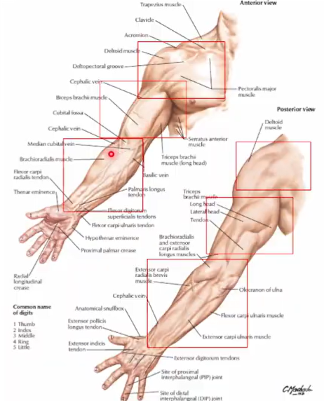
4 major segments of the upper extremity
Shoulder - consists of pectoral, scapular, and deltoid organs
Arm
Forearm
Hand
The bones of the upper limb (superior appendicular skeleton) are attached to the axial skeleton via _____
Pectoral girdle (clavicle and scapula)
Muscles that support, stabilize, and move the pectoral girdle. These muscles attached to the relatively fixed ribs, sternum, and vertebrae of the axial skeleton
Axioappendicular muscles

A long bone that has no medullary canal (composed of superficial compact bone and central spongy bone)
Shaft is described as a double curve in a horizontal line
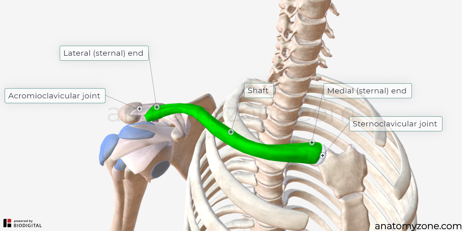
Clavicle (collarbone)
Describe the medial and lateral halves of the clavicle
Medial half - convex anteriorly; sternal end articulates with the manubrium of the sternum (Sternoclavicular joint)
Lateral half - concave anteriorly; acromial end articulates with the acromion of the scapula (Acromioclavicular joint)
Differentiate the superior surface and inferior surface of the pectoral girdle
Superior surface - smooth
Inferior surface - rough (strong ligaments bind to the 1st rib near its sternal end and suspend the scapula from its acromial end

Ligaments in inferior surface of the clavicle
Coracoclavicular ligaments - at the acromial end
Conoid ligament - inserts at the conoid tubercle
Trapezoid ligament - inserts at the trapezoid line
Groove for subclavius - attachment of subclavius muscle
A triangular flat bone that lies on the posterolateral aspect of the thorax (2nd to 7th ribs)
Scapula
Differentiate the posterior surface, costal surface, and body of scapula
Posterior surface - spine (small supraspinous fossa and large infraspinous fossa)
Costal surface - concave (subscapular fossa)
Body - translucent (thick borders)

Part of the scapula that continues laterally as the flat expanded acromion. It articulates with acromial end of the clavicle forming the acromioclavicular (AC) joint
Spine

Prominence indicating the medial point of attachment of the deltoid
Deltoid tubercle of the scapular spine
Shallow, concave, oval fossa located superolaterallly. It receives and articulates with the head of the humerus at the glenohumeral joint.
Glenoid cavity

Beak-like that projects anterolaterally. Superior to the glenoid cavity. Provides inferior attachments to the coracoclavicular ligaments
Coracoid process

Borders of scapula
Superior
Medial (vertebral)
Lateral (axillary)

Angles of the scapula
Superior - thinnest and shortest (Suprascapular notch)
Inferior
Lateral - thickest (Head and Neck)

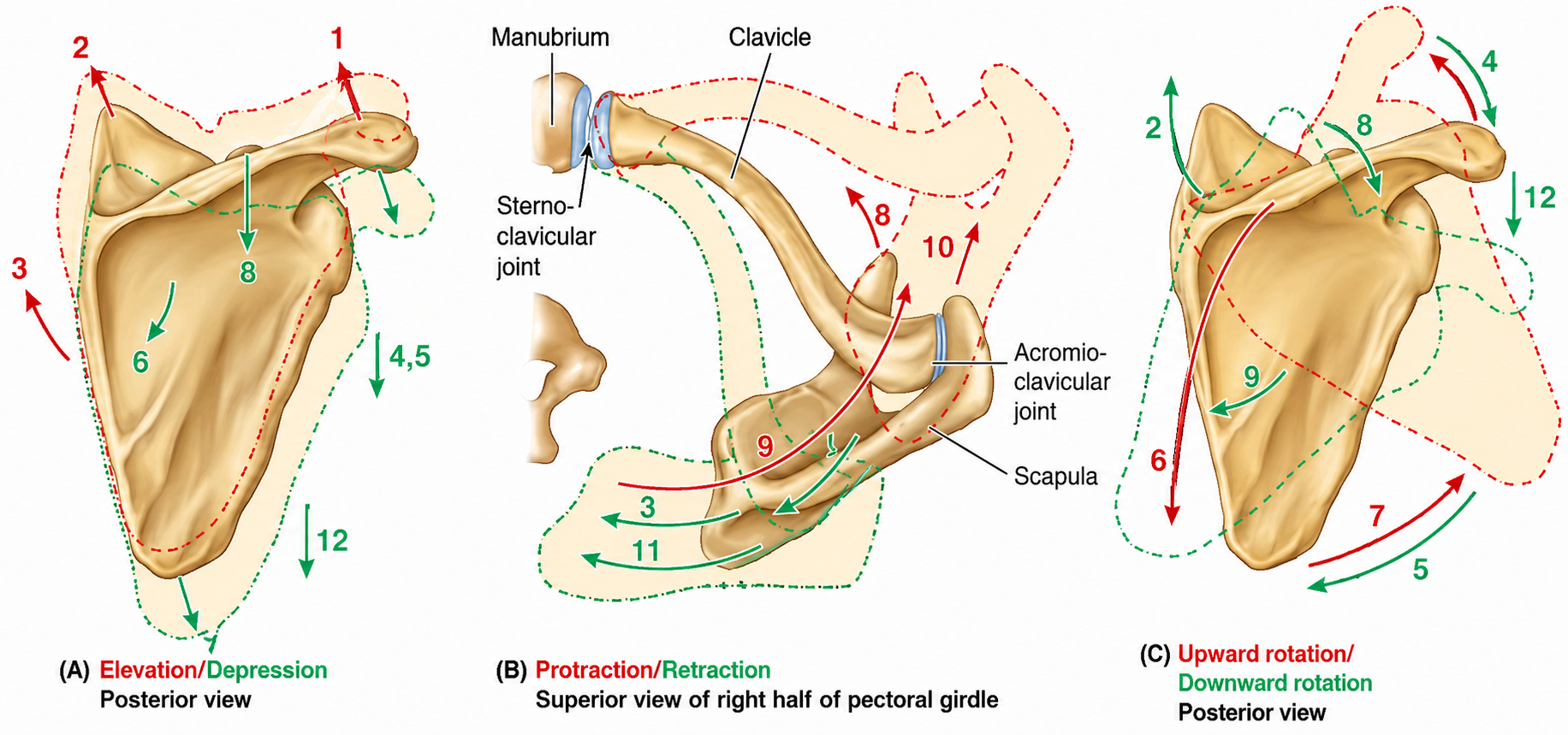
Movement of scapula
Elevation/Depression
Protraction/Retraction
Upward rotation/downward rotation
Largest bone in the upper limb
Humerus
2 joints articulated by the humerus
Glenohumeral joint
Elbow joint
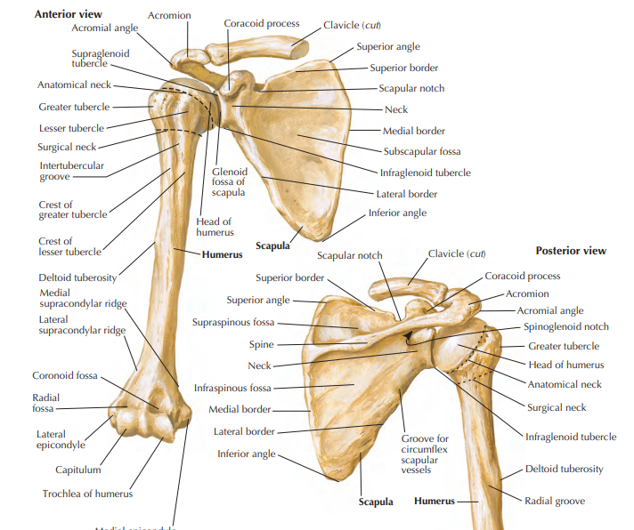
Structures of the Proximal Humerus
See attached photo

Structures of the Proximal Humerus
See attchached photo

What provides passage of long head biceps tendon in humerus
Intertubercular (bicipital) groove

Structure of the Shaft of Humerus
Deltoid tuberosity - lateral; location of the deltoid muscle
Radial groove - oblique and posterior; passage of the radial nerve and profunda brachii artery

Structure of the Distal HUmerus
Distal humerus – widens as the sharp medial and lateral supracondylar (supracondylar) ridges:
Medial epicondyle
Lateral epicondyle
These structures provide muscle attachment.

Articulates with the head of the radius
Capitulum (lateral)
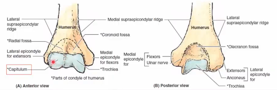
Medial, spool-shaped or pulley-like for articulation with the proximal end (trochlear notch) of the ulna
Trochlea

Located anteriorly. It receives the coronoid process of the ulna.
Coronoid Fossa

located anteriorly. It receives the radial head during full elbow flexion.
Radial fossa
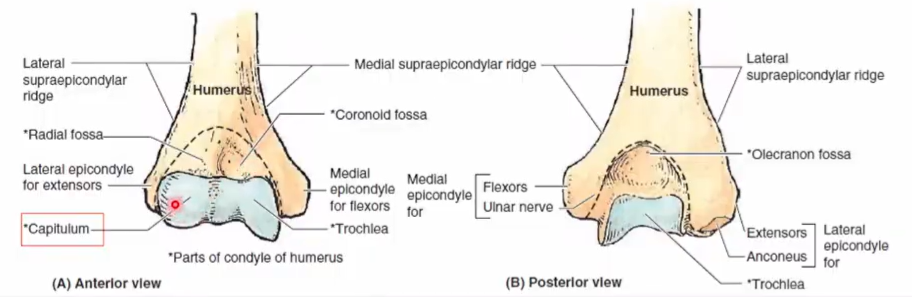
Located posteriorly. It receives the olecranon process of the
Olecranon fossa

2 parallel bones of the upper forearm
Ulna
Radius

Pivots around the ulna. Enables forearm supination and pronation
Radius
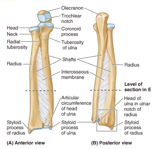
It is the stabilizing bone of the forearm. It is the medial and longer of the two forearm bones.
Ulna
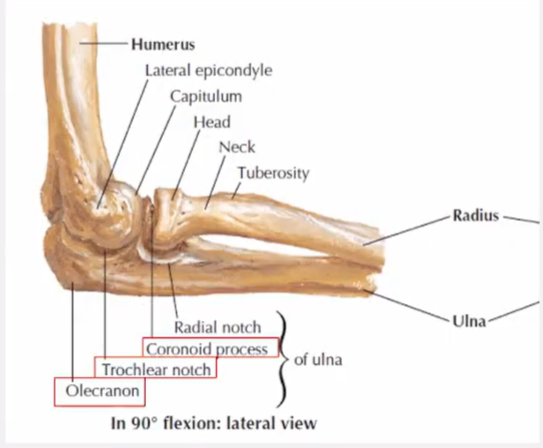
Structures of the Proximal Ulna
Olecranon process
Coronoid process
Trochlear notch
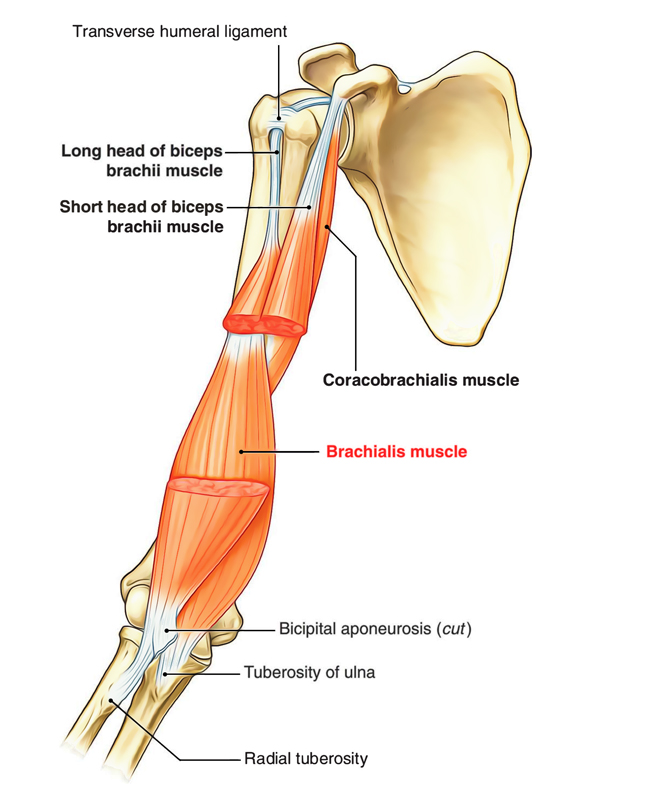
Ulnar tuberosity
Radial notch
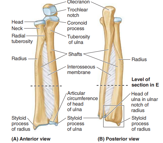
posterior prominent projection of the ulna
Olecranon process

Anterior prominent projection of the ulna
Coronoid process

articulates with the trochlea of the humerus. Formed by the olecranon and coronoid process.
Trochlear notch

located inferior to coronoid process and serves as attachment of the brachialis muscle.
Ulnar tuberosity
located laterally, receives the head of the radius.
Radial notch

Must know structures
…
Must know structures
….

Part of Ulna that is thick and cylindrical proximally
Shaft
Distal End of the Ulna
Ulnar Head
Ulnar Styloid Process
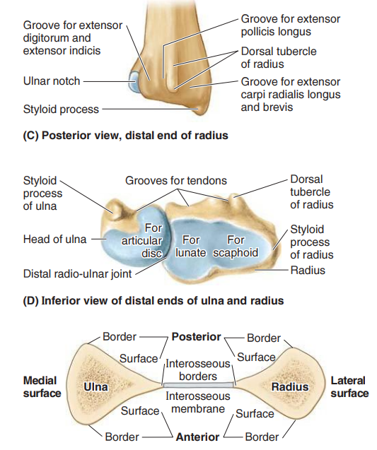
A small, abrupt enlargement, the disc-like structure at the narrow distal end of ulna
Ulnar head

Small, conical structure of the distal end of ulna
Ulnar styloid process
TRUE OR FALSE: The distal ulna articulates with the wrist bone and participates in joint movement
FALSE

The lateral and shorter of the two forearm bones
Radius
Proximal structures of the radius
Head
Neck
Radial tuberosity
It is the smooth, concave superior aspect. It articulates with the capitulum of the humerus.
Radial Head
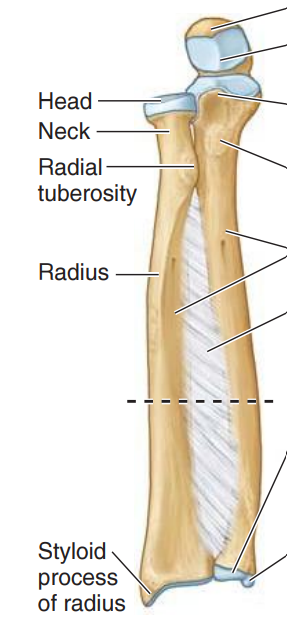
It is a constriction of the distal head.
Neck of Radius
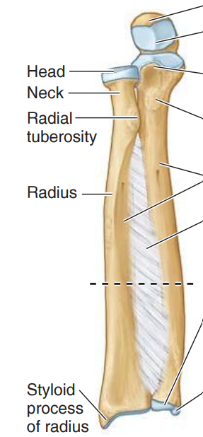
It is located medially and it demarcates the proximal aspect to the shaft.
Radial Tuberosity

Distal Structures of Radius
Ulnar notch
Radial styloid process
Dorsal tubercle
Scaphoid fossa
Lunate fossa
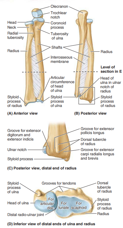
it accommodates the ulna
Ulnar notch
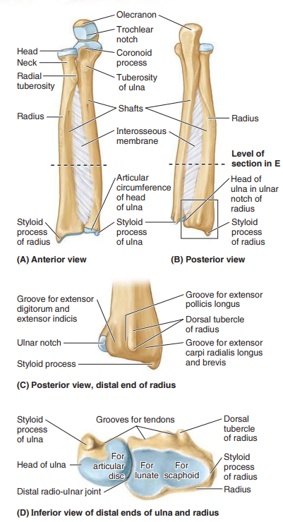
located on the lateral aspect of radius bone and increasingly becomes ridge-like.
Radial styloid process

articulates with scaphoid bone.
Scaphoid fossa
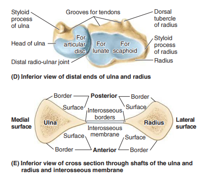
articulates with lunate bone.
lunate fossa

projects dorsally. Located in between the shallow grooves for the passage of the tendons of forearm muscles.
Dorsal tubercle
Describe the Shaft cross section of the forearm
The ulna and radius apices are lateral
Rounded, superficially directed base
Acute, deeply directed apex

A thin, fibrous membrane that connects the apices of the forearm bones
Interosseous membrane

It is the only articulation between the upper limb and the axial skeleton
Sternoclavicular joint

Formed by the articulation of the sternal end of the clavicle and manubrium and 1st costal cartilage
Sternoclavicular joint

serves as a shock absorber of the forces transmitted along the clavicle from the upper limb.
Articular disc
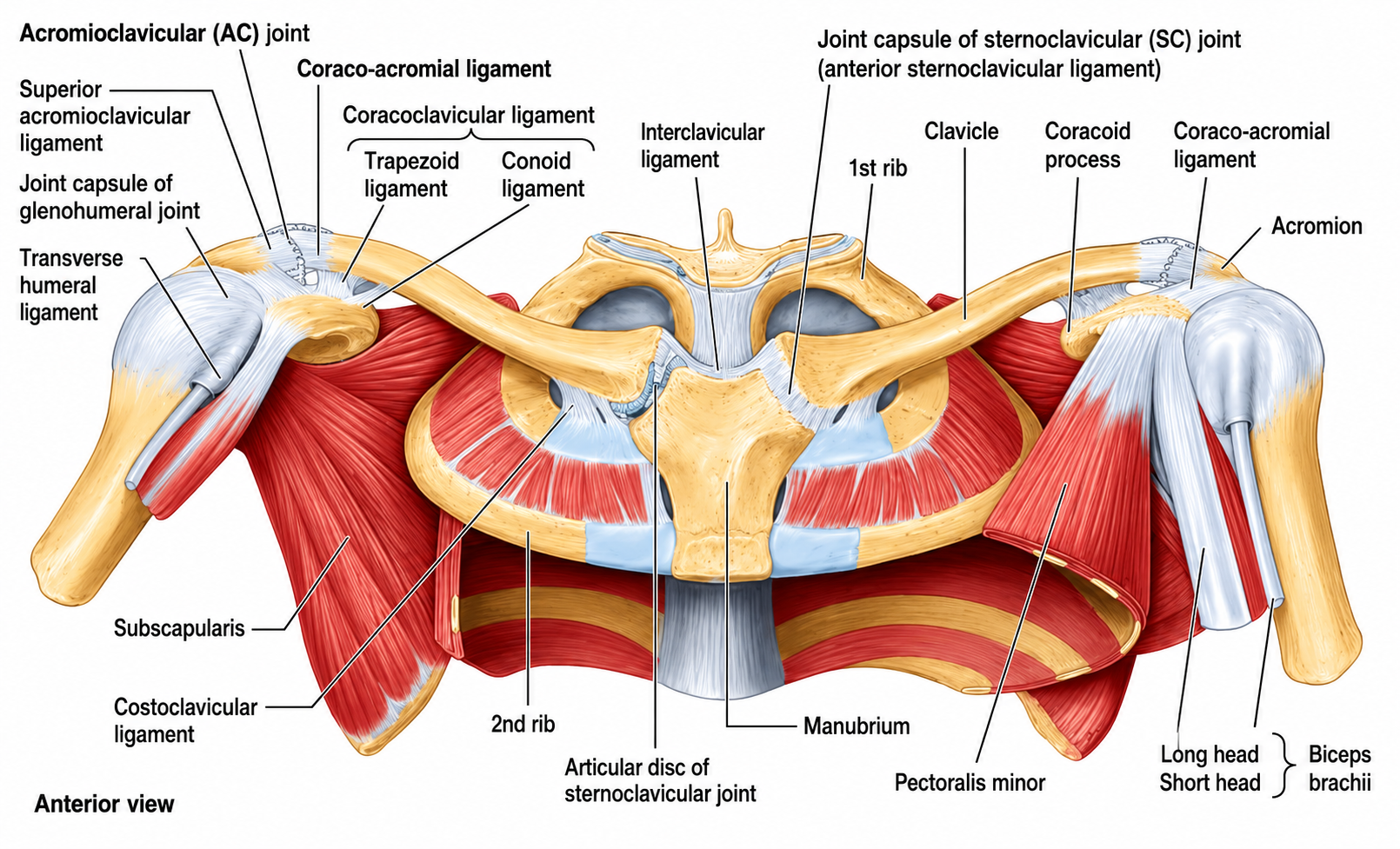
Covers the sternoclavicular joint
Joint capsule

reinforces the anterior and posterior capsule.
Anterior and posterior sternoclavicular ligament
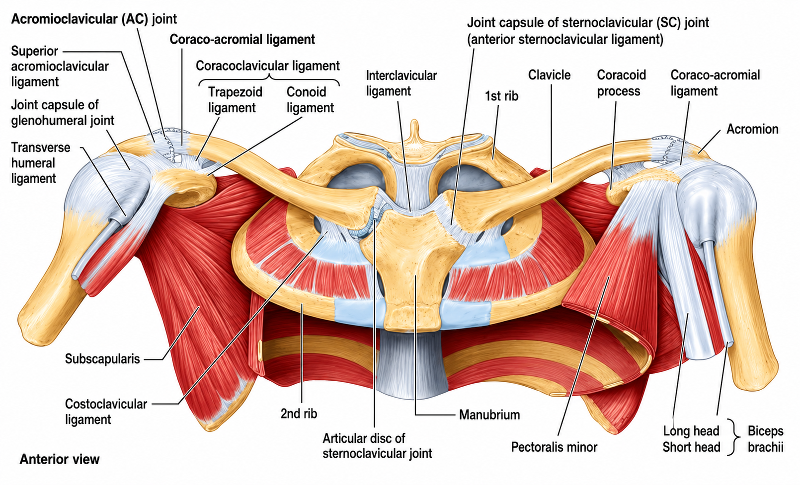
reinforces the superior capsule.
Interclavicular ligament
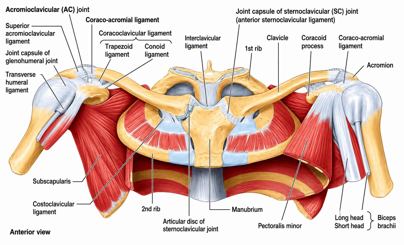
anchors the sternal end of the clavicle to the 1st rib, limiting its elevation.
Costoclavicular ligament

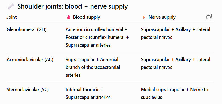
Blood supply of the sternoclavicular joint
Internal thoracic and suprascapular arteries.
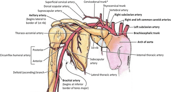
Nerve supply of sternoclavicular joint
Medial suprascapular nerve and nerve to subclavius.

Articulation between the head of humerus and glenoid fossa of scapula
ball and socket joint
Glenohumeral joint
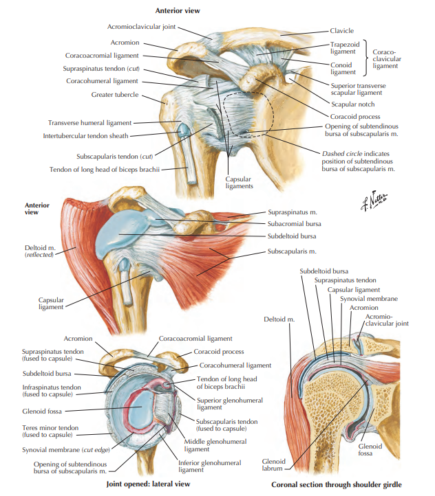
Fibrous, membranous structure that deepens the glenoid cavity
Glenoid labrum

loose and fibrous layer that surrounds the GH Joint
Joint capsule of GH joint
Parts of the Joint capsule of the GH joint
Medial - margin of glenoid cavity
Lateral - anatomical neck of humerus
Superiorly - coracoid process
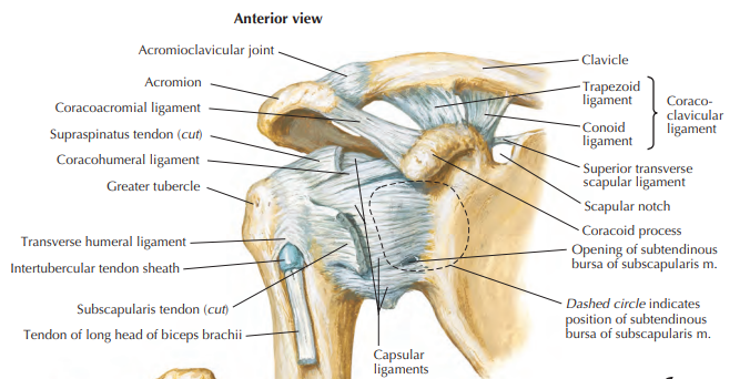
3 ligaments of the shoulder joint (Glenohumeral joint)
Glenohumeral ligaments
Coracohumeral ligament
Transverse humeral ligament

3 fibrous bands that reinforce the anterior aspect of the capsule
Glenohumeral ligament

Glenohumeral ligaments:
Origin:
Insertion:
Origin: Glenoid labrum
Insertion: anatomic neck of humerus

Coracohumeral ligaments:
Origin:
Insertion:
Origin: Base of coracoid process
Insertion: Greater tubercle of humerus

a small ligament that bridges the intertubercular (bicipital) groove of the humerus.
Holds the biceps long head tendon in place
Transverse humeral ligament
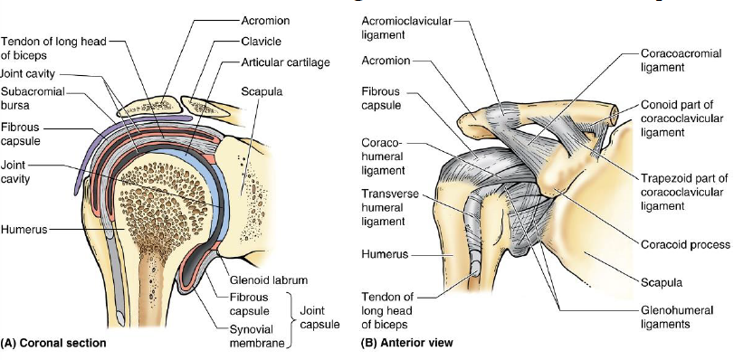
Transverse Humeral Ligament
Origin:
Insertion:
Origin: Greater tubercle of humerus
Insertion: Lesser tubercle of humerus

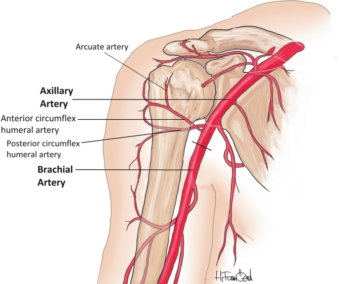
Shoulder Joint (GH Joint) blood supplies:
Anterior and posterior humeral circumflex arteries
Branches of suprascapular arteries
Shoulder Joint (GH Joint) nerve supplies:
Suprascapular nerve
Axillary nerve
Lateral pectoral nerve

Movement of glenohumeral joint
Abduction
Adduction
Flexion
Extension
Lateral rotation
Medial rotation
Tip: GH joint is a ball and socket joint capable of circumduction

Articulation between acromial end of clavicle and acromion of scapula (2–3 cm lateral acromion).
Acromioclavicular joint

sleeve-like, relatively loose fibrous layer but strengthened superiorly by the trapezius muscle fibers.
Joint capsule of the AC joint

What are the two ligaments of the AC joint?
Acromioclavicular ligament
Coracoclavicular ligament (Conoid ligament & Trapezoid ligament)

Fibrous band extending from the acromion to the clavicle that strengthens the AC joint superiorly.
Acromioclavicular ligament
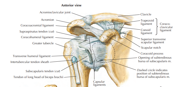
Strong pair of bands that unite the coracoid process of the scapula to the clavicle.
Coracoclavicular ligament
inverted triangle shaped. Attaches to the conoid tubercle of the clavicle.
Conoid ligament
from superior surface of the coracoid process and extends laterally to the trapezoid line on the inferior surface of the clavicle.
Trapezoid ligament
Blood supply of the AC joint
Suprascapular and thoracoacromial arteries

Nerve supple of the AC joint
Lateral pectoral nerves
Axillary nerve
(Suprascapular nerve)

Differentiate the blood and nerve supplies of the gh joint, ac joint, and sc joint.
See attached photo
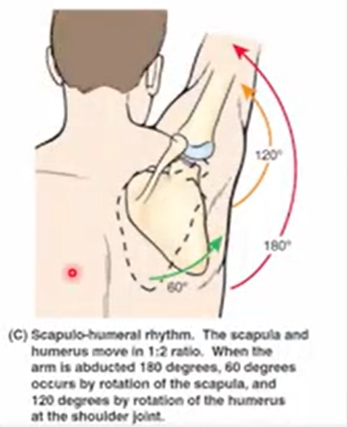
Joints responsible for the pectoral girdle range of motion (Scapulothoracic rhythm)
Scapulothoraric joint
Glenohumeral joint
Initial 30 degrees abduction
glenohumeral joint
Full elevation - for every 3 degrees
2 degrees from glenohumeral joint
1 degree from scapulothroracic joint
(2:1 ratio)
Clavicle - 60 degree
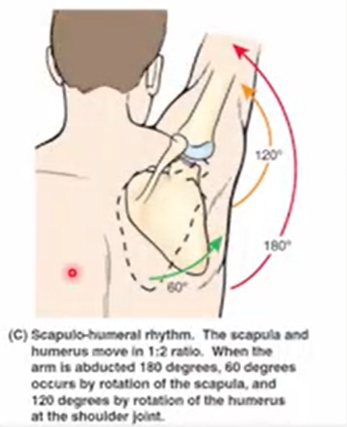
@180 degree abduction of the arm. What is the angle distribution of the glenohumeral joint and scapulathoracic joint
Glenohumeral joint - 120
Scapulathoracic joint - 60
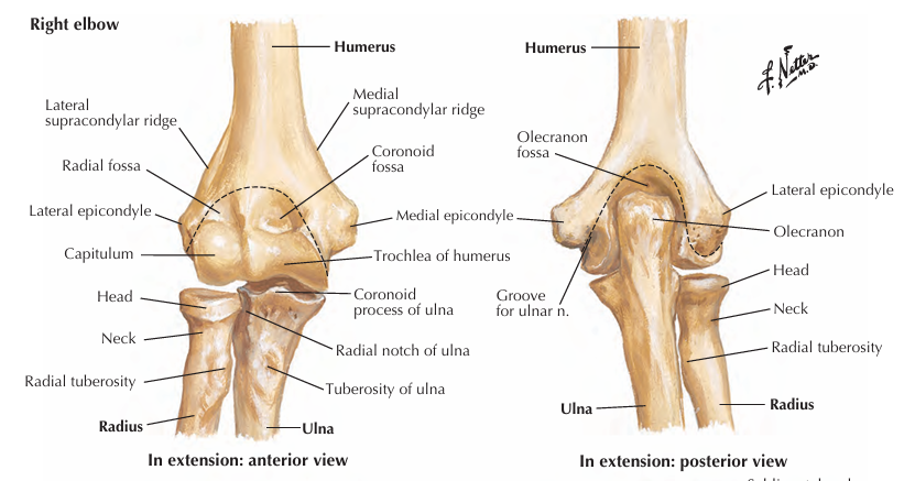
What are the articulations of the elbow joint?
Trochlea and Trochlear notch (Humeroulnar joint)
Capitulum and Radial Head (Humeroradial joint)
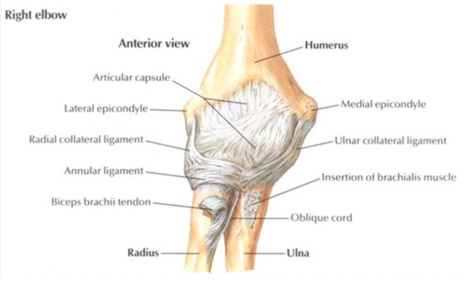
Joint capsule boundaries of the elbow joint
Lateral and medial ends - capitulum and trochlea
Anterior and posterior - coronoid and olecranon fossae (weak)
Sides: collateral ligaments (strong)
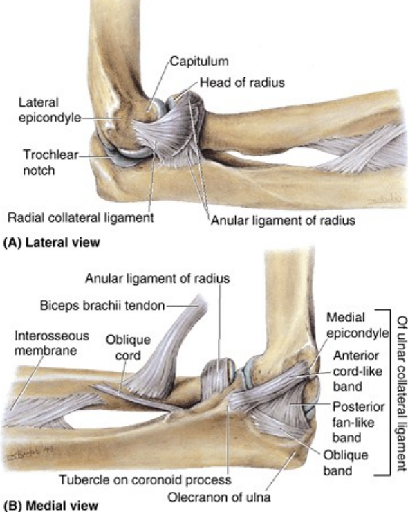
Collateral ligament of the humerus
Radial collateral ligament - from lateral epicondyle and inserts to annular ligament
Ulnar collateral ligament - from medial epicondyle and inserts to the coronoid and olecranon fossa
Anterior band - strongest
Posterior band - weakest
Oblique band
Movement of the elbow joint and the associated muscles
Flexion (Brachialis muscle, Biceps brachii muscle, Brachioradialis muscle)
Extension (Triceps brachii muscles, Anconeus muscle)

Blood supply of the elbow joint
Anastomosis arteries (no specific arteries)
Nerve supply of the elbow joint
musculocutaneous nerve
radial nerve
ulnar nerve

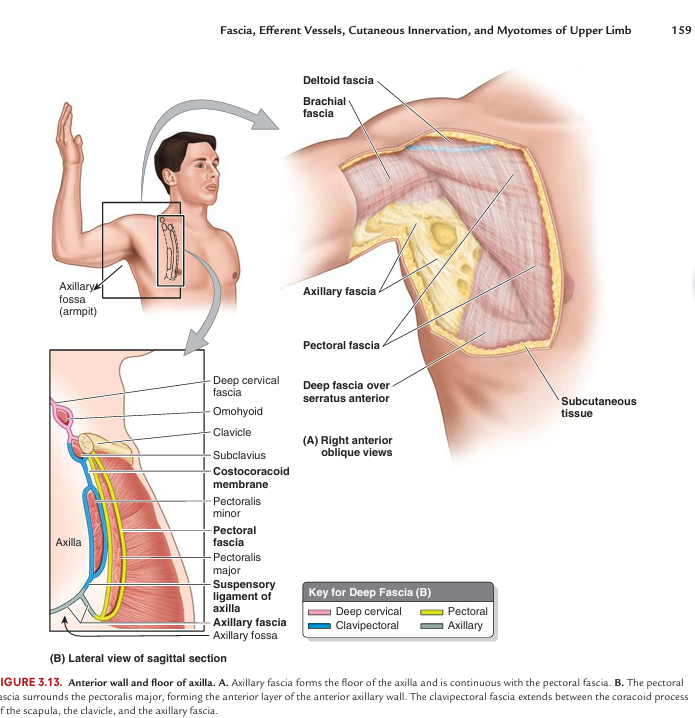
Subcutaneous (superficial fascia) vs Deep fascia
Subcutaneous fascia - fat containing
Deep fascia -wraps the muscle and compartmentalization