Congenial anomalies, cysts, and calcifications of Testicles
 Call Kai
 Learn
Learn Practice Test
 Spaced Repetition
 Match
Match Flashcards
 Knowt Play
1/39

There's no tags or description
Looks like no tags are added yet.
Name | Mastery | Learn | Test | Matching | Spaced | Call with Kai | Chat |
|---|
No analytics yet
Send a link to your students to track their progress
40 Terms
Polyorchidism
Three or more testicles present
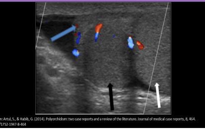
Polyorchidism
Testes normally descend into the scrotal sac between ____________ GA
26-34 weeks
Cryptorchidism
Undescended testes
Most undescended testes are palpable in inguinal canal area (70-80%)
Infertility and cancer are common complications
CT or MRI may be needed to locate the testes if they are in the abdomen
The undescended testicle is usually removed because of the risk of cancer
Orchiopexy → corrects cryptorchidism
Cryptorchidism USA
Identify echogenic mediastinum testis to distinguish cryptorchid testis
Absent testicle in scrotal sac
Usually found in ipsilateral inguinal canal
Hypoechoic, homogeneous
Almond shape
Well circumscribed
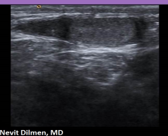
Cryptorchidism
Tunica albuginea cyst
Within the tunica surrounding testis
Located at the outer border of the testicle
Usually solitary and unilocular
Tunica albuginea cyst USA
Well defined margins
Through transmission and enhancement
Tunica vaginalis cyst
Rare
Single or multiple
May contain septations or hemorrhage
Difficult to differentiate from tunica albuginea cysts
Intratesticular cysts
Simple cysts
Usually <2cm
Thought to originate from the rete testis
Located within the testicular parenchyma
Intratesticular cysts USA
Well defined, smooth walls
Anechoic
Through transmission and posterior enhancement
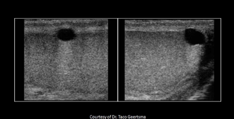
Tunica albuginea
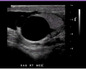
Intratesticular cyst
Tubular ectasia of rete testis
Associated with epididymal inflammation or trauma
Usually bilateral and seen with spermatocele
Vessels also course through the rete testis
If color is seen within the tiny cystic areas, an intratesticular varicocele is present and the scrotal veins should be evaluated for dilation and reflux
Tubular ectasia of rete testis USA
Multiple tiny cystic areas in the area of the mediastinum testis that do not fill with color
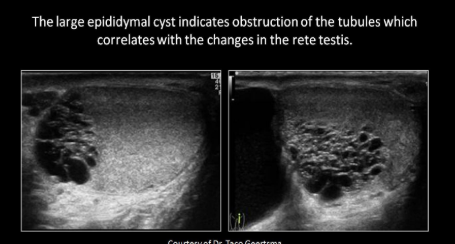
Dilated rete testis
Cystic dysplasia
Rare congenital malformation
Multiple cysts of different sizes with septations
Begins at mediastinum testis area and extends into parenchyma nearby
Frequently seen with renal agenesis or dysplasia
Epidermoid cyst
Benign teratoma
Walls are fibrous, may calcify
Described as having an onion or target appearance due to altering rings of hyperechoic tissues
Cyst contains thick keratin
USA Epidermoid cyst
Well defined hypoechoic mass
Thickened or calcified wall
May have “onion” appearance internally
Avascular
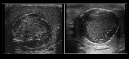
Epidermoid cyst
Simple epididymal cyst
Caused by dilation of the epididymal tubules
Found throughout the epididymis (head, body, tail)
Contain serous fluid
Simple epididymal cyst USA
Anechoic, well-circumscribed
Can be multiple
Most common scrotal mass
Spermatocele
Spermatocele
Caused by dilation of the epididymal tubules
Usually found ONLY IN THE HEAD OF EPI
Contain spermatozoa and sediment
Commonly seen in post-vasectomy patients
Spermatocele USA
Anechoic, well-circumscribed
Similar to simple cyst but may have low-level echoes
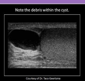
Spermatocele
Scattered calcifications seen with:
Tuberculosis, filariasis, previous trauma
Coarse calcifications seen with:
Sertoli cell tumors
Burned out germ cells
Microlithiasis
Germ cell tumors
Cryptorchidism
Trisomy 21 and Klinefelter’s syndrome
Previous radiotherapy
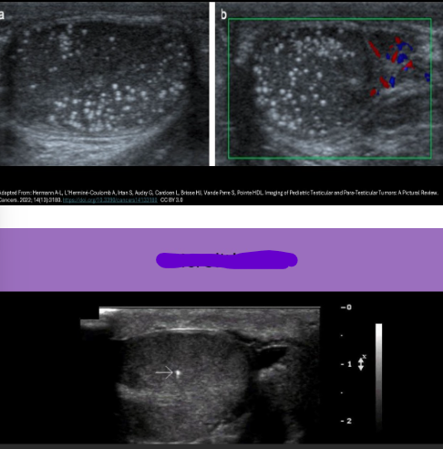
Microlithiasis
Extrascrotal calcifications
Scrotal pearls
Arise between the two layers of tunica vaginalis
May be loose within the scrotal sac
Extrascrotal calcifications USA
Echogenic foci with posterior shadowing outside the testicle, but within scrotal sac
Mobile
Twinkle artifact on doppler
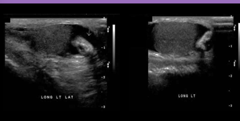
Extrascrotal calcifications/ Scrotal pearl
Post vasectomy changes
Epididymal enlargement
Tubular ectasia of epi
Spermatoceles and cysts
Dilated vas deferens
How can you differentiate an undescended testicle from an inguinal mass
Locate the mediastinum testis
How can you differentiate a tunica albuginea cyst from an intratesticular cyst
Location of the cyst
An epidermoid cyst us described as having the appearance of:
An onion
How can you differentiate an epidermoid cyst from a scortal tumor?
Activate color
A spermatocele is a common finding in patients with a Hx of:
Vasectomy
A scrotolith is identified:
Outside the testicle, within scrotal sac