PVCC BIO 201 EXAM 3
1/136
There's no tags or description
Looks like no tags are added yet.
Name | Mastery | Learn | Test | Matching | Spaced | Call with Kai |
|---|
No study sessions yet.
137 Terms
4 functional characteristics of muscle?
E-CEE
Excitability- responds to stimuli
Contractility- can shorten
Extensibility- can extend
Elasticity- can recoil to resting length
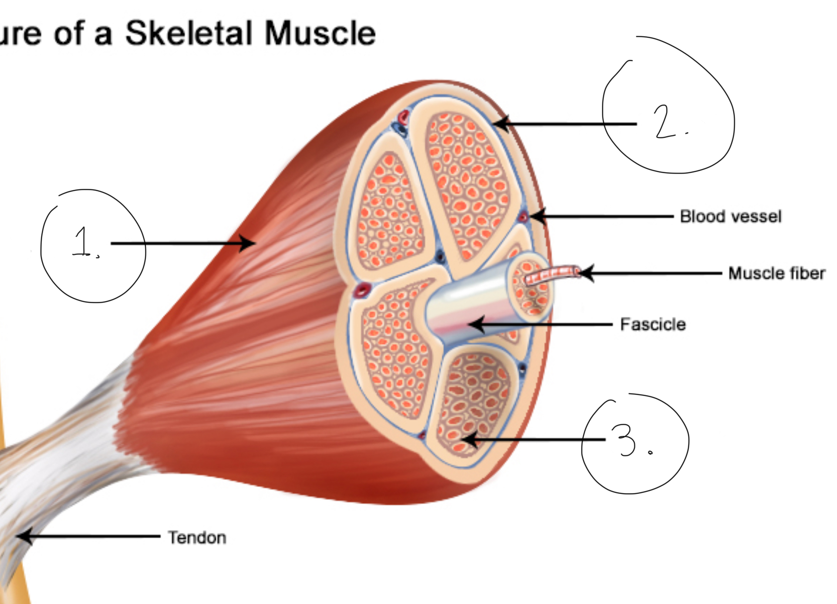
Epimysium
Outermost connective sheath- surrounds bundles of fascicles
Perimysium
Middle connective layer, surrounds individual fascicle
Endomysium
Innermost connective layer, surrounds individual muscle cell
Epimysium
Perimysium
Endomysium
Fascicle
Made of multiple muscle cells
Sarcolemma
Cell membrane of muscle cells
Myofibril
A strand of the muscle cell, has actin and myosin
Muscle fiber
One muscle cell- elongated and multi-nucleate
Advantage of the structure of -mysium and tendons?
All continuous, transfers force more efficiently to bone
How does tendon elasticity convert kinetic energy—> potential energy?
Tendons absorb kinetic energy and store it, turning it into potential energy. This allows for a rapid recoil if needed.
Glycosomes
Granules of stored glycogen, stored in muscle cells to provide fast energy
Glycogen
Made of many glucose molecules- glucose can be converted to ATP
Myoglobin
Carries oxygen
How does the body create large muscle cells?
Many cells fuse during development, creates large cell
How does body coordinate muscles?
The sarcolemma is electrically excitable like a nerve cell, allows for fast communication
How does body distribute gene products in a large cell
Many nuclei = shorter distances for gene products to be distributed
How do muscle cells contract?
Using actin+myosin and ATP

A.
Thick filament (myosin)

B.
Thin filament (actin)

C.
Z disk
D.
H zone

E.
M line

F.
I band (on either side of A band)

G.
A band

A.
Myosin
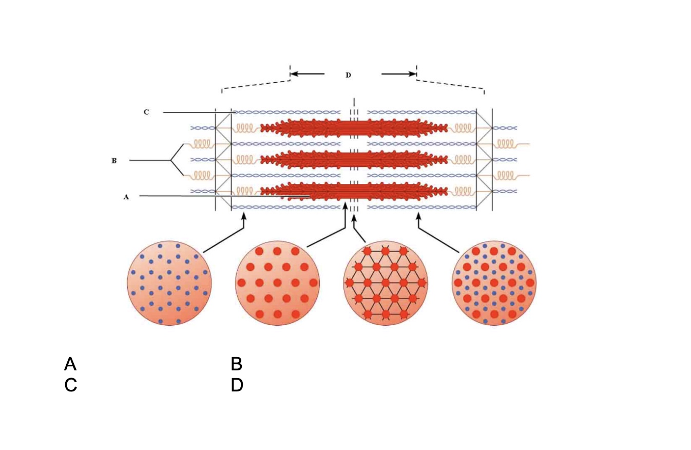
B.
Elastic filaments

C.
Actin

D.
Sarcomere
Describe how the myosin binding sites are blocked in muscle’s resting state
The troponin complex blocks binding sites on actin, the long tropomyosin protein must be moved by troponin for myosin to bind to actin
What is the neurotransmitter molecule released by motor neuron?
Acetylcholine
What causes the actin and myosin to relax?
The ATP binds to myosin, which releases the myosin from the myosin binding sites (on the actin)
What causes a ligand gated channel to open?
The ligand (acting molecule) binds to the receptor channel and opens it
What causes a voltage gated channel to open?
Voltage of the cell becomes positive, which opens the channel
Describe distribution of Na+ and K+ inside vs outside cell, before and after action potential
The cell has lower Na+ and higher K+, and the outside of the cell has higher Na+ and lower K+. When triggered by an action potential, Na+ flows into the cell and creates a positive charge (Depolarizes). Then K+ leaves the cell to return the cell to negative charge.
Where is acetylcholine contained?
In synaptic vesicles of the nerve cell
Describe the 3 states of the voltage gated sodium channel
Rest = closed
Open = due to positive voltage
Inactivated = after opening, channel closes by itself and recovers, will cycle back to resting
How does calcium affect acetylcholine at neuromuscular junction?
Increase in calcium causes the synaptic vesicles to exocytose ACh onto muscle cell. This calcium enters the nerve cell through voltage gated calcium channels (activated by action potential)
Purpose of acetylcholinesterase
Acetylcholinesterase destroys acetylcholine so that muscle can relax and is not re-triggered.
Effect of acetylcholine on muscle receptors
ACh binds to the ACh receptor (ligand gated) and opens the channel. This allows Na+ to flow into the cell and K+ to flow out of the cell. This starts the process of creating positive charge in the cell, and triggers voltage gated sodium channels to open and initiate action potential.
What is the job of the T tubule?
Relay action potential signal to sarcoplasmic reticulum, causes release of calcium ion into the muscle cell
Job of sarcoplasmic reticulum
Stores and releases calcium
What do the calcium ions in the muscle cell bind to?
Calcium ions bind to troponin, moving tropomyosin, allowing the myosin to bind to actin
Sequence of events that causes muscle contractions
Action potential arrives at neuromuscular junction
Nerve cell releases acetylcholine, which triggers voltage gated sodium channels, creating action potential along surface of sarcolemma
Action potential descends through T tubules into the muscle cell
T tubules signal sarcoplasmic reticulum to release calcium ions
Calcium binds to troponin complex, which moves tropomyosin from binding sites, allowing actin and myosin to interact (contraction)
Calcium is removed from the sarcoplasm and troponin complex blocks binding sites, stops muscle contraction
Motor unit
Consists of all muscles stimulated by a single motor neuron, small motor units allow for finer movements. More motor units allows for larger stronger movements
Isotonic vs isometric contraction
Isotonic= tension develops, load is moved, muscle fibers shorten
Isometric= tension develops but load doesn’t move, muscle fibers stay the same length
What is a muscle twitch?
Response of motor unit to single action potential. Faster twitch = greater accuracy but lesser strength
What occurs during muscle’s latent period?
latent period = delay between action potential and tension observed in muscle
On molecular level, calcium is diffusing out of SR, binding to tropinin complex, and moving tropomyosin
2 ways to change muscle tension response
Changing frequency of nerve stimulation, multiple action potential = stronger contraction
Changing strength of stimulus, more intense stimulus causes other motor units to contract (recruitment)
Describe recruitment
Over time more/larger motor units are being activated due to stimulus gaining strength. Small motor units are recruited before larger ones
Describe summation
Changing frequency, but not strength, of nerve stimulation. Treppe effect = repeated equal stimuli produce progressive increase in tension generated. Caused by accumulation of calcium ions within muscle fiber. Repeated stimuli = not enough time for cell to move all calcium back to SR
Duration of energy from ATP vs glycogen vs mitochondria+oxygen
ATP= 4 to 6 seconds
Glycogen= broken down into glucose, 30-40 seconds
Mitochondria= if oxygen present, lasts for hours
Job of creatine kinase in muscle cell
Creatine Kinase is an enzyme that transfers phosphate to ADP to create ATP, catalyzes the reaction of creatine phosphate + ADP → creatine + ATP
Slow twitch vs fast twitch muscle fibers
Slow twitch: oxidative, slow contractions but resistant to fatigue. High oxygen demand (<mitochondria), thinner and weaker muscles
Fast twitch: glycolytic, rapid contractions and fatigues easily. No oxygen used (>mitochondria) and primarily anaerobic, larger and stronger muscles
Fast oxidative/glycolytic fibers
Intermediate type of muscle fiber, intermediate properties/strength/size
smooth muscle location and junctions
Smooth muscle lines the body’s hollow organs, excluding the heart
Use gap junctions that allow electrical signals to pass between cells
Purpose of varicosities
Varicosities = nerve endings that secrete hormones onto muscles to control contractions. Hormones are released into diffuse junctions. Effect can be short or long lasting (hours)
Single unit smooth muscle vs multiunit smooth muscle
single unit smooth muscle: waves of contraction in sheets of tissue, muscles connected by gap junctions, contracts as a single unit. In hollow organs
Multiunit smooth muscle: local control of small motor units, fibers act independently, few gap junctions. In iris, arrector pili, airways
In smooth muscle, what do calcium ions released from sarcoplasmic reticulum bind to?
In smooth muscle, calcium ions bind to calmodulin
Process of actin and myosin movement in skeletal muscle
Calcium ions bind to troponin, moved the tropomyosin off of the myosin binding site. Myosin heads bind to site on actin strand and an ADP molecule is released. After pulling the myosin filament, the myosin head releases from actin, binds to ATP, and resets potential energy using ATP. Cycle can now repeat with myosin binding with actin again.
How do muscles move quickly despite short length of sarcomere?
Multiple sarcomeres end to end amplify small movements
How do muscles generate high force despite small sarcomere size?
Multiple sarcomeres stacked top to bottom create stronger forces then an individual sarcomere
Muscles and job of rotator cuff
Supraspinatus
Infraspinatus
Teres minor
Subscapularis
Form a cuff that holds head of humerus in glenoid cavity
describe compartment syndrome
Compartment syndrome = inflammation of the fascia
This inflammation creates pressure and compresses muscles,nerves,bloodvessels
Can cause ischemia (poor blood flow). Treated by cutting the muscle to relieve pressure
Describe ALS
Motor neurons progressively die. Muscles and brain neurons are unaffected. Causes paralysis
Describe myasthenia gravis
Antibodies attack acetylcholine receptors at neuromuscular junction, causes them to be unresponsive to ACh. Presents as muscle weakness, drooping eyelids, and rapid muscle fatigue
Describe Duchenne muscular dystrophy
Caused by defective dystrophin protein. Dystrophin acts as a shock absorber and without proper function the muscle cells destroy their own cell membranes when contracting. Kills muscle cells
Describe flaccid paralysis due to toxins that block calcium channels in motor neurons
If calcium channels in motor neurons are blocked, the trigger to release ACh isn’t reached and muscle won’t comtract
Ryanodine receptor
Opens channel that allows calcium ions out of sarcoplasmic reticulum. Located at junction of T tubule and SR
Describe flaccid paralysis due to failure of choline re-uptake
Without re-uptake of choline into motor neuron, it can’t be recycled back into ACh and nerve terminal runs out of neurotransmitter
Hyper- vs hypo-kalemia
Hyperkalemia= elevated extracellular potassium levels
Hypokalemia= decreased extracellular potassium levels
Both cause poor excitability of T tubule due to charge imbalance
Describe acetylcholinesterase
Inactivates ACh. When defective/destroyed, causes rigid paralysis due to overactivity of ACh
Describe calcium pump SERCA
SERCA=sarcoplasmic reticulum calcium transport ATPase
Found only in skeletal and cardiac muscles, when damaged leads to high calcium events/excessive muscle contractions and eventually rigid paralysis
Describe Parkinson’s disease symptoms
Continuous shaking of the limbs but no muscle fatigue. Also causes specific gait when walking
Describe clonus
Rhythmic pattern of contractions, 3-7 per second, due to alternating stretching-unloading of muscle spindles in spastic muscle
Describe Guillain Barre syndrome
Caused when antibodies attack a virus and myelin is collateral damage. Symptoms include loss of sensation, unsteady walking, difficulty with face/throat movements and difficulty breathing
Muscle insertion point vs muscle origin
Muscle insertion= end that moved when contracting
Muscle origin= end that doesn’t move during contraction
Forearm flexion muscles and opposing muscle
Flexion =Biceps, brachioradialis, brachialis
Opposition =triceps brachii
Muscle action and antagonist of pectoralis major
Flexes the arm
Antagonist = latissimus dorsi
Muscle action and antagonist of the deltoid
Abducts the arm (lifts away from body)
Antagonist = teres major
Agonist
Prime mover in muscle action
Synergist vs antagonist
Synergist =aids prime mover, can modify direction of movement
Antagonist =opposes direction of prime muscle movement
Fixator
Holds bones in place
Which muscles close the jaw?
Temporalis and masseter
Genioglossus vs hypoglossus vs syloglossus muscle
Genioglossus =protracts and retracts tongue
Hypoglossus =depresses tongue
Syloglossus =retracts and elevates tongue
Iliocostalis and longissimus muscle function (same for both)
Extends and laterally flexes vertebral column
Spinalis movement
Extends vertebral column
External intercostals movement
Elevates rib cage during inspiration
Rectus abdominis movement
Compresses abdomen
Levator ani function
Maintains position of pelvic viscera
Which muscles protract the scapula?
serratus anterior, pectoralis minor, pectoralis major (all synergists)
Which muscles retract the scapula?
Trapezius and rhomboids
Describe structure of trapezius
Has 3 heads that can activate independently, acts as its own antagonist, can raise or lower scapula
Afferent vs Efferent signals
Afferent =input,towards the brain
Efferent =output, towards the body
Think cAuse and Effect
How does spinal fluid differ from blood?
Contains no red blood cells and has low protein content
Significance of Louveau finding
Discovered antibodies in brain
Could cause problems with inflammation leading to multiple sclerosis and alzheimers, but knowing the cause creates opportunities for a cure
Job of blood brain barrier
Protects brain from cells, large proteins, and amino acids that would kill neuronal cells
Structures in CNS
Brain and spinal cord
4 types of glial cells in CNS
Astrocytes, microglia, ependymal cells, ogliodendrocytes
AMEO
2 types of glial cells in PNS
Satellite cells, schwann cells