Anatomy of the Lower Body
 Call Kai
 Learn
Learn Practice Test
 Spaced Repetition
 Match
Match Flashcards
 Knowt Play
1/255

There's no tags or description
Looks like no tags are added yet.
Name | Mastery | Learn | Test | Matching | Spaced | Call with Kai | Chat |
|---|
No analytics yet
Send a link to your students to track their progress
256 Terms
tibialis anterior
extensor digitorum longus
fibularis tertius
extensor longus hallucis
muscles in the anterior compartment of the leg

proximal 2/3 of the anteriorlateral tibia
tibialis anterior origin

medial + inferior surfaces of medial cuneiform and base of 1st metatarsal
tibialis anterior insertion
anterior fibula and interosseous membrane
extensor digitorum longus origin
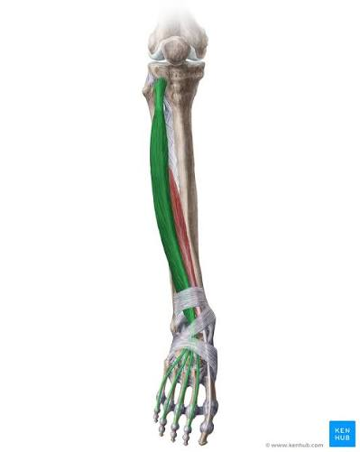

middle and distal phalanges of lateral four digits
extensor digitorum longus insertion
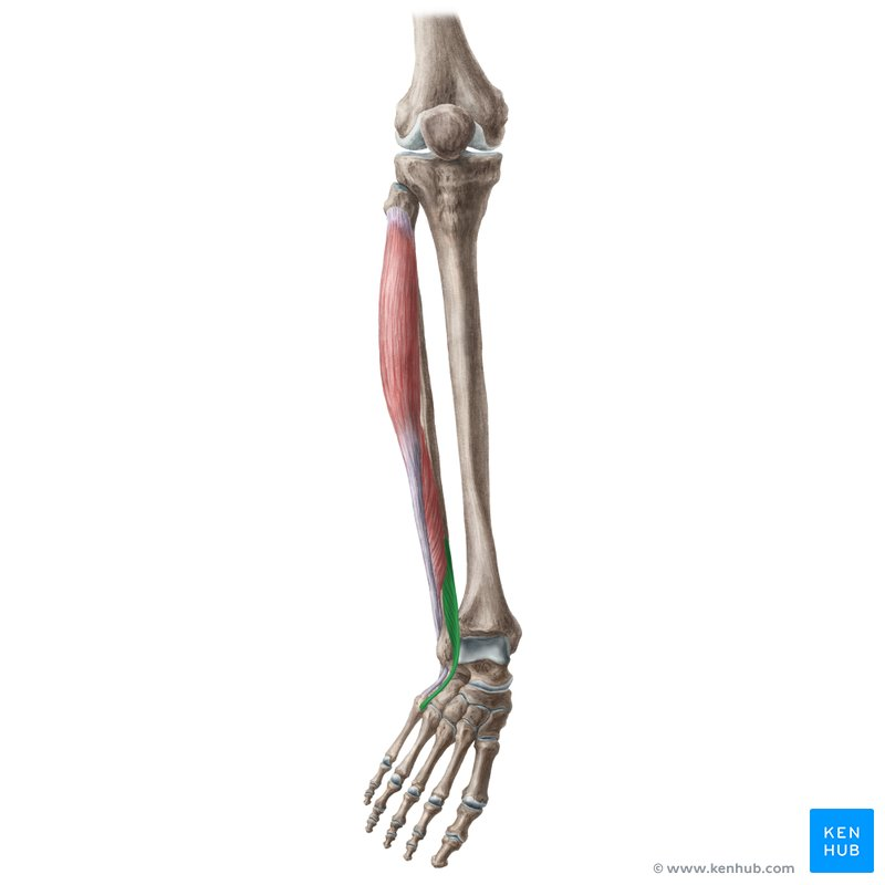
distal 1/3 of anterior fibula and interosseous membrane
fibularis tertius origin

base of 5th metatarsal
fibularis tertius insertion

anterior fibula and interosseous membrane
extensor hallucis longus origin
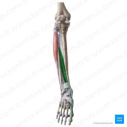
dorsal aspect of base of distal phalanx of great toe
extensor hallucis longus insertion
L4-L5
nerve roots for tibialis anterior innervation

deep fibular nerve
nerve that innervates tibialis anterior
L5-S1
nerve roots for extensor digitorum longus

deep fibular nerve
nerve that innervates extensor digitorum longus
L5-S1
nerve roots for extensor hallucis longus innervation

deep fibular nerve
nerve that innervated extensor hallucis longus
L5-S1
nerve roots for fibularis tertius

deep fibular nerve
nerve that innervates fibularis tertius
dorsiflexion of ankle joint and foot inversion
main action of tibialis anterior
extends digits 2-5, ankle dorsiflexion
extensor digitorum longus main action
ankle joint dorsiflexion, foot eversion
fibularis tertius main action
great toe extension, ankle dorsiflexion
extensor hallucis longus main action
fibularis longus, fibularis brevis
muscles of the lateral compartment of the leg
head of the fibula and lateral fibula
fibularis longus origin


base of 1st metatarsal and medial cuneiform
fibularis longus insertion
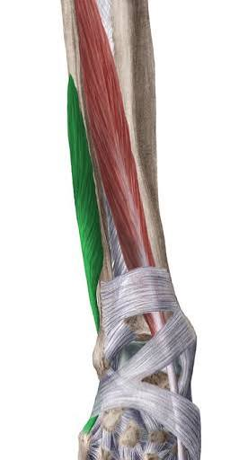
distal 1/3 of lateral fibula
fibularis brevis origin

dorsal surface of lateral tuberosity on 5th metatarsal
fibularis brevis insertion
foot eversion, weak ankle plantar flexion, resist foot inversion
fibularis longus main action
foot eversion, weak ankle plantar flexion, resist foot inversion
fibularis brevis main action

superficial fibular nerve
nerve that innervates fibularis longus
L5, S1, S2
nerve branches for fibularis longus innervation

superficial fibular nerve
nerve that innervates fibularis brevis
L5, S1, S2
nerve branches for fibularis brevis
anterior tibial artery
artery that supplies blood flow for the anterior compartment of the leg
fibular (peroneal) artery
artery that supplies blood flow for the lateral compartment of the leg
anterior tibial veins
veins of the anterior compartment of the leg
proximally to the dorsal aspect of the ankle joint
superior extensor retinaculum location

anterior aspect of lateral distal fibula
superior extensor retinaculum lateral attachment
anterior aspect of medial distal tibia
superior extensor retinaculum medial attachment
house muscles of anterior compartment of the leg
superior extensor retinaculum function

lateral aspect of calcaneus around peroneal trochlea
inferior peroneal retinaculum insertion

Posterior border and tip of the lateral malleolus
superior peroneal retinaculum origin
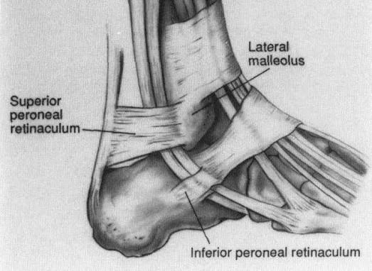
lateral aspect of the calcaneus and deep fascia of the leg
superior peroneal retinaculum insertion
Secures fibular tendons as they split and travel toward their attachment points.
inferior peroneal retinaculum function
primary anchor for fibular tendons
superficial peroneal retinaculum function
extensor hallucis brevis, extensor digitorum brevis
muscles of the dorsum of the foot
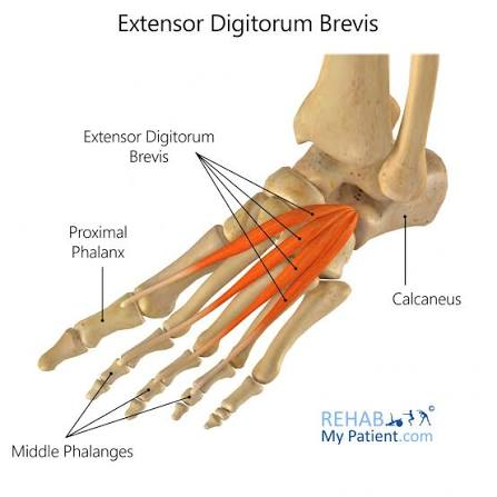
calcaneus, floor of tarsal sinus
extensor digitorum brevis origin
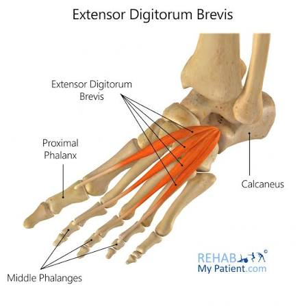
base of proximal phalanx of toes 2-4
extensor digitorum brevis insertion

calcaneus, first of the tarsal sinus
extensor hallucis brevis origin

proximal phalanx of great toe
extensor hallucis brevis insertion
extends digits 2-4
extensor digitorum brevis action
extends great toe
extensor hallucis brevis action

deep fibular nerve
nerve that innervates extensor digitorum brevis
L5, S1
nerve roots for extensor digitorum brevis
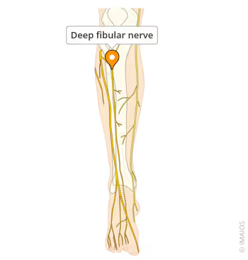
deep fibular nerve
nerve that innervates extensor hallucis brevis
L5-S1
nerve roots for extensor hallucis brevis
dorsalis pedis
artery that supplies blood to the dorsum of the foot
anterior inferior tibiofibular ligament (AITFL)
posterior inferior tibiofibular ligament (PITFL)
transverse tibiofibular ligament (TTFL)
interosseous tibiofibular ligament (IOL)
ligaments in the distal tibiofibular joint
fibrous (syndesmosis)
distal tibiofibular joint type

deep fibular nerve
cutaneous innervation of the yellow section


saphenous nerve
cutaneous innervation of the medial posterior foot (green) section
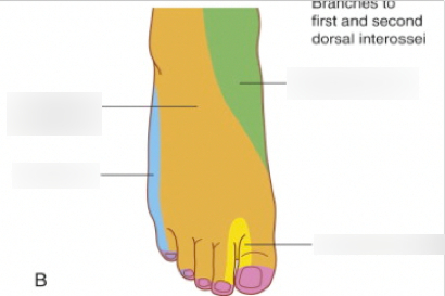

superficial fibular nerve
cutaneous innervation of the midfoot (orange) section

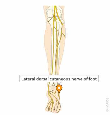
dorsal lateral cutaneous nerve
cutaneous innervation of the lateral foot (blue section)

flexor digitorum brevis
abductor hallucis
abductor digiti minimi
muscles in the first layer of the plantar foot

calcaneal tuberosity and plantar aponeurosis
flexor digitorum brevis origin

middle phalanges of toes 2-5
flexor digitorum brevis insertion
flexes toes 2-5
flexor digitorum brevis action

medial plantar nerve
nerve that innervates flexor digitorum brevis
L5, S1
nerve branches for flexor digitorum brevis
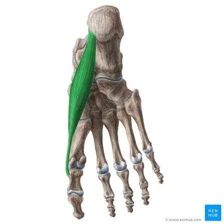
medial process of tuberosity of calcaneus, flexor retinaculum, and plantar aponeurosis
abductor hallucis origin

medial side of base of proximal phalanx of big toe
abductor hallucis insertion
abducts and flexes big toe
abductor hallucis action
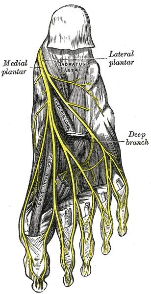
medial plantar nerve
nerve that innervates abductor hallucis
L5, S1
nerve branches for abductor hallucis

medial and lateral processes of tuberosity of calcaneus, plantar aponeurosis, and intermuscular septa
abductor digiti minimi origin

lateral side of base of proximal phalanx of 5th digit
abductor digiti minimi insertion
abducts and flexes 5th digit
abductor digiti minimi action

lateral plantar nerve
nerve that innervates abductor digiti minimi
S1-S3
nerve roots for abductor digiti minimi
quadratus plantae
lumbricals
muscles in the 2nd layer of the plantar foot

medial surface and lateral margin of plantar surface of calcaneus
quadratus plantae origin

posterolateral margin of tendon of flexor digitorum longus
quadratus plantae insertion
flexes lateral four digits
quadratus plantae action

lateral plantar nerve
nerve that innervates quadratus plantae
S1-S3
nerve branches for quadratus plantae

tendons of flexor digitorum longus
lumbricals origin

medial aspect of extensor expansion of lateral four digits
lumbricals insertion
flex proximal phalanges and extends middle and distal phalanges of digits 2-5
lumbricals action
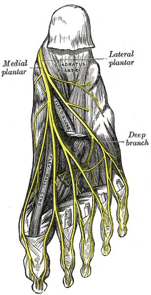
medial plantar nerve
nerve innervation for medial lumbrical

lateral plantar nerve
nerve innervation for lateral three lumbricals
L5, S1
nerve branches for medial lumbrical
S1-S3
nerve branches for lateral three lumbricals
flexor hallucis brevis
adductor hallucis
flexor digiti minimi
muscles in the 3rd layer of the plantar foot

plantar surface of cuboid and lateral cuneiforms
flexor hallucis brevis origin
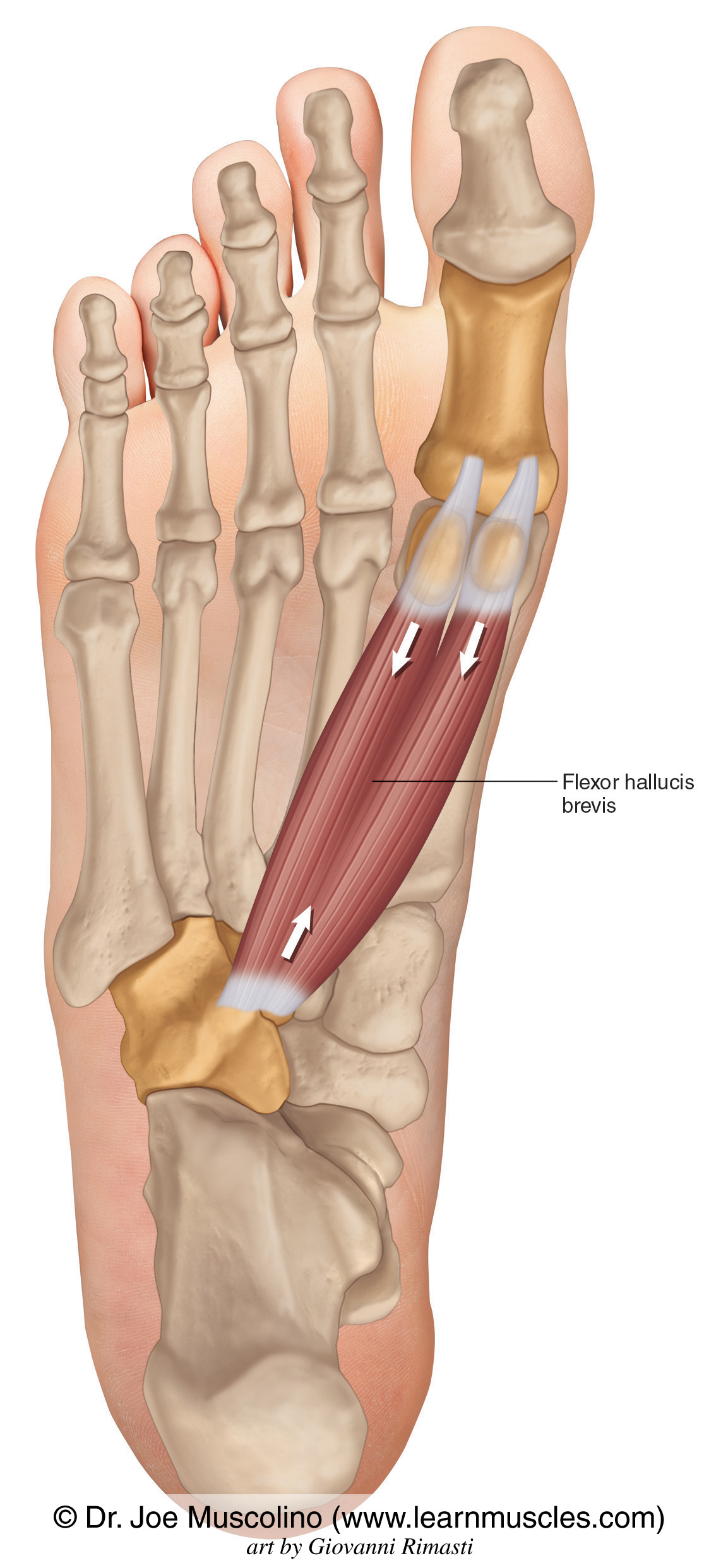
both sides of base of proximal phalanx of big toe
flexor hallucis brevis insertion
flexes proximal phalanx of first digit
flexor hallucis brevis action

medial plantar nerve
nerve that innervates flexor hallucis brevis
L5, S1
nerve branches for flexor hallucis brevis

bases of metatarsals 2-4
adductor hallucis oblique head origin

plantar ligaments of MTP joints
adductor hallucis transverse head origin

lateral side of base of proximal phalanx of big toe
adductor hallucis insertion (for both heads)