MRI Lecture 2
 Call Kai
 Learn
Learn Practice Test
 Spaced Repetition
 Match
Match Flashcards
 Knowt Play
1/153

Earn XP
Description and Tags
Non-Ionising
Name | Mastery | Learn | Test | Matching | Spaced | Call with Kai | Chat |
|---|
No analytics yet
Send a link to your students to track their progress
154 Terms
How does MRI produce an image?
B₀ (main magnetic field) aligns hydrogen nuclei.
Hydrogen nuclei become either:
Parallel (low energy)
Anti-parallel (high energy)
An RF pulse at the Larmor frequency excites the nuclei.
Hydrogen nuclei move into the transverse plane (B₁).
RF pulse stops.
Relaxation begins:
T1 longitudinal relaxation
T2 transverse decay
Relaxing nuclei release RF energy.
Receiver coils detect this energy.
The computer reconstructs these signals into an MRI image.
How does MRI produce an image?
MRI image formation occurs in this order:
The patient enters the scanner.
The main magnetic field (B₀) aligns hydrogen nuclei.
An RF pulse is applied at the hydrogen Larmor frequency.
Hydrogen nuclei absorb energy.
They move into the transverse plane (B₁).
The RF pulse is switched off.
Relaxation begins:
T1 (longitudinal recovery)
T2 (transverse decay)
Relaxing nuclei emit RF energy.
Receiver coils detect this signal.
The computer reconstructs the detected signals into an MRI image.
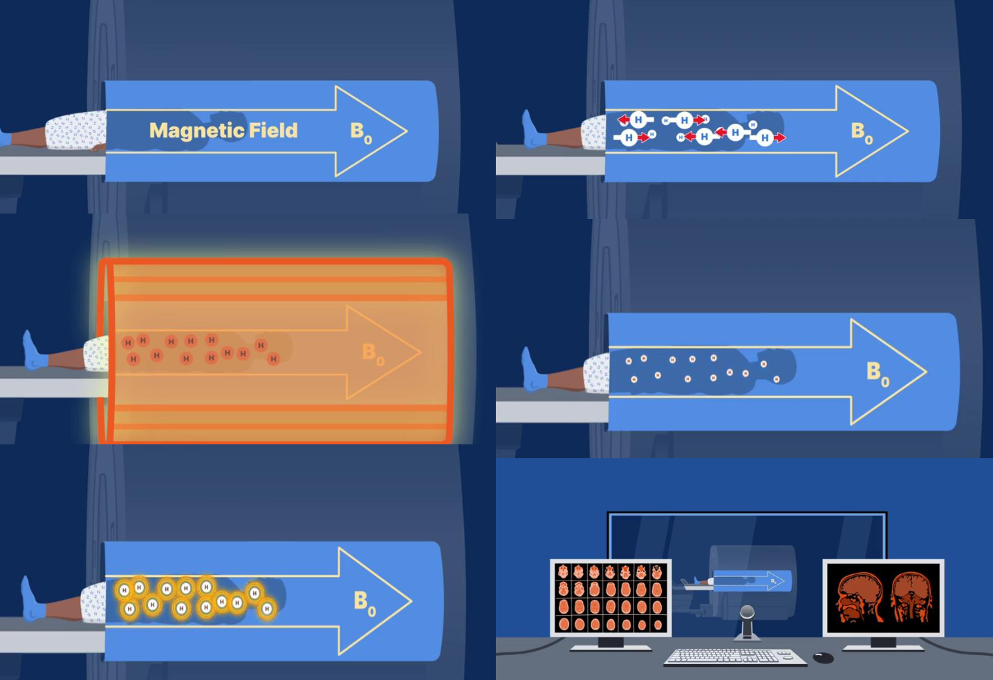
What happens to hydrogen nuclei before entering the MRI scanner?
Outside the MRI scanner:
Hydrogen nuclei are naturally precessing.
They oscillate in many different directions.
Their magnetic effects cancel one another.
There is no net magnetisation.
Because they are randomly oriented, no usable MRI signal exists.
What happens when hydrogen nuclei enter the MRI scanner?
When hydrogen nuclei enter the scanner:
They experience the main magnetic field (B₀).
They align either:
Parallel to B₀ (lower energy)
Anti-parallel to B₀ (higher energy)
Slightly more nuclei align parallel than anti-parallel.
This difference creates the net magnetisation required for MRI.
What is the purpose of the RF pulse?
The RF pulse:
is applied at the Larmor frequency of hydrogen,
supplies energy to hydrogen nuclei,
excites the nuclei,
tips the net magnetisation away from B₀,
moves magnetisation into the transverse plane (B₁).
This excitation is essential for producing a measurable MRI signal.
What happens after the RF pulse is switched off?
Once the RF pulse stops:
Hydrogen nuclei begin relaxing.
Two processes occur simultaneously:
T1 longitudinal relaxation
T2 transverse decay
Although both occur together, MRI sequences generally emphasise one relaxation process at a time.
How is the MRI signal detected?
During relaxation:
Hydrogen nuclei release the absorbed RF energy.
Receiver coils detect these tiny RF signals.
The scanner processes these signals.
Image reconstruction software converts the signals into diagnostic MR images.
What are the three main MRI magnet types?
The three major MRI magnet types are:
Permanent magnets
Resistive magnets
Superconducting magnets
Of these:
superconducting magnets are by far the most common in modern clinical MRI.Which MRI magnet type is most commonly used?
Which MRI magnet type is most commonly used?
The superconducting magnet is the standard clinical MRI system.
It is used because it can generate:
strong magnetic fields,
stable magnetic fields,
high-quality images,
whole-body imaging,
advanced MRI sequences.
Permanent and resistive magnets are much less common.
Where are permanent MRI scanners more commonly found?
Permanent MRI scanners are more common in:
developing countries,
low-resource healthcare systems,
some rural locations.
The lecturer specifically notes that if working overseas (e.g. parts of Africa), permanent magnets are encountered more frequently because they are cheaper to purchase and operate.
How does a permanent MRI magnet produce B₀?
A permanent MRI scanner produces its magnetic field using:
permanently magnetised blocks,
or assemblies of permanently magnetised material.
Unlike other magnet types:
it does not require a powered superconducting coil,
it does not require electrical current to maintain B₀.
Instead:
The magnetised material itself produces the static magnetic field.
Why is a permanent magnet described as "always on"?
A permanent magnet is always on because:
the magnetic field comes directly from the magnetised material,
no electricity is required to maintain magnetisation,
the magnetic field cannot simply be switched off.
Therefore:
if a ferromagnetic object becomes attached,
the magnet cannot be de-energised,
removal requires brute force.
This is an important MRI safety consideration.
Does a permanent MRI scanner require electricity?
The main magnetic field does not require electricity.
However, electricity is still needed to operate:
gradient coils,
RF systems,
computer systems,
patient table,
cooling systems,
supporting hardware.
Only the main B₀ field is maintained without electrical input.
How are permanent MRI magnets manufactured?
Permanent MRI magnets are made using ferromagnetic materials, including:
Alnico,
ferrites,
rare-earth magnetic materials.
Manufacturing process:
The material is exposed to a very strong external magnetic field.
Microscopic magnetic domains align.
The external field is removed.
Much of the alignment remains.
The material becomes permanently magnetised.
This permanent magnetisation generates the scanner's B₀ field.
This permanent magnetisation is why these scanners remain magnetised continuously.
Why are permanent MRI magnets generally low-field systems?
Permanent magnets are limited because the permanently magnetised material has a relatively small magnetic moment, meaning it cannot generate extremely strong magnetic fields.
As a result:
They cannot achieve conventional high field strengths.
They produce lower magnetic field strengths than superconducting magnetsWhat magnetic field strengths are typical for permanent MRI scanners?
What magnetic field strengths are typical for permanent MRI scanners?
Typical permanent MRI field strengths are:
Portable MRI scanners: 0.05–0.08 Tesla
Open-bore MRI scanners: 0.20–0.44 Tesla
For comparison:
Conventional hospital MRI scanners are typically 1.5 T or 3 T
Why does a lower magnetic field strength reduce image quality?
Lower field strength results in:
lower signal-to-noise ratio (SNR),
weaker MR signal,
reduced image quality,
lower spatial resolution.
The lecturer notes that lower Tesla = lower SNR, which directly affects diagnostic image quality.
What scanner designs are possible with permanent magnets?
Because permanent magnets are not long superconducting solenoids, they can be manufactured in several designs, including:
Open MRI
C-shaped scanners
H-shaped scanners
Single-pillar scanners
Extremity MRI systems
Portable MRI scanners
This flexibility is a major design advantage.
What are the clinical advantages of open permanent MRI scanners?
Open MRI scanners can:
reduce patient anxiety,
reduce claustrophobia,
improve patient comfort,
make patient positioning easier,
accommodate larger patients,
allow weight-bearing imaging,
enable portable MRI systems.
Because they are open, they can perform examinations that may be difficult inside a conventional tunnel-bore scanner.
Why are permanent MRI scanners commonly used in veterinary medicine?
Permanent MRI scanners are common in veterinary clinics because:
open designs make positioning animals easier,
scanners are less expensive,
lower operating costs make them economically attractive,
many veterinary applications do not require ultra-high field strengths.
The lecturer specifically highlights veterinary clinics as a common use of permanent magnets.
What are the major advantages of permanent MRI magnets?
Advantages include:
No electrical power required to maintain B₀.
Simpler magnet design.
Lower maintenance costs.
Lower operating costs.
Open scanner configurations.
Improved patient comfort.
Portable MRI options.
Weight-bearing imaging capability.
These advantages explain why they remain useful despite their lower image quality.
What are the disadvantages of permanent MRI magnets?
Permanent magnets have several important limitations:
Limited to approximately 0.4 T.
Lower signal-to-noise ratio.
Lower image quality.
Longer scan times.
Cannot perform many advanced MRI applications.
Cannot easily produce highly homogeneous magnetic fields.
Magnet is always on.
Cannot be quenched or powered down during emergencies.
Why do permanent magnets struggle to produce homogeneous magnetic fields?
Permanent magnets rely entirely on permanently magnetised material.
Although this material produces B₀:
it cannot easily generate extremely uniform magnetic fields,
maintaining homogeneity across the imaging volume is difficult,
image quality therefore suffers compared with superconducting systems.
High magnetic homogeneity is essential for high-quality MRI.
How do scan times compare between permanent MRI and 3 Tesla MRI?
The lecturer gives the example of a routine brain MRI:
3 Tesla scanner
Approximately 12–15 minutes
0.44 Tesla permanent magnet
Approximately 14 minutes
Although the scan duration is similar in this example, the permanent magnet still produces noticeably poorer image quality.
Why is image quality poorer on permanent MRI scanners?
Compared with conventional MRI:
Permanent magnets produce:
larger voxels,
reduced spatial resolution,
poorer tissue definition,
reduced visibility of fine anatomical structures,
poorer lesion conspicuity,
lower signal-to-noise ratio.
Small pathological findings may therefore be more difficult to detect.
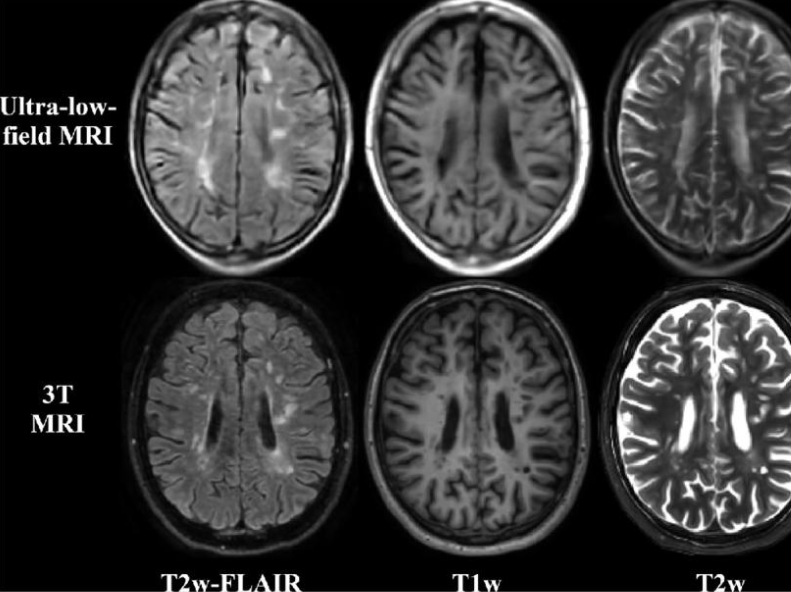
What image quality differences did the lecturer highlight between ultra-low-field MRI and 3T MRI?
Compared with 3T MRI:
Ultra-low-field MRI demonstrates:
blurred cortical anatomy,
difficulty identifying gyri and sulci,
poorer white matter definition,
larger pixel size,
reduced visibility of blood vessels,
poorer depiction of lesions,
reduced ability to diagnose conditions such as vascular dementia or multiple sclerosis.
Why are MRI scanner rooms kept cold?
MRI hardware performs best in a cool environment.
A controlled temperature helps:
maintain hardware performance,
protect electronic components,
prevent overheating,
improve scanner reliability.
The lecturer notes this is similar to CT scanners—radiology equipment generally performs best in cool rooms.
Why can permanent MRI magnets create safety problems?
Permanent magnets are always magnetised.
If an unsafe ferromagnetic object becomes attached:
the magnet cannot be switched off,
there is no method to ramp down the magnetic field,
the object must be removed using physical force.
This is an important MRI safety consideration
What is a resistive MRI magnet?
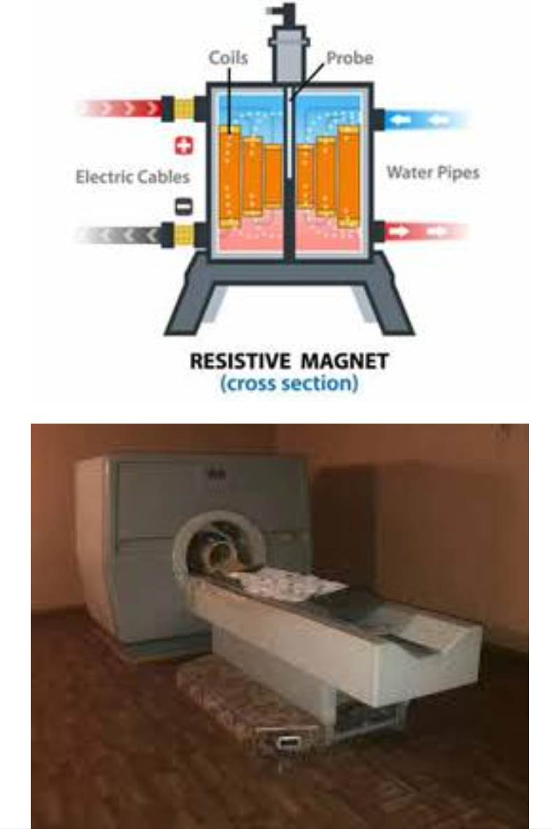
A resistive magnet is an electromagnet that produces the main magnetic field (B₀) by passing an electrical current through coils of wire.
Unlike permanent magnets:
It requires electricity to generate the magnetic field.
The magnetic field only exists while current is flowing.
When the power is switched off, the magnetic field disappears.
How does a resistive MRI magnet generate a magnetic field?
A resistive magnet works according to the principles of electromagnetism:
Electrical current passes through coils of copper wire.
The moving electric charges generate a magnetic field.
The more current flowing through the coils, the stronger the magnetic field.
When current stops, the magnetic field immediately disappears.
This makes resistive magnets fundamentally different from permanent magnets.
Why are resistive magnets called electromagnets?
They are called electromagnets because:
the magnetic field is produced entirely by electricity,
no permanent magnetic material is involved,
the field strength depends on the electrical current.
Without electrical current, there is no magnetic field.
What is the main advantage of a resistive MRI magnet?
The greatest advantage is that the magnet can simply be turned off.
If electricity is removed:
the magnetic field disappears,
projectile hazards are eliminated,
maintenance is easier,
emergencies are easier to manage than with permanent magnets.
The lecturer contrasts this with permanent magnets, which remain magnetised continuously.
Why are resistive MRI magnets expensive to operate?
Resistive magnets require:
continuous electrical current,
high power consumption,
electricity 24 hours a day to maintain B₀.
Because electricity is constantly being used, operating costs become extremely high.
Why do resistive magnets generate heat?
When electrical current flows through copper wire:
electrical resistance is present,
electrical energy is converted into heat,
this is known as Joule heating.
The stronger the current:
the more heat is produced.
This heat becomes one of the major limitations of resistive MRI systems.
How is excess heat removed from resistive MRI magnets?
Because large amounts of heat are produced, resistive magnets require cooling systems.
Cooling may involve:
circulating water,
heat exchangers,
other cooling mechanisms to remove excess thermal energy.
Without cooling, the coils would overheat and become damaged.
Why can't resistive magnets produce very high magnetic field strengths?
To produce stronger magnetic fields:
larger electrical currents are required.
However:
larger currents create much more heat,
cooling requirements become excessive,
electricity costs increase dramatically,
practical engineering limits are reached.
This limits resistive magnets to relatively low field strengths.
What field strengths are typically achieved by resistive MRI magnets?
Most resistive MRI scanners operate at approximately:
0.3 Tesla
Some specialised systems have reached:
around 0.6 Tesla
These values are much lower than modern superconducting MRI scanners (1.5–3 T).
What are the advantages of resistive MRI magnets?
Advantages include:
Magnetic field can be switched off.
Easier maintenance.
Simpler emergency management.
No permanently magnetised components.
Less complex than superconducting systems.
No liquid helium required.
These advantages are outweighed by their major operating disadvantages.
What are the disadvantages of resistive MRI magnets?
Major disadvantages include:
High electricity consumption.
Expensive to operate.
Large heat production.
Continuous cooling required.
Limited field strength.
Lower signal-to-noise ratio.
Lower image quality than superconducting MRI.
For these reasons, resistive magnets are now uncommon.
Why are resistive MRI scanners rarely used today?
Modern hospitals generally avoid resistive MRI because:
electricity costs are extremely high,
cooling requirements are significant,
field strengths are relatively low,
image quality is inferior,
superconducting magnets provide much better performance.
Consequently, most clinical MRI scanners today are superconducting systems.
How do permanent and resistive MRI magnets compare?
Permanent Magnet | Resistive Magnet |
|---|---|
Uses permanent magnetic material | Uses electrical current |
Always on | Can be switched off |
No electricity for B₀ | Continuous electricity required |
Low operating cost | High operating cost |
Difficult emergency shutdown | Easy emergency shutdown |
Lower field strength | Lower field strength |
Both are now much less common than superconducting magnets.
What is the next major development in MRI magnet technology?
After permanent and resistive systems, MRI technology advanced to superconducting magnets.
These provide:
very high magnetic field strengths,
excellent field uniformity,
superior image quality,
higher signal-to-noise ratio,
faster imaging,
support for advanced MRI techniques.
Superconducting magnets became the standard for modern clinical MRI.
Why are superconducting magnets considered the gold standard in MRI?
Superconducting magnets combine:
high magnetic field strength,
excellent field homogeneity,
stable magnetic fields,
high signal-to-noise ratio,
superior spatial resolution,
reliable clinical performance.
Because of these advantages, they have replaced permanent and resistive magnets in the vast majority of modern MRI departments.
What is a superconducting MRI magnet?
A superconducting MRI magnet is the standard high-field MRI system used in most imaging departments.
Instead of using:
a permanent magnet block, or
a normal resistive electromagnet,
it produces the main magnetic field (B₀) using a coil made from superconducting wire.
In simple terms:
the wire is cooled to an extremely low temperature,
it can then carry electrical current with essentially no electrical resistance,
allowing a very strong and stable magnetic field to be maintained efficiently.
Why are superconducting MRI scanners the clinical standard?
Superconducting MRI systems dominate modern clinical imaging because they provide the best combination of:
high magnetic field strength,
excellent image quality,
high signal-to-noise ratio,
faster scan times,
clinical versatility,
stable magnetic fields,
efficient operation.
Most hospitals therefore use superconducting magnets rather than permanent or resistive systems.
What magnetic field strengths are used in superconducting MRI?
Typical superconducting MRI field strengths include:
1.5 T – the most common clinical scanner
3 T – increasingly common clinical scanner
7 T – specialised clinical and research scanner
11–11.5 T – research only
The lecturer notes that although magnets up to 42 T have been developed experimentally, routine clinical imaging almost always uses 1.5 T or 3 T.
Why are 1.5 T and 3 T considered the "clinical workhorses"?
The lecturer describes 1.5 T and 3 T as the clinical workhorses because they offer the best balance of:
image quality,
scan speed,
patient compatibility,
operating cost,
clinical versatility.
Higher field strengths exist but are generally reserved for research.
What limitations are associated with ultra-high-field MRI (7–11 T)?
Ultra-high-field MRI provides exceptional image detail but also has limitations.
The lecturer explains that at around 11 T:
the magnetic field can significantly affect the vestibular (inner ear) system,
patients may experience severe vertigo,
rapidly moving the head into or out of the bore may even cause collapse.
For this reason, these scanners remain largely confined to research facilities.
What material is most commonly used for superconducting MRI coils?
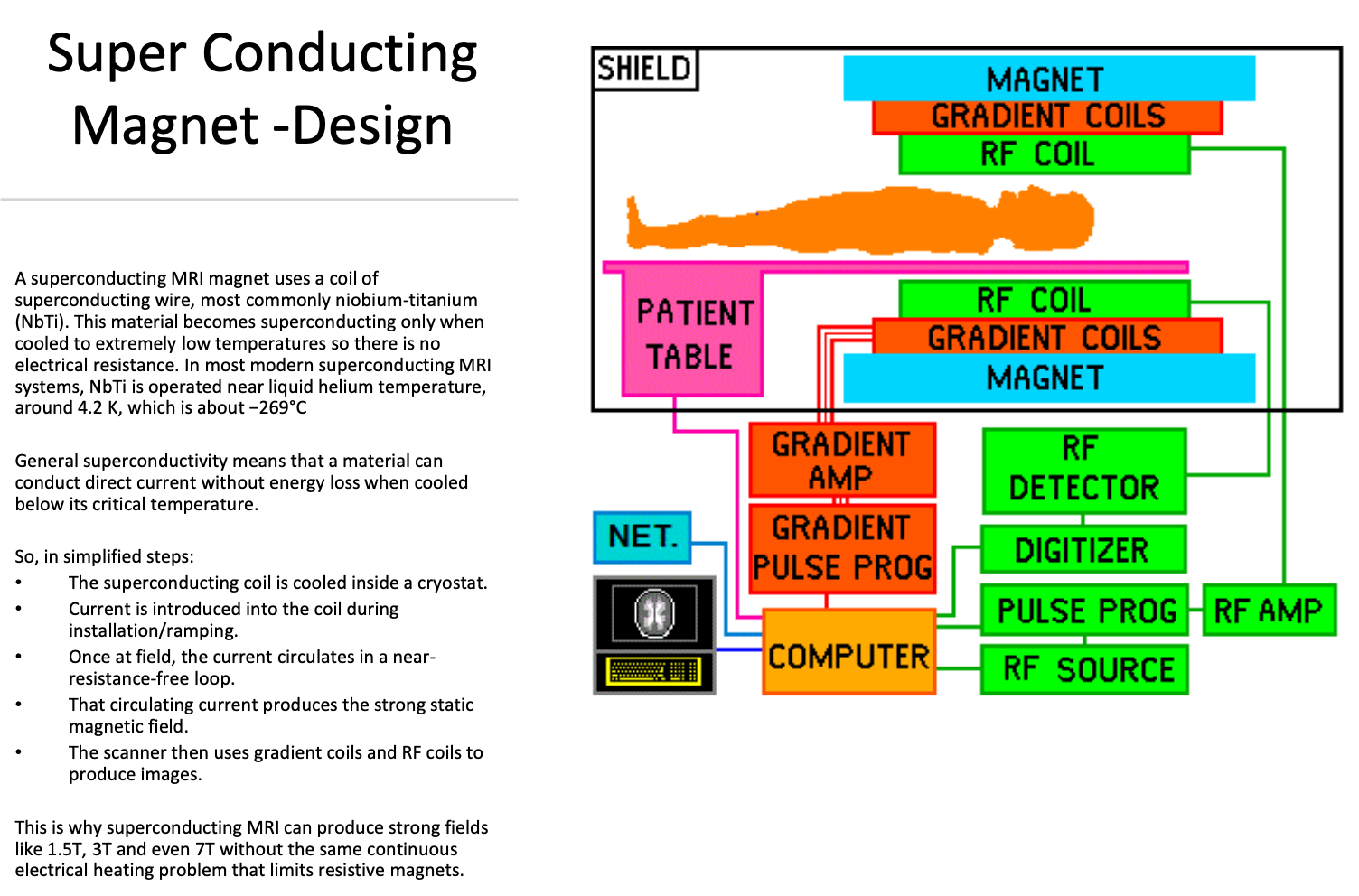
Most superconducting MRI magnets use niobium-titanium (NbTi) wire.
This material:
becomes superconducting only below its critical temperature,
loses essentially all electrical resistance when cooled sufficiently,
allows large electrical currents to circulate without significant energy loss.
What is superconductivity?
Superconductivity is the ability of a material to conduct direct electrical current with essentially no electrical resistance when cooled below its critical temperature.
In MRI this means:
almost no electrical energy is lost,
almost no heat is produced,
extremely large currents can circulate continuously.
This allows MRI scanners to generate very strong magnetic fields efficiently.
Why must superconducting wire be cooled to extremely low temperatures?
Cooling is essential because:
normal electrical resistance disappears only below the material's critical temperature,
without cooling, the wire behaves like an ordinary conductor,
ordinary conductors generate heat when large currents flow,
excessive heat would prevent production of strong MRI magnetic fields.
Modern MRI systems cool NbTi to approximately:
4.2 K
−269°C
using liquid helium.
How is a superconducting MRI magnet cooled?
According to the lecturer:
The system is initially cooled using liquid nitrogen (approximately −180°C).
The nitrogen is then removed.
Liquid helium is added.
Helium cools the magnet to approximately −269°C (4.2 K).
The helium is sealed within the magnet system for normal operation.
This ultra-low temperature enables superconductivity.
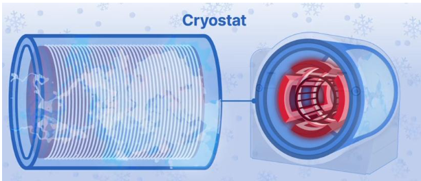
What is the cryostat?
The cryostat is the insulated vessel surrounding:
the superconducting coils,
the helium reservoir.
Its purpose is to:
minimise heat transfer,
keep the coils extremely cold,
reduce helium boil-off,
maintain superconductivity.
It achieves this using:
vacuum insulation,
thermal shields,
specialised cooling systems.
How is a superconducting MRI magnet energised ("ramped up")?
During installation:
The superconducting coil is cooled inside the cryostat.
Electrical current is introduced into the coil.
The magnet is gradually ramped up to the desired field strength (e.g. 1.5 T or 3 T).
Once the target field is reached, the current continues circulating within the superconducting loop.
The scanner then uses gradients and RF coils to produce images.
What is persistent mode?
After ramping:
the superconducting coil forms a closed electrical loop,
the external power supply is disconnected,
the current continues circulating because there is essentially no electrical resistance.
This is known as persistent mode.
It allows the magnetic field to remain stable without continuously supplying electrical power.
Why is a superconducting MRI magnet described as "always on"?
Even when:
no patient is being scanned,
no gradient noises are heard,
imaging has stopped,
the superconducting current continues circulating in persistent mode.
Therefore:
the magnetic field remains present continuously,
the magnet remains energised until deliberately ramped down or quenched.
This is why MRI safety rules apply at all times.
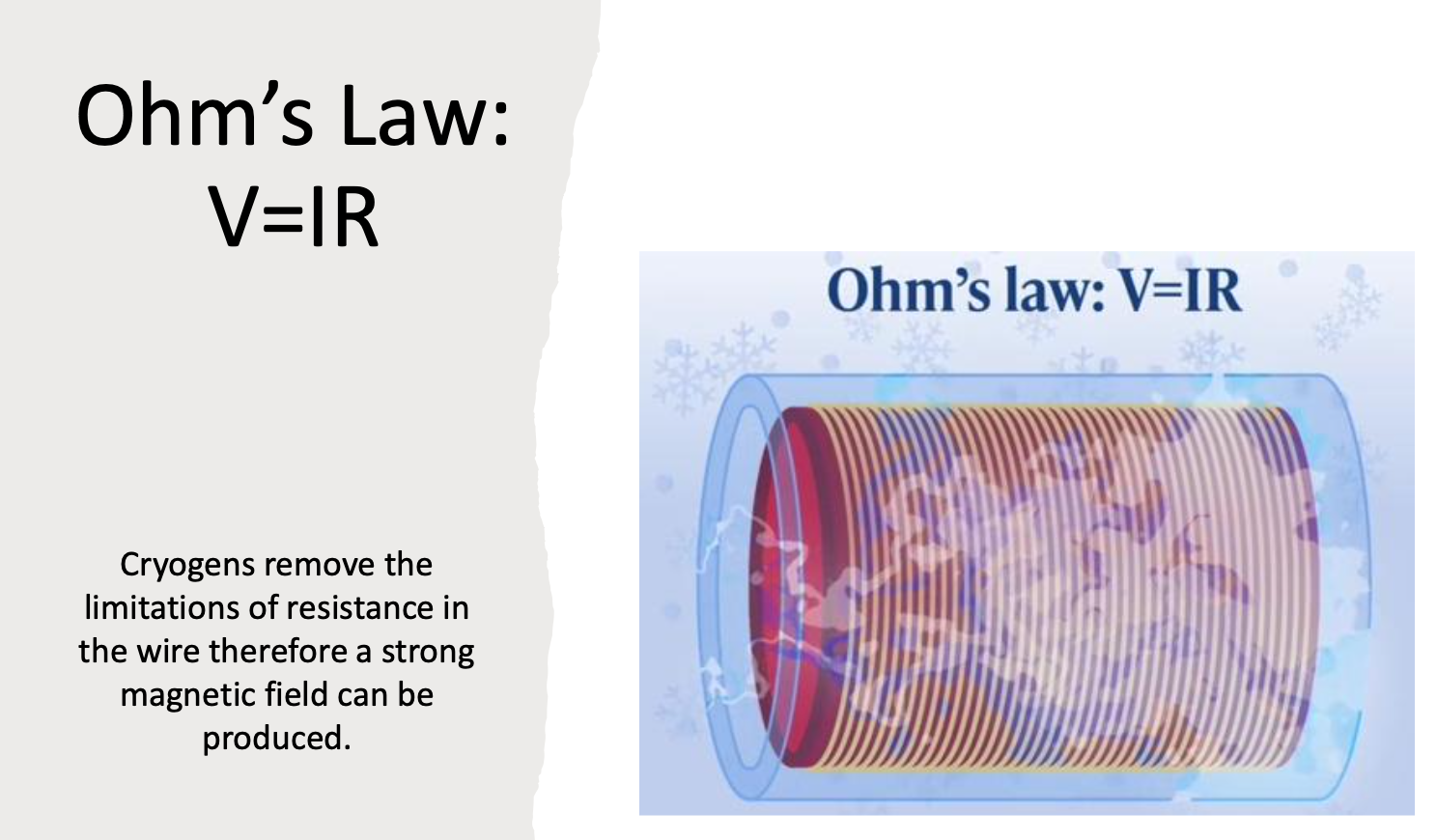
How does Ohm's law explain the advantages of superconducting MRI?
The lecturer emphasises the importance of Ohm's law.
In a normal conductor:
electrical resistance opposes current,
large currents produce large amounts of heat,
continuous electrical power is required.
In a superconducting conductor:
resistance is essentially zero,
almost no heat is generated,
large currents circulate efficiently,
much stronger magnetic fields become possible than with resistive magnets.
Why can superconducting MRI scanners achieve much higher Tesla strengths than permanent or resistive magnets?
Superconducting MRI scanners can produce very high magnetic field strengths because:
the superconducting wire has essentially zero electrical resistance,
very large electrical currents can circulate continuously,
virtually no energy is lost as heat,
continuous high-power electrical input is unnecessary once persistent mode is reached.
This overcomes the major limitation of resistive magnets, where electrical resistance generates excessive heat.
What is a quench in MRI?
A quench is the sudden loss of superconductivity within the MRI magnet.
During a quench:
the superconducting wire rapidly becomes a normal conductor,
electrical resistance suddenly returns,
the circulating current stops,
the magnetic field rapidly collapses,
a large amount of stored magnetic energy is released as heat.
This heat causes the liquid helium to boil extremely rapidly into helium gas.
What causes an MRI magnet to quench?
A quench occurs when the superconducting wire rises above its critical temperature.
Possible causes include:
equipment malfunction,
failure of the cooling system,
damage to the superconducting coil,
emergency manual quench,
accidental warming of the magnet.
Once the wire is no longer sufficiently cold, superconductivity is lost.
What happens during an MRI quench?
The sequence of events during a quench is:
Superconductivity is lost.
Electrical resistance suddenly appears.
Electrical current stops circulating.
The magnetic field collapses.
Stored magnetic energy becomes heat.
Liquid helium boils rapidly.
Large volumes of helium gas are produced.
The gas is vented outside the building through the quench pipe.
This process protects patients and staff from excessive pressure inside the scanner room.
Why does liquid helium boil during a quench?
When superconductivity is lost:
electrical resistance returns,
large amounts of heat are generated almost instantly,
the heat is transferred to the liquid helium,
the helium rapidly changes from liquid to gas.
The lecturer explains that this phase change happens very quickly because helium has an extremely low boiling point.
Why is a quench pipe essential?
The quench pipe safely removes helium gas from the MRI room.
Without a functioning quench pipe:
helium gas would fill the scan room,
oxygen concentration could fall,
there would be a serious risk of asphyxiation,
visibility could decrease due to condensation,
pressure inside the room could increase.
For this reason, every superconducting MRI installation includes a quench vent.
When might an MRI magnet be intentionally quenched?
Although quenching is generally avoided because it is expensive, it may be performed in an emergency.
Examples include:
a life-threatening projectile incident,
a patient trapped by a ferromagnetic object,
severe equipment failure,
situations where rapidly removing the magnetic field is essential for safety.
The lecturer notes that emergency quenching is uncommon because of the cost and disruption involved.
Why is an MRI quench expensive?
A quench is costly because:
large quantities of liquid helium are lost,
the magnet must be cooled again,
new helium must be supplied,
engineers must inspect the scanner,
the magnet must be re-ramped,
clinical service is interrupted.
Helium itself is expensive, making unnecessary quenches highly undesirable.
What is the difference between a "wet" magnet and a "dry" magnet?
Wet magnet
Contains a large reservoir of liquid helium.
Requires ongoing helium management.
Traditional superconducting design.
Dry magnet
Uses a much smaller quantity of helium.
Employs advanced cryocooling technology.
Greatly reduces helium consumption.
Lower maintenance requirements.
The lecturer explains that many modern MRI systems are now designed as dry magnets.
Why were dry MRI magnets developed?
Dry magnets were introduced because:
helium is expensive,
helium is a limited natural resource,
worldwide helium shortages have occurred,
reducing helium consumption lowers operating costs,
modern cooling technology allows magnets to operate with much less helium.
How do dry MRI magnets stay cold?
Instead of relying on a large bath of liquid helium, dry magnets use:
a small sealed amount of helium,
cryocoolers (refrigeration systems),
efficient thermal insulation.
The cryocoolers continuously remove heat, allowing the helium to remain cold enough for superconductivity.
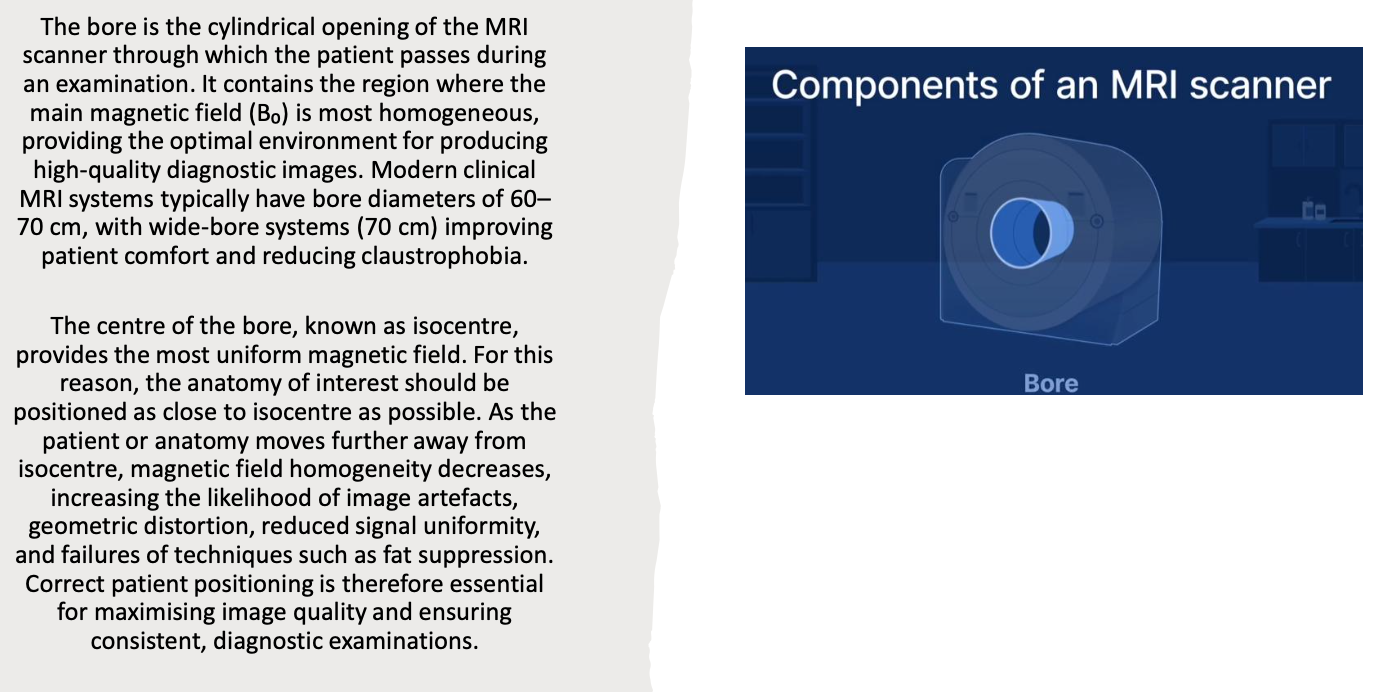
What is the MRI bore?
The bore is the opening through the centre of the MRI magnet where:
the patient lies,
the patient table moves,
imaging occurs.
Modern scanners generally have bores approximately 70 cm in diameter, although wider bores are available.
What is the MRI isocentre?
The isocentre is the exact centre of the MRI magnet.
It is important because:
the magnetic field is most homogeneous here,
gradients are most accurate,
image quality is highest,
patients should be positioned so the anatomy of interest is as close to the isocentre as possible.
Correct positioning is essential for optimal imaging.
Why is patient positioning at the isocentre important?
Placing the anatomy of interest at the isocentre:
maximises magnetic field uniformity,
improves signal quality,
reduces image distortion,
improves gradient accuracy,
produces the highest image quality.
Poor positioning away from the isocentre can reduce image quality.
What is the main magnet (B₀)?
The main magnet generates the static magnetic field known as B₀.
Its functions are to:
align hydrogen nuclei,
create net magnetisation,
provide the magnetic environment required for MRI,
establish the Larmor frequency,
enable excitation by RF pulses.
Without B₀, MRI would not be possible.
What are the characteristics of the MRI main magnetic field (B₀)?
The main magnetic field:
is static (does not change during imaging),
is very strong,
is highly homogeneous within the imaging volume,
remains present continuously in superconducting MRI systems,
provides the foundation for all MRI signal generation.
The lecturer emphasises that every other MRI hardware component (gradients, RF coils, shim systems, and computer processing) depends on the presence of a stable and uniform B₀ field.
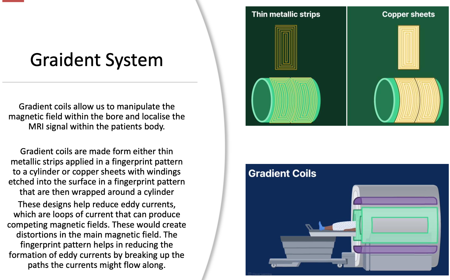
What are gradient coils?
Gradient coils are electromagnets located inside the main magnet that temporarily alter the strength of the main magnetic field (B₀).
Unlike the main magnet:
they can be rapidly switched on and off,
they produce small changes in the magnetic field,
they are essential for determining where the MRI signal originates.
Without gradients, MRI could detect a signal but could not determine its location within the body.
Why are gradient coils necessary in MRI?
The main magnetic field (B₀) is intentionally very uniform.
Because every hydrogen nucleus experiences almost the same magnetic field:
they all resonate at approximately the same Larmor frequency,
the scanner cannot identify where individual signals originate.
Gradient coils solve this problem by creating controlled changes in magnetic field strength across the patient, allowing spatial localisation.
How do gradient coils localise MRI signals?
When a gradient is applied:
one end of the patient experiences a slightly stronger magnetic field,
the opposite end experiences a slightly weaker magnetic field,
hydrogen nuclei therefore precess at different frequencies depending on their location.
The scanner can then determine where each signal originated.
This process is called spatial encoding or spatial localisation.
What are the three gradient coils in an MRI scanner?
MRI scanners contain three independent gradient coils, each acting along a different axis:
X-gradient – left to right
Y-gradient – anterior to posterior
Z-gradient – head to foot (superior to inferior)
Each gradient can be activated independently or in combination to localise MRI signals in three dimensions.
What is slice selection?
Slice selection is the first stage of spatial encoding.
During slice selection:
A gradient is applied.
The magnetic field varies across the patient.
Only hydrogen nuclei within a specific location resonate at the RF pulse frequency.
Only those nuclei are excited.
As a result, only the selected slice produces an MRI signal.
How does slice selection work?
The process is:
A slice-selection gradient is switched on.
Different body positions experience different magnetic field strengths.
Different magnetic field strengths produce different Larmor frequencies.
An RF pulse with a specific frequency is transmitted.
Only nuclei with the matching Larmor frequency absorb energy.
Only that slice is excited and later produces signal.
This allows MRI to image one slice at a time.
What determines slice thickness?
Slice thickness depends on:
the bandwidth of the RF pulse,
the strength of the slice-selection gradient.
General relationships:
Stronger gradient → thinner slice
Weaker gradient → thicker slice
Broader RF bandwidth → thicker slice
Narrower RF bandwidth → thinner slice
The scanner adjusts these parameters to achieve the desired slice thickness.
What is frequency encoding?
Frequency encoding is the second stage of spatial localisation.
During frequency encoding:
a gradient is applied while the MRI signal is being received,
nuclei in different positions precess at different frequencies,
the scanner identifies position by analysing these frequencies.
Frequency encoding localises signal along one axis of the image.
How does frequency encoding identify position?
When the frequency-encoding gradient is active:
stronger magnetic fields produce higher Larmor frequencies,
weaker magnetic fields produce lower Larmor frequencies,
every location has a unique frequency.
The computer analyses these frequencies to determine where each signal originated.
What is phase encoding?
Phase encoding is the third stage of spatial localisation.
Unlike frequency encoding:
the gradient is switched on only briefly,
hydrogen nuclei temporarily change their precession speed,
when the gradient is switched off, they return to the same frequency,
however, they retain different phase positions.
The scanner uses these phase differences to determine location along the second imaging axis.
How does phase encoding differ from frequency encoding?
Frequency Encoding
Gradient remains on during signal acquisition.
Position determined by frequency differences.
Phase Encoding
Gradient applied briefly before signal acquisition.
Position determined by phase differences.
Frequency returns to normal after the gradient is removed.
Both are essential for producing a two-dimensional image.
What is spatial encoding?
Spatial encoding is the process of determining exactly where each MRI signal originates.
It requires three steps:
Slice selection
Frequency encoding
Phase encoding
Together these allow the scanner to assign every signal to its correct location within the image.
What is gradient strength?
Gradient strength describes how much the magnetic field changes over a given distance.
Higher gradient strength means:
greater change in magnetic field,
larger frequency differences,
more precise localisation,
thinner slices,
higher spatial resolution.
Gradient strength is commonly measured in mT/m (millitesla per metre).
What is gradient slew rate?
The slew rate is the speed at which a gradient reaches its maximum strength.
It is usually measured in:
T/m/s (Tesla per metre per second).
Higher slew rates allow:
faster imaging,
quicker switching between gradients,
more advanced pulse sequences,
shorter scan times.
However, rapid switching also contributes to MRI acoustic noise.
Why do MRI scanners make loud knocking sounds?
he loud knocking noises are produced by the gradient coils.
When gradients rapidly switch on and off:
electrical current changes rapidly,
strong magnetic forces act on the gradient coils,
the coils vibrate,
vibrations are transmitted to the scanner housing,
these vibrations create the characteristic knocking and banging sounds heard during MRI examinations.
This is why patients wear hearing protection during MRI scans.
What are RF coils?
Radiofrequency (RF) coils are specialised antennas used to transmit and/or receive radiofrequency energy during MRI.
Their functions are to:
transmit RF energy into the patient,
excite hydrogen nuclei,
receive the RF signal emitted during relaxation,
convert RF energy into electrical signals,
send these signals to the computer for image reconstruction.
Without RF coils, MRI images cannot be produced.
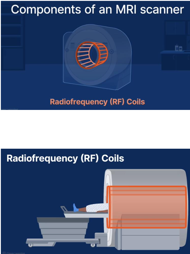
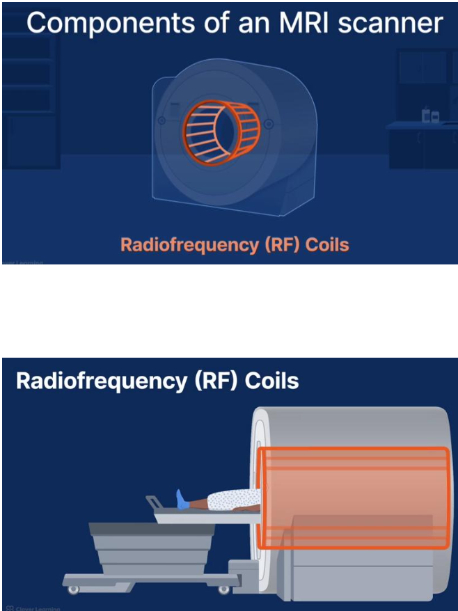
Where are RF coils located within the MRI scanner?
RF coils are positioned inside the bore of the MRI scanner, closer to the patient than the gradient coils.
The arrangement from outside to inside is generally:
Main superconducting magnet
Gradient coils
RF body coil
Patient
Additional specialised RF coils may be placed directly on or around the anatomy being imaged.
What are the two main functions of RF coils?
RF coils perform two essential roles:
1. Transmission
Produce the B₁ radiofrequency field.
Deliver RF pulses at the Larmor frequency.
Excite hydrogen nuclei.
2. Reception
Detect RF signals emitted during relaxation.
Convert these signals into electrical signals.
Send the signals to the computer for image formation.
Some coils perform both functions, while others are receive-only.
What is a transmit RF coil?
A transmit RF coil produces the B₁ magnetic field.
Its purpose is to:
generate the RF pulse,
excite hydrogen nuclei,
tip the net magnetisation away from B₀,
initiate MRI signal production.
Without RF transmission, no hydrogen nuclei would be excited.
What is a receive RF coil?
A receive RF coil detects the weak RF energy released by relaxing hydrogen nuclei.
It:
receives the MR signal,
converts RF energy into electrical current,
sends this information to the MRI computer,
determines much of the final image quality.
The received signal is extremely small, making coil sensitivity very important.
Can one RF coil both transmit and receive?
Yes.
Some RF coils perform both functions.
These coils:
transmit the RF pulse,
receive the returning MR signal,
are known as transmit/receive coils.
Other systems use separate transmit and receive coils depending on the examination.
What is a volume coil?
A volume coil surrounds the anatomy being examined.
It provides:
uniform RF transmission,
uniform RF reception,
homogeneous signal throughout a relatively large volume.
Examples include:
body coil,
head coil,
knee volume coil.
Volume coils are commonly used when imaging larger anatomical regions.
What are the advantages of volume coils?
Advantages include:
Uniform RF field.
Excellent signal uniformity.
Good image homogeneity.
Ability to image an entire anatomical region.
Suitable for transmission and reception.
These characteristics make volume coils ideal for routine MRI examinations.
What is a surface coil?
A surface coil is a small receive coil placed directly over the anatomy of interest.
Unlike a volume coil:
it covers only a limited area,
it is positioned very close to the patient,
it provides much higher signal close to the coil.
Examples include:
wrist coil,
ankle coil,
shoulder coil,
temporomandibular joint (TMJ) coil.
Why do surface coils produce higher signal-to-noise ratio (SNR)?
Surface coils are positioned very close to the anatomy.
Because the signal travels only a short distance:
more signal is detected,
less signal is lost,
noise remains relatively unchanged,
signal-to-noise ratio increases.
The closer the anatomy is to the coil, the stronger the received MR signal.
What are the disadvantages of surface coils?
Although surface coils provide excellent SNR, they have limitations:
Small field of view.
Limited depth of signal penetration.
Signal decreases rapidly with increasing distance from the coil.
Non-uniform signal across larger anatomical regions.
They are therefore unsuitable for imaging large body areas.
How do volume coils and surface coils compare?
Volume Coil | Surface Coil |
|---|---|
Surrounds anatomy | Placed on surface |
Uniform signal | Highest signal near coil |
Larger field of view | Small field of view |
Lower SNR | Higher SNR |
Better homogeneity | Less uniform signal |
Images large regions | Best for small structures |
The choice depends on the anatomical region and the clinical question.