respiration
 Call Kai
 Learn
Learn Practice Test
 Spaced Repetition
 Match
Match Flashcards
 Knowt Play
1/70

There's no tags or description
Looks like no tags are added yet.
Name | Mastery | Learn | Test | Matching | Spaced | Call with Kai | Chat |
|---|
No analytics yet
Send a link to your students to track their progress
71 Terms
types of respiration
external and internal (cellular respiration)
external respiration
ventilation or breathing: exchange of air between atmosphere and lungs
exchange of O2 & CO2 beteen lungs and blood
- transport of O2 and CO2 by blood
exchange of O2 and CO2 beteen blood and cells
internal or cellular respiration
intracellular reaction of O2 ith nutrients to produce CO2, ater and energy
functions of the respiratory system
regulation of blood pH
additional route for water loss and heat elimination (not as effective in humans)
enhances venous return via respiratory pump
enables vocalisation and smell
defends against inhaled foreign matter
CO2 content
more concerned about CO2 homeostasis rather than O2
as it determines blood pH, CO2 generates H+ in the blood
changes in pH are disastrous for cells
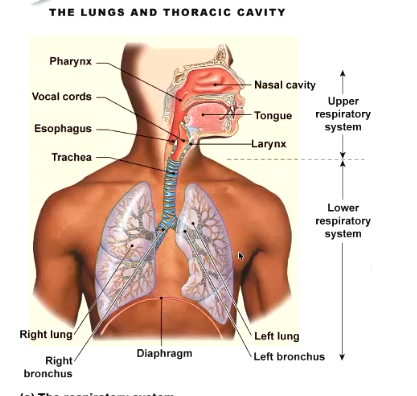
the parts of the respiratory system
upper (everything in the head) and lower tract
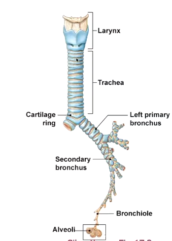
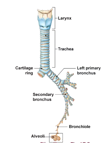
branching of airays
branch in a fractal pattern
every alveolus is equidistant from the trachea
why is every alveolus equidistant from the trachea?
ensures they all have the same resistance and thus inflate equally
if they weren’t equidistant, some wouldn’t inflate during quiet breathing
hat affects resistance?
length of vessel
diameter of vessel
viscosity
viscosity of air
increased by humidity
cartilage of the airays
blue, asthma usually involves constriction down at the levels of the bronchioles here there is smooth muscle. cartilage keeps the airways open everyhere else
total cross sectional area
greatest at the alveoli
resistance decreases as e move through the system
here all the exchange takes place
hat keeps the lung inflated?
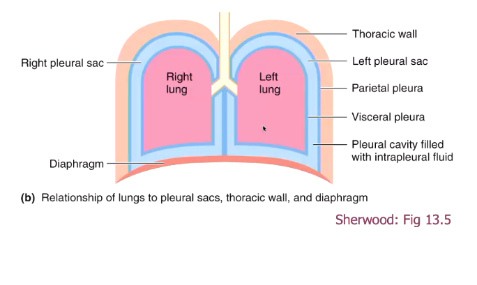
pleural sac attaches the lung to the thoracic all (prevents lung from collapsing hen e exhale)
pressure differences
pleural sac
to membrane structure surrounding the lungs,
inner membrane attached to lungs, outer membrane attached to thoracic all
filled ith fluid

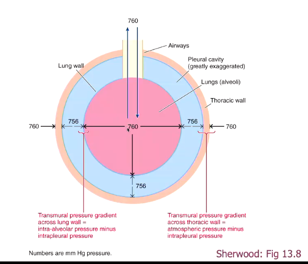
pressure difference beteen lung and pleural sac
for the lung to remain inflated, the pressure in the pleural sac must be less than the pressure ithin the lung
atmospheric pressure at sea level is 760 mm Hg, hich is the same as the pressure of the lungs. pressure in the pleural sac is 756 Hg mm
transmural pressure gradient
pressure gradient across the chest all
if the pressure becomes loer in the lungs, air ill rush into the lungs
pneumothorax
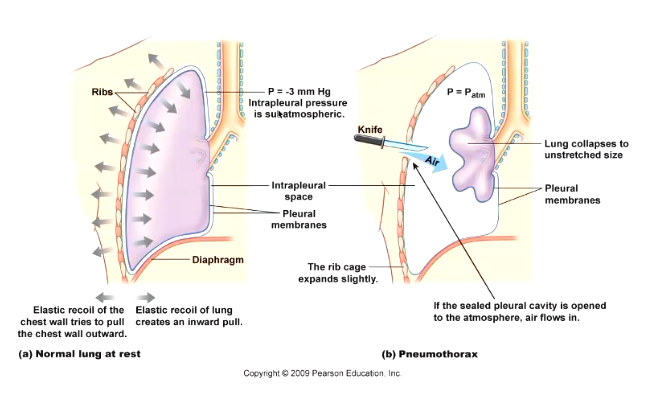
disruption of pleural space can lead to collapsed lung
if air enters the fluid layer, it loses its pressure and the lung collapses inwards
can occur in lung diseases here inflammation causes blisters in the pleural sac, if one blister ruptures it disrupts the membrane
tension pneumothorax
hen air entering the pleural space cannot exit so every time you breathe in the pressure gets higher and higher
pressure on organs and heart, compress the heart
need to pull the air out of the pleural space
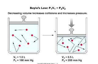
Boyle’s la
if pressure is halved, volume must be doubled
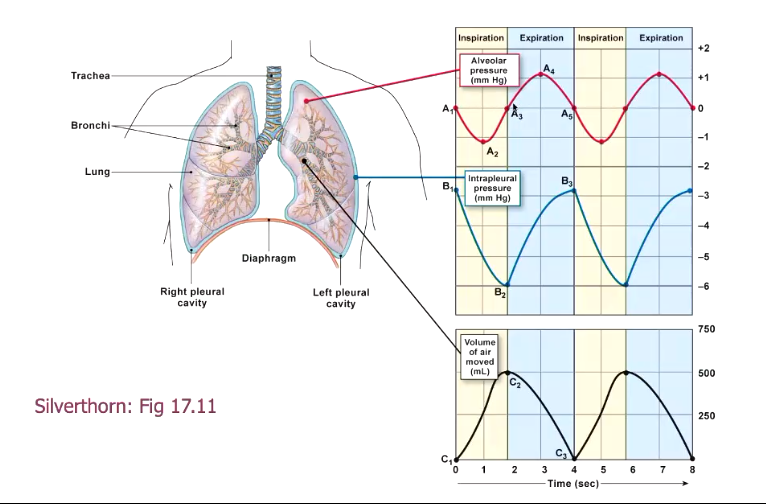
pressure changes during inhalation
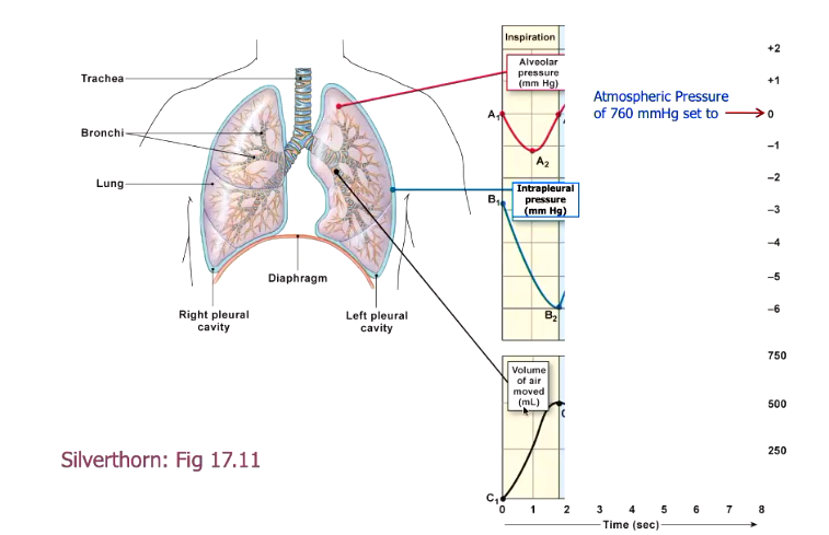
the volume of the lungs increases, decreasing the pressure
intra-alveolar pressure decreases
intrapleural pressure decreases (as the chest all is expanding, this pulls the intrapleural space ith it, increasing volume)
air flows into the lungs

graph of pressure changes during inspiration
V shape in alveolar pressure is due to initial drop in pressure when you first breathe in, but as air moves in, it re-equilibrates ith atmospheric pressure
pressure changes during expiraton
lung volume must decrease
intra-alveolar pressure must increase
intrapleural pressure must increase
graph of pressure changes during quiet breathing
muscles used for breathing
diaphragm
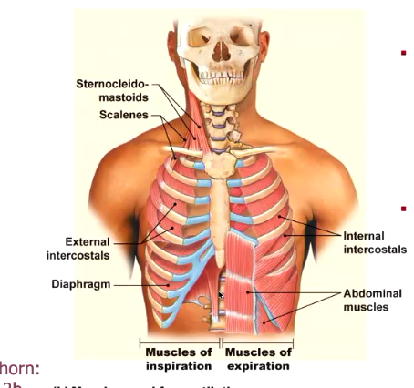
quiet breathing (stage 1 breathing)
should be able to breathe just using your diaphragm ithout the ribs expanding
stage 2 breathing
recruit external intercostals (pull ribs out)
stage 3 breathing
recruit sternocleidomastoids and scalenes hich lift ribs up and out
expiration
normally passive, occurs due to passive elastic recoil of lungs and thoracic cage, saves energy
muscles of expirationused only in forced breathing or voluntary exhalation, internal intercostals pull ribs in
reflex control of ventilation
chemoreceptors in the brain detects CO2 levels (primary mechanism)
other sensors detect O2 and pH
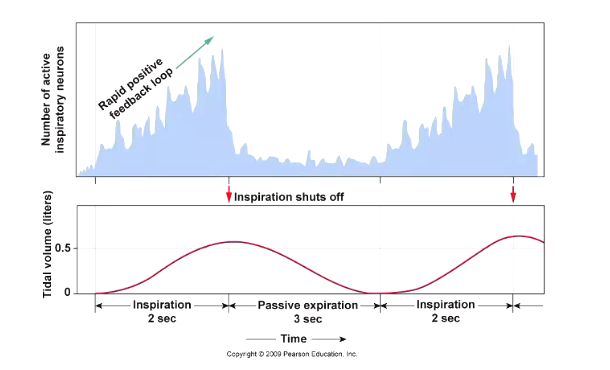
high CO2 triggers respiratory centres in the brainstem to trigger outputs to respiratory muscles
neural networks that control ventilation
higher brain centres
medullary chemoreceptors
other chemoreceptors and mechanoreceptors
very complex as you have to regulate depth vs rate, vocalising, eating and swallowing
neural activity cycles during quiet breathing
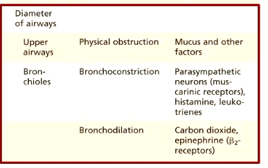
factors that affect airay resistance
upper airays more affected by physical obstruction because the cartilage keeps them open
resistance in bronchioles dependent on bronchoconstriction and bronchodilation
formula for resistance
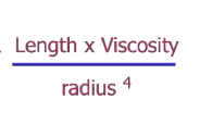
bronchoconstriction
parasympathetic neurons (muscarinic receptors)
hat affects the ork of breathing?
airay resistance (decreased airay diameter leads to increased airay resistance hich increases ork of breathing)
lung compliance
lung compliance
ability of lung to stretch
decreased lung compliance makes it harder to stretch the lungs, increasing ork of breathing
lung compliance vs elastance
lung compliance = ability of lung to stretch (inspiration)
lung elastance = ability of lung to spring back after being stretched (ho ell they can do passive expiration)
relationship between lung compliance and lung elastance
independent of each other
here is lung compliance affected?
decreased in diseases such as pulmonary fibrosis hen inelastic scar tissue is formed hich reduces the ability of alveoli to stretch
here is lung elastance affected?
decreased in diseases such as emphysema hen elastin fibres around alveoli are destroyed
ho does alveolar surface tension affect lung compliance?
surface tension due to hydrogen bonds beteen ater molecules hich are more attracted to each other than gases at air-fluid interface
attraction beteen ater molecules resists any force that increases its surface area, thus opposes expansion of alveolus creating surface tension
liquid surface area tends to shrink due to attraction beteen ater molecules, thus reducing alveolus size

law of LaPlace
the smaller the alveolus, the more inward pressure from the surface tension
to prevent smaller alveoli from collapsing:
surfactant
alveolar interdependence
surfactant
disrupt cohesive forces between water molecules and replace water at the surface, decreases surface tension and resistance of lung to stretch
surfactant is more concentrated in smaller alveoli, decreasing surface tension and pressure
produced by type 2 alveolar cells, higher proportion in smaller alveoli to make all the alveoli of the same compliance
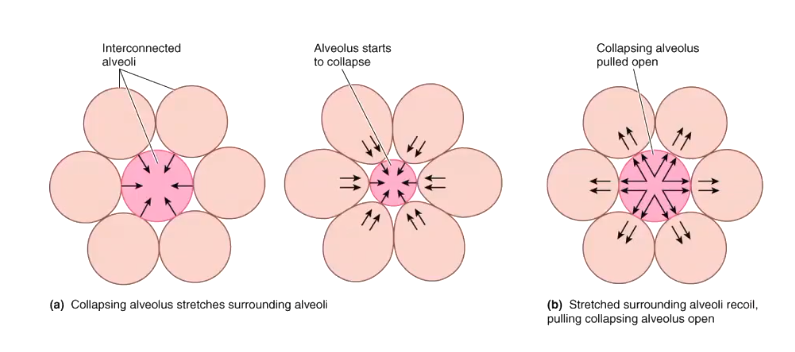
alveolar interdependence
the alveoli are all connected
if one collapses, the others start pulling it outards
types of respiratory diseases
obstructive
restrictive
obstructive lung diseases
narrowing of lower airways
increased airway resistance
more difficult to expire
issue ith the airays themselves which is reducing rate that you can move air, issue ith airflow
e.g. asthma, emphysema, chronic bronchitis, COPD
restrictive lung diseases
decrease in lung compliance, reduces volume you can breathe in as the lungs can’t stretch as well
more difficult to inflate lung
pulmonary fibrosis
spirometry
measurement of lung volumes

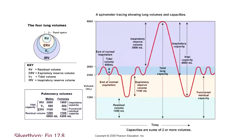
tidal volume
amount of air breathed in a normal quiet breath, around 500 mL.
less than 10% of the air in your lungs is exchanged in one breath (the capacity is 5800 mL)
left of graph

expiratory reserve
the breath that you can continue to breathe out after a quiet expiration
yellow
inspiratory reserve volume
extra amount that you can inhaleafter a normal inhalation
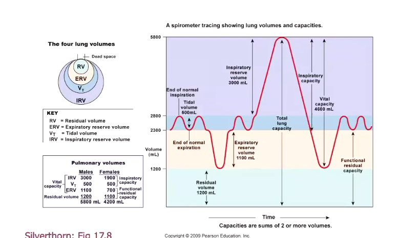
vital capacity
biggest breath you can take
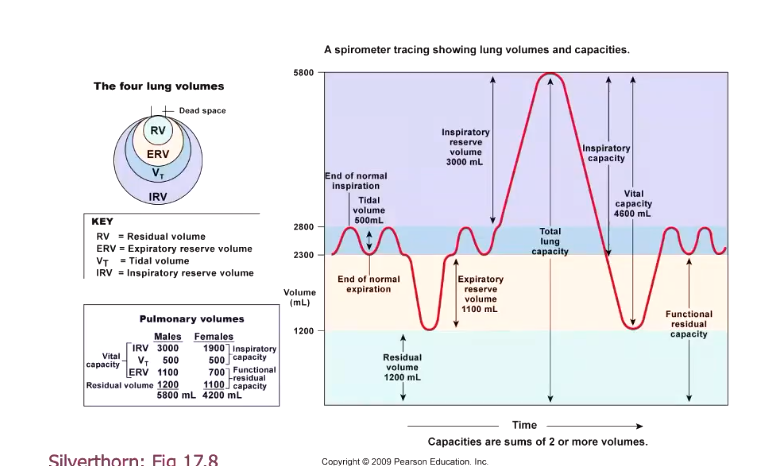
residual volume
you can breathe out all you ant, but your lungs are still held open by your rib cage
even when breathing our vital capacity, we do not exchange 100% of our breath, so the oxygen and carbon dioxide content ill never be equal to that of the atmosphere

can e measure residual volume?
yes, using the helium dilution method
fill the bell ith helium and oxygen, if you kno the intial concentration of helium and volume of the bell and the person breathes out of it for an extended period of time and the helium concentration equilibrates between their lung and the bell you can remeasure the concentration of helium and calculate the overall volume of the lung

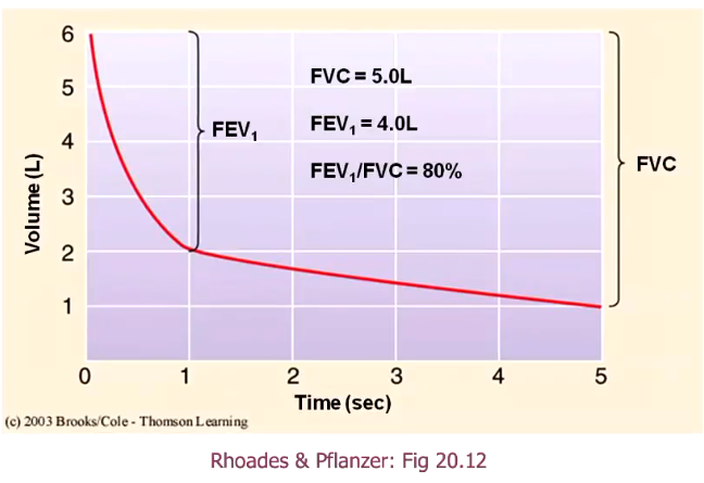
forced expiratory volume in one second (FEV1)
breathe all the ay in, and breathe all the air out as quickly as you can
measure what percentage of the air you exhaled in the first second
set up a FEV1/FVC ratio
FEV1 = how much you breathe out in the first second
FVC = total amount breathed out
80% is normal
if there’s something wrong, there is an issue with the airways rather than the lungs as it is an issue with airflow
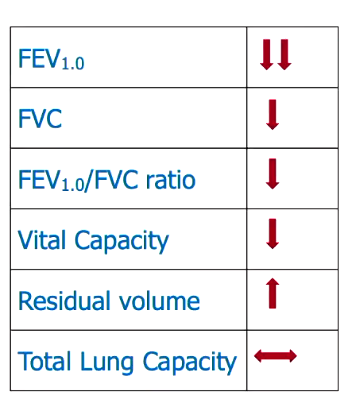
hallmarks of obstructive lung disease?
FEV1 and FEV1/FVC are affected as there is a flow issue
exhalation is usually more affected than inhalation, as exhalation causes increased pressure in the thoracic cage, so if your airays are already constricted to begin ith, this causes microcollapses of the airays hich traps air in the lungs
vital capacity drops, trapping air in the lungs as it is harder to exhale because of the microcollapses of the airays, this means that residual volume therefore increases
types of COPD
chronic bronchitis, asthma, emphysema
all cause inflammation of the airways, causing them to narrow
bronchitis has mucus
emphysema = elastance is decreased
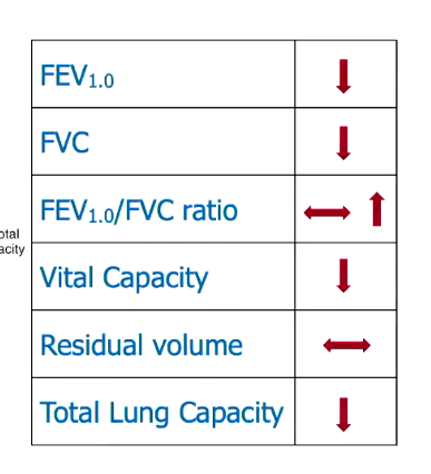
hallmarks of restrictive lung disease?
total lung capacity decreases, as residual volume can’t decrease, this decrease comes out of the vital capacity
therefore FVC also decreases, as does FEV1 as you have less air in total to be breathing out, so also less in the first second
as FVC drops so much, there might sometimes be an increase in FEV1/FVC ratio
reduction in surfactant
decreases compliance
artificial surfactant can be given to babies ho don’t produce surfactant
stimulate endocrine system to promote surfactant
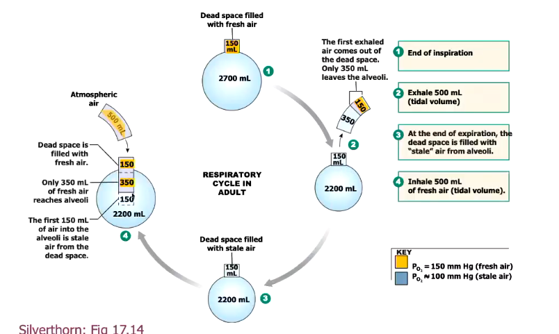
anatomic dead space
alveoli are the only sites in your lungs for air exchange
conducting airays knon as anatomical dead space as the air there doesn’t reach the interface for exchange
hen e breathe out, the atmospheric air gets kicked out, stale air fills the airays, hen e breathe in, e re-breathe in the stale air folloed by atmospheric air
this volume is around 150mL
as the total inhalation during quiet breathing is usually 500mL, the effective air moved is 350 mL
minute ventilation (Ve)
total amount of air breathed in a minute
tidal volume x respiratory rate
alveolar ventilation (Va)
better measure of effective rbeathing
(tidal breath - deadspace) x respiratory rate
air reaching alveoli every minute

is there a functional difference beteen these situations?
yes, an increase in alveolar ventilation is achieved more efficiently by increasing TV than frequency
normal breathing rate
500mL x 12 per minute, just uses diaphragm instead of other muscles such as intercostals, most efficient rate of breathing
usually results in pulmonary ventilation 6 L/min, alveolar ventilation 4.2 L/min
maximum voluntary ventilation
125-170 L/min
eupnea
normal quiet breathing
hyperpnea
increased respiratory rate and/or volume in response to increased metabolism e.g. during exercise
hyperventilation
increased respiratory rate or volume ithout increased metabolism, if you breathe off too much CO2 you become alkalotic
dyspnea
difficulty breathing
apnea
cessation of breathing
breathing through a tube underater
transmural pressure gradient: if pressure from ater is greater than air pressure breathed in, the lungs on’t be able to expand properly
in scuba diving, the air is pressurised to match the ater pressure
thinner tubes increase resistance
ider tubes increase dead space