musculoskeletal system
 Call Kai
 Learn
Learn Practice Test
 Spaced Repetition
 Match
Match Flashcards
 Knowt Play
1/114

There's no tags or description
Looks like no tags are added yet.
Name | Mastery | Learn | Test | Matching | Spaced | Call with Kai | Chat |
|---|
No analytics yet
Send a link to your students to track their progress
115 Terms
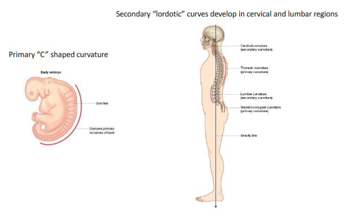
vertebral column curvatures
primary C-shaped curvature, primary curvatures are kyphotic
secondary “lordotic” curves develop in cervical and lumbar regions, in opposition to the C-curve
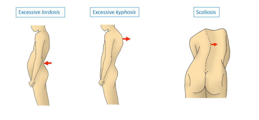
atypical curvatures
major anatomical landmarks of typical vertebra
vertebral body
vertebral arch
spinous & transverse processes
articular processes
vertebral body
eight bearing
articulate superiorly and inferiorly
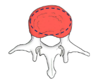
epiphyseal ring (dotted circle), helps ith attachment to intervertebral disk
vertebral arch
pedicles (1) + lamina (2)
forms vertebral foramen
protects spinal cord

spinous and transverse processes
levers for muscle attachment
levers for ligamentous attachment
spinous (in the middle), transverse (sides)
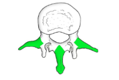
articular processes
projections (yello) ith articular facets (blue)
articulate superiorly (big yello) and inferiorly (small yello), hich connect beteen vertebra
blue: smooth surface for articulation, surface here the bones connect: articular facet.

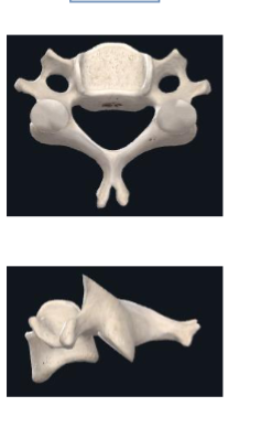
cervical vertebrae
transverse processes have extra holes
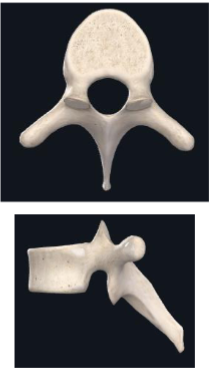
thoracic vertebrae
long spinous process hich projects donards
lumbar vertebrae
large vertebral body
thick spinous process

sacrum
fused vertebrae

vertebral foramen
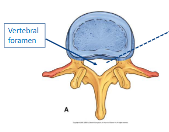
vertebral canal
multiple foramen in a ro form a canal
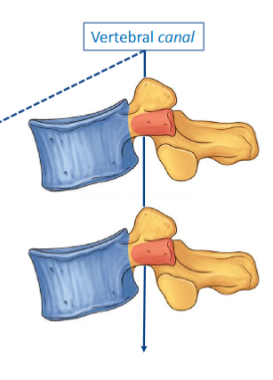
vertebral notch
incomplete hole
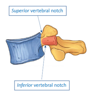
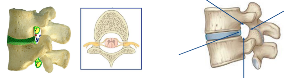
intervertebral foramen
a foramen created from to vertebrae stacked together
inferior notch of upper vertebra + superior notch of loer vertebra = foramen
spinal nerve roots leave through these foramen
intervertebral joints
vertebral body + disk + vertebral body
secondary cartilaginous joints (fibrocartilaginous)
disks in aging
loer is in a younger adult, here it appears more atery
disk dries out over time
disk bulges are more likely in younger population here the atery substance can move out and cause herniations

annulus fibrosus
attaches to epiphyseal ring
- keeps the vertebra together
concentric lamellae of collagen
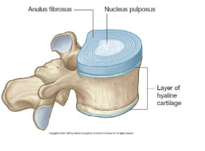
nucleus pulposus
pushes compressive force in all directions to spread the load, shock absorber
surrounded by annulus fibrosus
keeps the vertebra apart
gelatinous consistency, deforms but not compressible
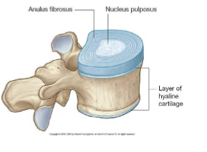
layers of annulus fibrosus
concentric lamellae of collagen
permits movement in all directions
resists excessive rotation
some layers are going in diffrent directions to limit tisting - if e tist too far, some layers ill get taught

zygapophyseal joints
plane synovial joints
movement dependent on the orientation of the articular surfaces involved, differs along the vertebral column
beteen superior and inferior articular processes hich have articular surfaces or “facets” on them
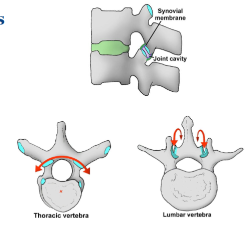
thoracic vs lumbar plane joints
thoracic more specialised for coronal movement
lumbar for sagittal movement (flexing and extending in the lumbar region)
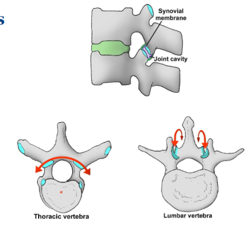
longitudinal ligaments
thick at the front, smaller at the back

interspinous ligament
beteen spinous processes

supraspinous ligament
superficial to spinous processes

ligamentum flavum
project from lamina
extra elastin tissue hich assists us in big movements

extrinsic muscles of the back
attach to and act on the upper limb
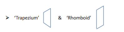
trapezium and rhomboid
major and minor
lattisimus (broadest) and dorsi (back)
levator (elevate) and scapulae (shoulder blade)
superficial
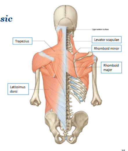
trapezium vs rhomboid
intrinsic muscles of the back
primarily attach to and act on the back
erector spinae
transversospinalis

erector spinae
prime movers of the back, particularly in the sagittal plane
concentrically extend the trunk
eccentrically control trunk flexion (e.g. sloly bending over)
commonly implicated ith postural issues
transversospinalis
deep to the erector spinae
short muscles
control more fine movements at vertebral segments
important in rehabilitation for improving smaller segmental movements
multifidus is the name of these muscles in the lumbar spine

bony framework of upper limb
attaches to anterior aspect of axial skeleton
distinct pattern of 1 proximal (arm) and 2 distal bones (forearm)
pentadactyl hand
thumb is rotated 90 degrees in relation to the hand
shoulder girdle
clavicle (collar bone)
scapula (shoulder blade)
clavicle
less smooth on the inferior vie as there are ligaments and muscles that attach
long bone
provides the only upper limb articulation between the appendicular and axial skeletons
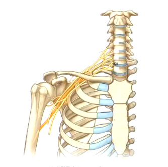
projects underlying neurovasculature
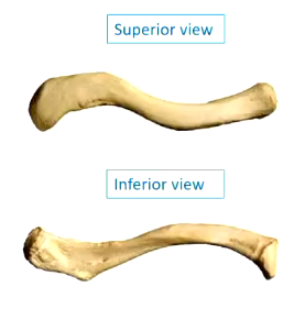
key features of the clavicle
sternal (one end to sternum) and acromial (one end to acromion) ends
distinct S curvature
roughened inferior surface
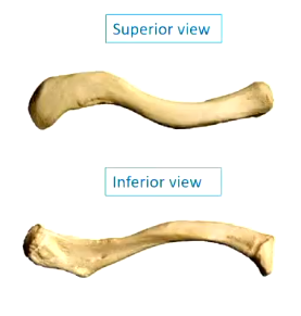
acromial end vs sternal end
acromial end is usually flatter (left), sternal end is more like a circle (right)
scapula
large and flat, good for muscles to anchor onto (significant muscle attachment)
flat bone
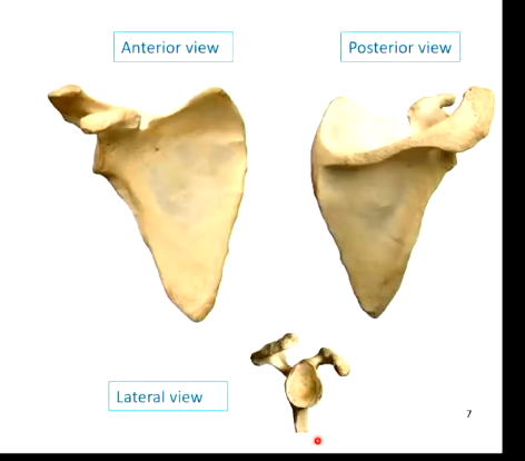
key features of the scapula
bony ridge is called the spine of the scapula
processes: acromion and coracoid
acromion is on the top right of the posterior view, top left of lateral view
coracoid is the other smaller bit that sticks out
fossae for muscle attachment
glenoid fossa
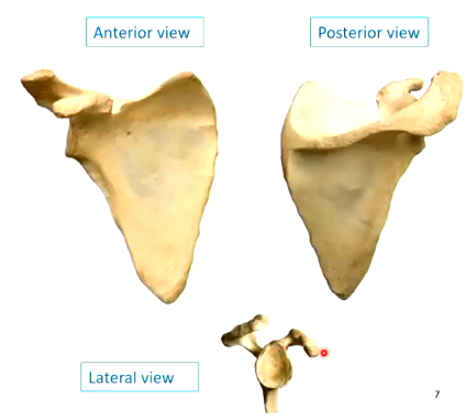
glenoid fossa
flat surface for bone attachment, here the humerous attaches
flat surface on lateral vie
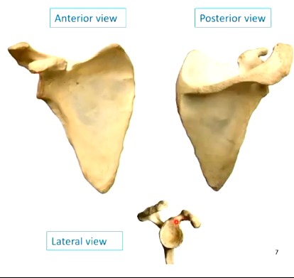
clavicular joints
sternoclavicular
acromioclavicular
sternoclavicular joint
rolling your shoulders hile keeping your humerous still
intra-articular disk
from clavicle to first rib: costoclavicular ligament
saddle joint: shrugging up and don to forwards and back is similar in ROM
strong capsule

why does the sternoclavicular joint have to be strong?
the only synovial joint that is connecting the appendicular skeleton to the axial skeleton
movement of the sternoclavicular joint
elevation and depression
pro- (shoulder pushing forards) and retraction (shoulders back, chest out)
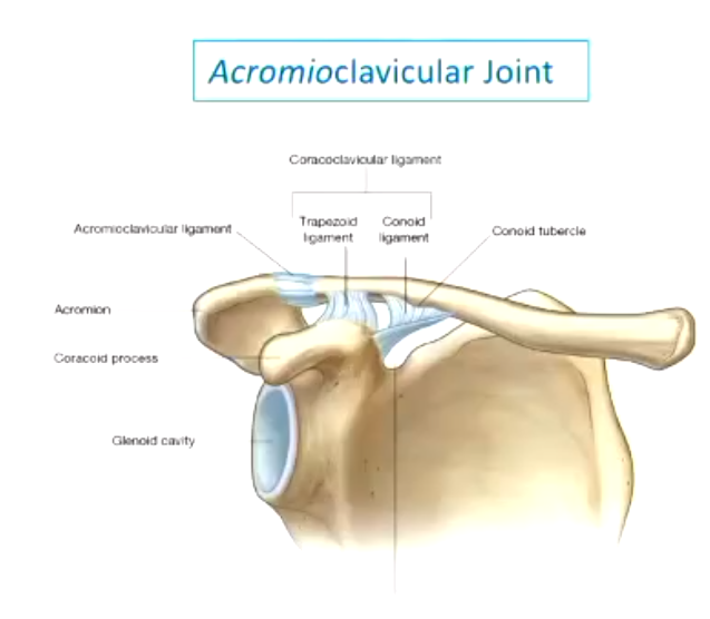
acromioclavicular joint
plane joint
strong coracoclavicular ligament but weak capsule
coracoclavicular ligament comes off the coracoid process and attaches to the inferior surface of the clavicle
acromioclavicular ligament is quite weak - injury often occurs here
neurovasculature near the clavicle
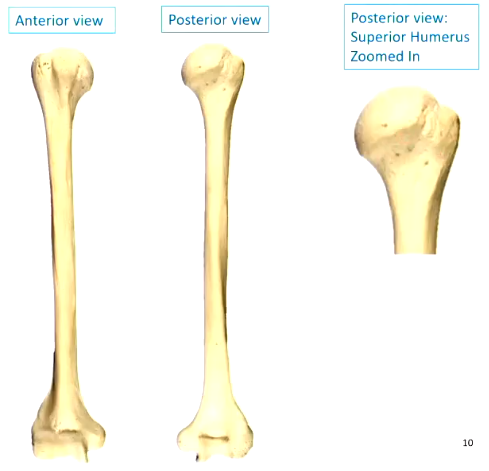
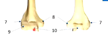
key features of the humerus
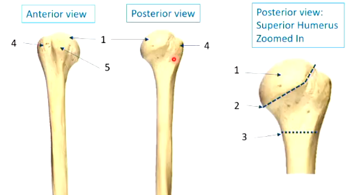
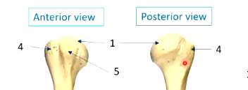
head (1)
neck - anatomical (2) and surgical (3)
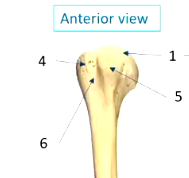
tubercles - greater (4) and lesser (5)
bicipital/intertubecular groove (6)
epicondyles - lateral (7) and medial (8)
condyles - capitulum (9) and trochlea (10)
head
smooth surface (1)
neck
anatomical = clear demarcated line in beteen the smooth and rough surface on the head (2)
surgical = here fractures normally happen (3)

tubercles
greater (4)
lesser (5)
sites for attachments of muscles
greater tubercule looks smaller but actually continues around the lateral aspect to the posterior aspect
bicipital/intertubecular groove
6
epicondyles
lateral (7) and medial (8)
muscle attachment

condyles
capitulum (9): lateral forearm bone attachment
trochlea (10): medial forearm bone attachment
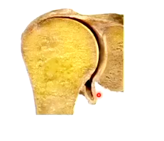
glenohumeral joint
triaxial
ball and socket
incongruent tissues - the articular surfaces don’t fit ell together
large humeral head + shallow glenoid fossa
loose joint capsule = unstable joint (loose bit toards the bottom extends hen you reach out to the side)
supportive features of the glenohumeral joint
specialised fibrocartilage
muscles
ligaments
muslces of the glenohumeral joint
rotator cuff (blend into the capsule)
long head of biceps brachii
ligaments of the glenohumeral joint
glenohumeral & coracohumeral (both blend into capsule)
fibrocartilage of the glenohumeral joint
glenoid labrum
extra rim - continues past the glenoid fossa to deepen the socket

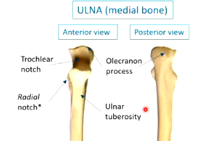
ulna and radius
ulna has a big divot here it connects to the trochlea
radius has a disk at the top, allowing rotation
long bones
connected via interosseous membrane

articulations of the ulna and radius
synovial joints ith humerous = flexion and extension
x2 synovial joints ith each other, one at each end = rotation
radius ith carpal bones (rist)
olecranon process

trochlear notch
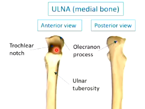
tuberosities of the ulna and radius
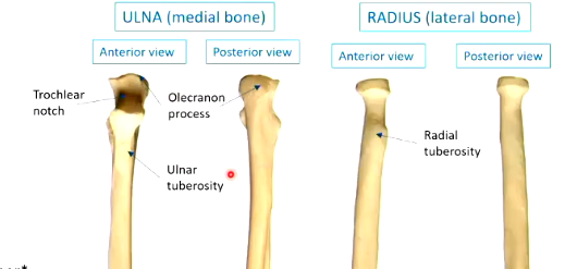
radial notch
for the radius to fit into on the ulna - head of the radius articulates here
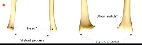
ulnar notch
here the ulna bone articulates on the radius

styloid process
bony projections
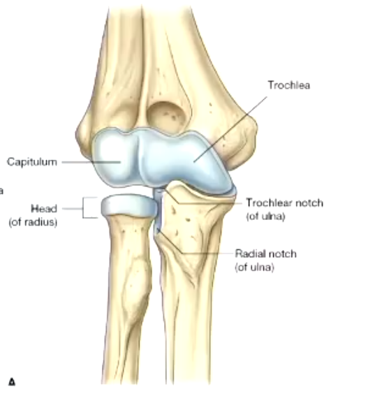
elbo joint
hinge joint
strong collateral ligament support to prevent medial and lateral movements
collateral = ligaments going don laterally
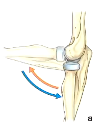
movement in the elbo joint
the ulnar bone is the primary bony contact, only hen e get into flexion the radius has a bit more articulation
humero-ulnar shares synovial cavity ith proximal radio-ulnar joint
proximal radioulnar joint
pivot joint
radial head —> radial notch on the ulna
annular ligament encircles the radial head

distal radioulnar joints
ulnar head —> ulnar notch on the radius
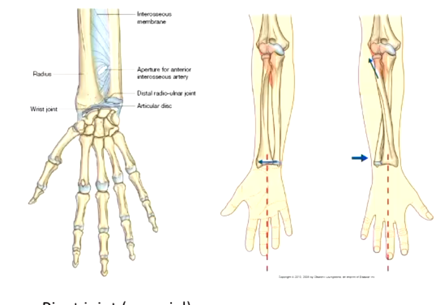
supernation
uncrosses the forearm bones, palm upards
pronation
crosses the forearm bones, palm donards
bone groups of the rist and hand
metacarpals and phalanges are long bones
carpals are short bones

sesamoid bone
encapsulated by tendons, e.g. patella, there are also some in the fingers
radiocarpal joint
ulna does not make a direct bony articulation, there is an articular disk in beteen
joint is the articulation beteen the radius and the lunate and scaphoid
condyloid
ulnar and radial deviation = movement of the rist in the coronal plane toards either of the bones
complex ligamentous support

scaphoid fractures
scaphoid has a unique blood supply, fractures can lead to bony necrosis
can occur from a fall on an outstretched hand (FOOSH)
joints of the hand
8 carpal bones and 5 metacarpal bones
thumb is 1st digit, pinky is 5th
intercarpal joints: here the carpals join ith each other, plane joints
carpometacarpal joints
carpometacarpal joints
1st CMC joint - saddle joint
vital for the unique functions of the thumb and the most degenerative joint in the body
unique movement: opposition - moving to touch the thumb to the pad of the pinky finger, specific muscle associated ith this movement
2nd-5th - plane joints
interphalangeal joints
only one on the first digit
2 each on the other digits (proximal and distal)
14 phalanges
hinge joints
commonly subluxated or dislocated

metacarpophalangeal joints
condyloid joints
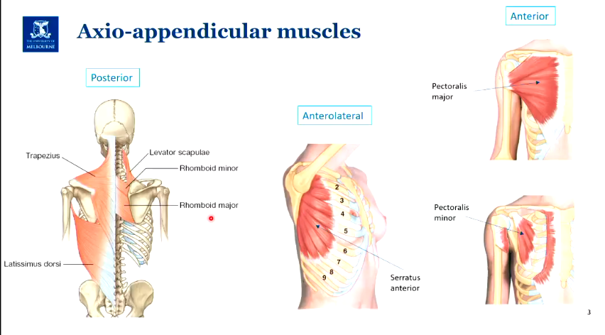
axio-appendicular muscles
includes the extrinsic back muscles
serratus anterior
pectoralis major (superficial, attaches to humerous) and minor (deeper, attaches to scapula)
scapulohumeral muscles
rotator cuff muscles
deltoid and teres major
rotator cuff muscles
attach to the head of the humerus, to the greater and lesser tubercules, rapping around the glenohumeral joint for support
supraspinatus (above the spine of the scapula)
infraspinatus
teres minor
subscapularis
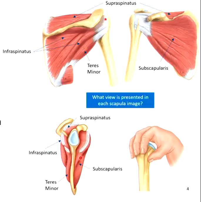
ho do the rotator cuff muscles relate to the joint capsule?
they blend ith the joint capsule to compress the humeral head into the glenoid fossa
movements of the rotator cuff muscles
abduction (supraspinatus)
external rotation (infraspinatus and teres minor)
internal rotation (subscapularis)
deltoid and teres major
attach more distally on the humerus than the rotator cuff

anterior vie of the deltoid and teres major

deltoid
triangle
broad fan from humerus to the scapula and clavicle
produces shoulder flexion, abduction and extension
teres major
larger than teres minor
attaches more anteriorly to the humerus
internal rotation at the shoulder
upper limb compartments and deep fascia
muscles are separated into compartments largely by intermuscular septa
deep fascia can be specialised
intermuscular septa
commonalities beteen muscles ithin these compartments: share a nerve, collective movements
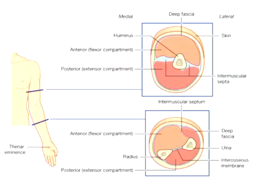
specialised deep fascia
interosseous membrane: in beteen bone
retinacula: hold don tendons, creates pulley system
aponeurosis: broad sheet of connective tissue

arm anterior compartment
biceps brachii (long head and short head)
coracobrachialis (connects to coracoid process)
brachialis
all cross over the anterior aspect of at least one joint and produce flexion
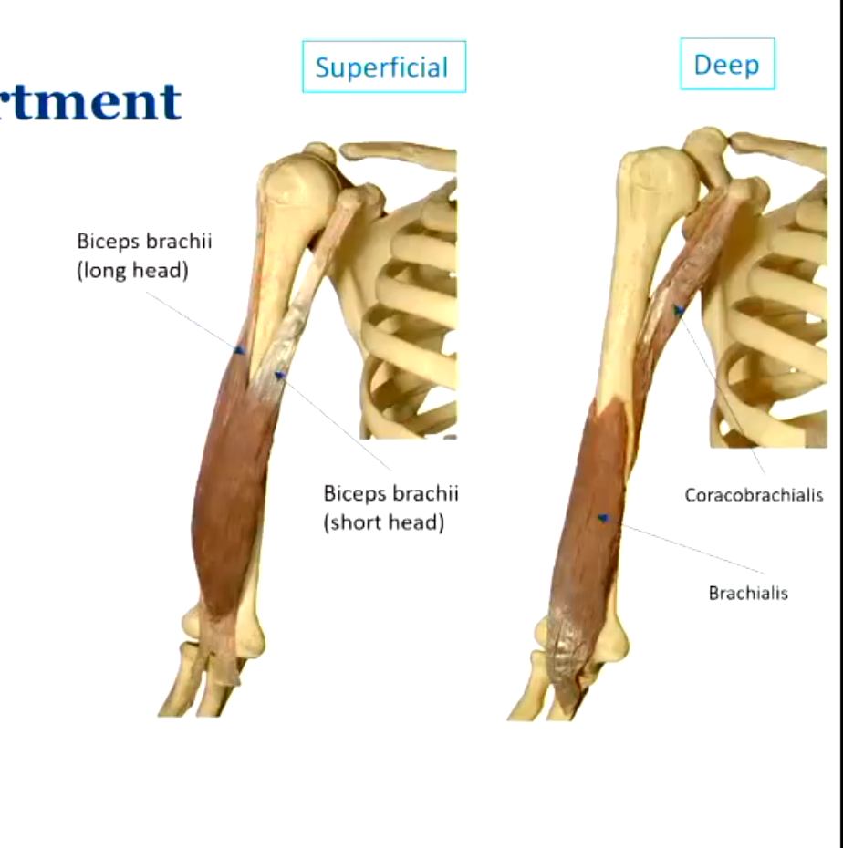
long head of biceps brachii
tendon enters the glenohumeroid joint
hat muscle is responsible for supination?
the biceps brachii as it attaches to the radius
brachialis
just causes flexion and extension
arm posterior compartment
triceps brachii
triceps brachii
all aspects of the triceps brachii cross over the posterior aspect of at least one joint (the elbo) to attach onto the olecranon process and produce extension
if it crosses the back of the shoulder, it causes shoulder extension, if it crosses the back of the elbo, it causes elbo extension

forearm muscles - anterior compartment
superficial, intermediate and deep
the medial epicondyle of the humerus provides attachment for the superficial and intermediate muscle groups
if the muscle attaches to the carpals/metacarpals = rist flexor
attaches to digits = finger flexor
distal attachment into radius = pronator

forearm muscles - posterior compartment
musculature attaches to lateral epicondyle of the humerus

grip strength
extensors allo rist flexors to be at a good length to contract (not too shortened)