IPFC 1 - Anemia
 Call Kai
 Learn
Learn Practice Test
 Spaced Repetition
 Match
Match Flashcards
 Knowt Play
1/231

There's no tags or description
Looks like no tags are added yet.
Name | Mastery | Learn | Test | Matching | Spaced | Call with Kai | Chat |
|---|
No analytics yet
Send a link to your students to track their progress
232 Terms
Transferrin is a ____________ acute phase protein while ferritin is a ________________ acute phase protein
Transferrin is a negative acute phase protein (decreases under a state of inflammation, trauma or infection)
while ferritin is a positive acute phase protein (increases under a state of inflammation, trauma or infection)
What is anemia?
• When blood has a reduced capacity to deliver oxygen
Most common lab values indicating anemia
Reduced amount of Hematocrit, Hemoglobin, or RBC count
Anemia Triad
- inadequate production of normal RBCs
- Blood loss
- RBC destruction
What conditions can lead to the anemia triad?
• Heavy periods (blood loss)
• Ulcers (blood loss)
• Surgery (blood loss)
• A diet low in iron, folic acid, vitamin B12 (inadequate RBC production)
• Sickle cell anemia (inadequate production of normal RBCs)
• Lack of erythropoietin (inadequate RBC production)
• Pregnancy (blood ‘loss’, inadequate production)
• Kidney disease requiring dialysis (RBC destruction)
• Hemolytic anemia (RBC destruction)
Acute anemia symptoms
– Tachycardia (fast heart rate), palpitations
– Hypotension, lightheadedness
– Dyspnea (shortness of breath)
Chronic anemia symptoms
- Weakness, fatigue
- Headache, vertigo, faintness
- Sensitivity to cold, pallor, loss of skin tone
What symptoms can occur when Hgb falls to 90 g/L or less
– Tongue pain, smooth tongue, pica, pagophagia (ice eating)
WHO definition of anemia
- Males: Hgb < 130 g/L, Females: Hgb < 120 g/L
What is erythropoiesis and what hormone is involved?
• The production of red blood cells
• An important catalyst for this process is the hormone erythropoietin (EPO)
Explain in detail the process of erythropoiesis
1) Pluripotent stem cells
2) Pro-erythroblast (using EPO) and most of the iron gets incorporated in this stage
3) Basophilic erythroblast (increaseed ribosomes to make Hgb)
4) Polychromatic erythroblast (Hgb present)
5) Pyknotic erythroblast (small dense nucleus extruded)
6) Bone marrow reticulocyte
7) Peripheral (immature) erythrocyte (indicates bone marrow activity
8) Mature erythrocyte
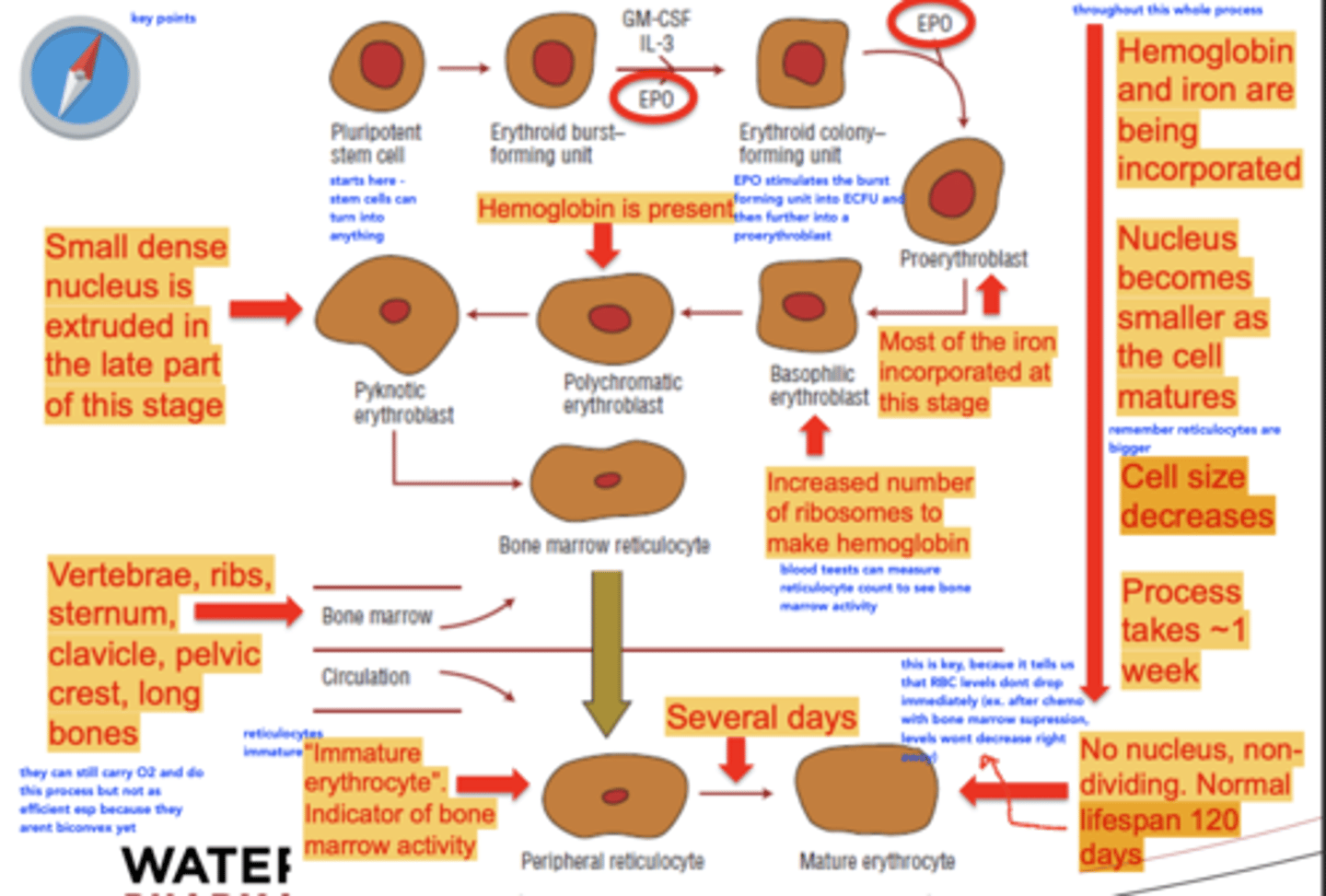
What events happen in the stages of erythropoiesis
1) Hgb and iron get incorporated
2) Nucleus becomes smaller as the cell matures
3) Cell size decreases
Process takes about 1 week
How long does it take for Peripheral (immature) erythrocytes to mature?
Several days
What does a normal mature erythrocyte look like and how long is its life span
No nucleus, non- dividing. Normal lifespan 120 days
Importance of Erythropoietin (EPO)
1. Stimulate stem cells to differentiate into proerythroblasts
2. Increase the rate of mitosis at each cell maturation stage
3. Prevent apoptosis of erythroid precursor cells
4. Increase release of reticulocytes into circulation
5. Increase hemoglobin formation
Where is EPO produced and how is it regulated?
• 90% produced by the kidneys
• Regulated by a negative feedback loop (if more RBCs needed, more EPO will be released)
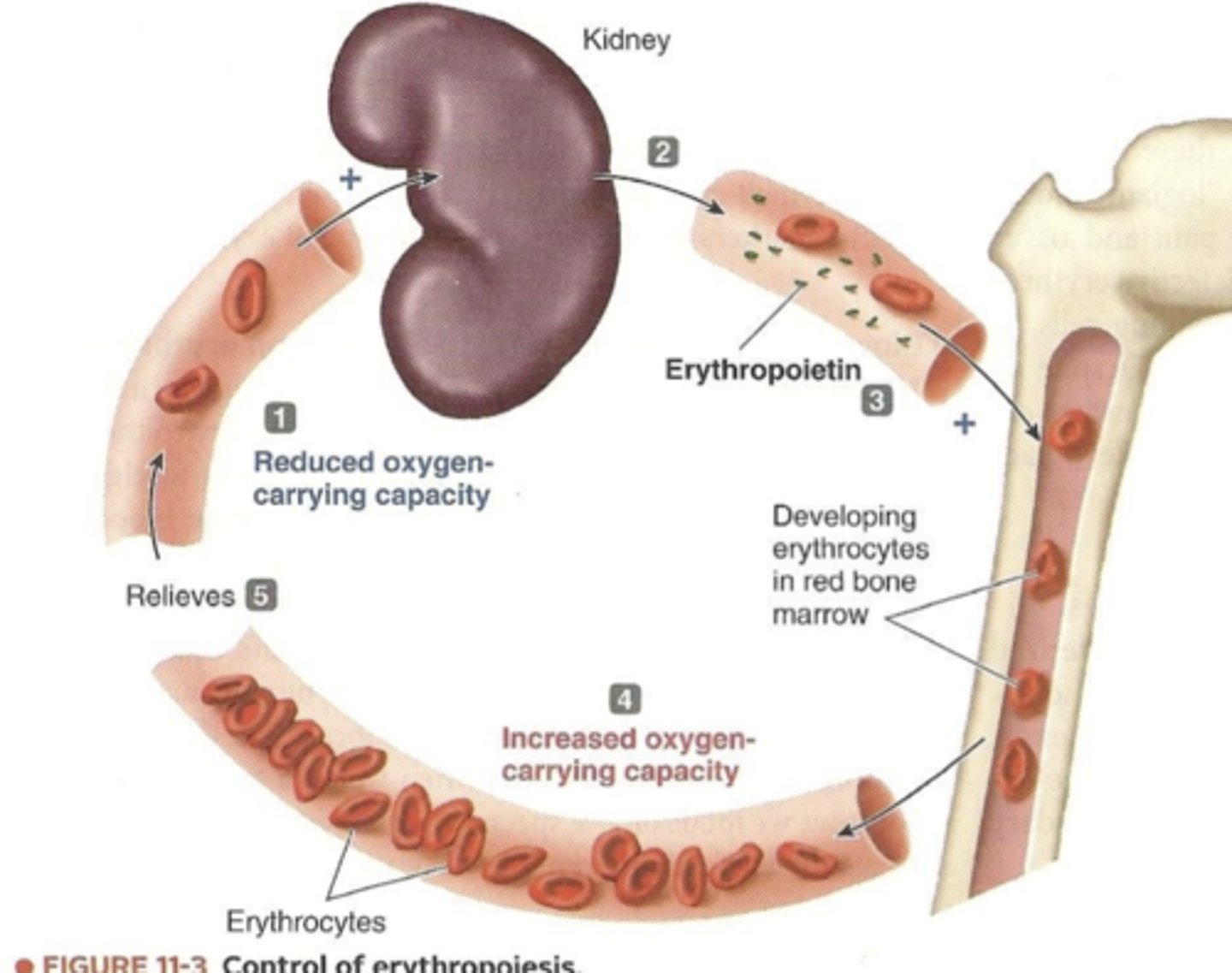
Explain EPO production
1) kidneys detect reduced O2 carrying capacity of blood
2) When less O2 is delivered to kidneys, they secrete EPO into blood
3) EPO stimulates erythropoiesis by bone marrow
4) The additional circulating erythrocytes increase O2 carrying capacity of blood
5) Increased O2-carrying capacity relieves initial stimulus
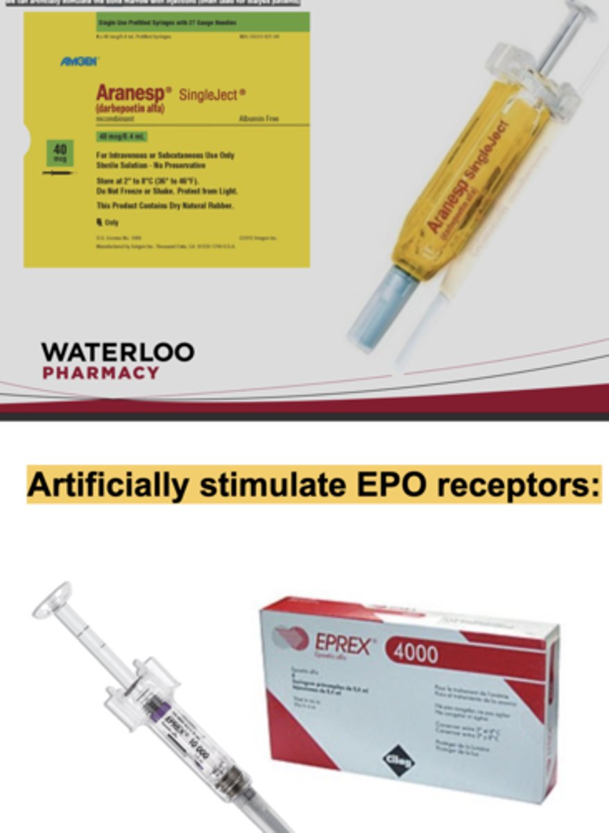
What if EPO receptors don't work?
we can artificially stimulate the bone marrow with injections (often used for dialysis patients)
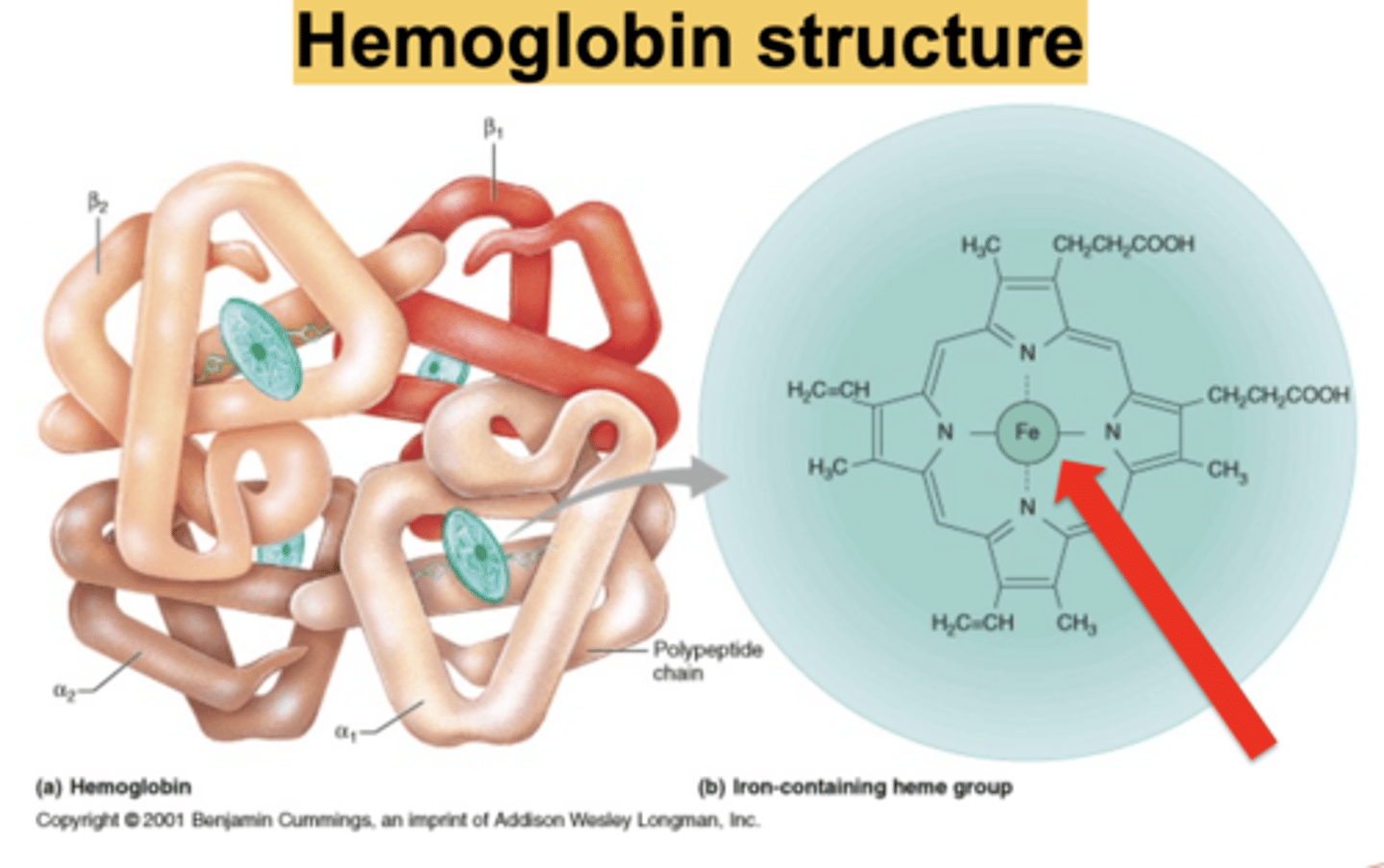
Composition of hemoglobin
Hemoglobin (Hgb) is the oxygen carrying unit in RBCs
• Protein consists of 2 α-chains and 2 b-chains
• Each chain links to a heme group
• Each Hgb molecule can hold 4 oxygen molecules
• Expressed as a concentration of hemoglobin per 100 mL of whole blood (g/dL) or per liter of whole blood (g/L)
Heme group composition
porphyrin ring with a Fe2+ atom chelated in the center - this iron atom is necessary to binding oxygen
How does iron get where it needs to be (in the bone marrow) for erythropoiesis?
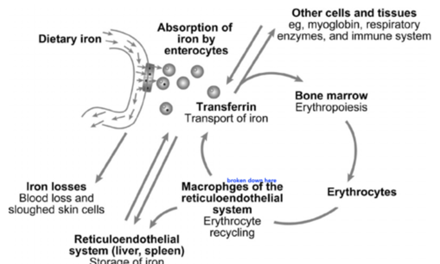
Transferrin
Transferrin
- Transferrin is a transport protein in the plasma
- delivers iron to the bone marrow to be incorporated into maturing TBCs
- Circulating transferrin normally ~30% saturated with iron (usually between 20-50%)
• Transferrin delivers extra iron to storage sites for later use: macrophages in the liver, marrow, spleen (also known as the RES)
Ferritin and hemosiderin
• Transferrin delivers extra iron to storage sites for later use: macrophages in the liver, marrow, spleen (also known as the RES)
– This body storage iron is also called ferritin
– Hemosiderin is a form of compacted ferritin molecules – a less available form of storage iron than ferritin
Reticuloendothelial system
- Iron gets absorbed in the intestines by enterocytes.
(Most gets lost and not absorbed)
- What's taken up is bound by transferrin and the majority gets sent to the bone marrow. Some is send elsewhere for a variety of metabolic functions.
Iron is not only for erythropoiesis
Hemoglobin normally exists in 2 states:
- Oxyhemoglobin (oxygen saturated)
- Deoxyhemoglobin (deoxygenated)
Hemoglobin releases oxygen to tissues when these changes occur in tissues / RBCs:
- High Concentration of CO2
- High Concentration of H+ ( lower pH)
- Increased Temperature
- 2,3-diphosphoglycerate (an oxygen affinity regulator)
2,3 Diphosphoglycerate (2,3-DPG)
Produced in the RBC which regulates the affinity of hemoglobin for oxygen.
An increase in 2,3-DPG causes ...
decreased Hgb affinity for oxygen
- Increases desaturation of hemoglobin
- Increases oxygen delivery to the tissues
- Causes "right shift" in O2 dissociation
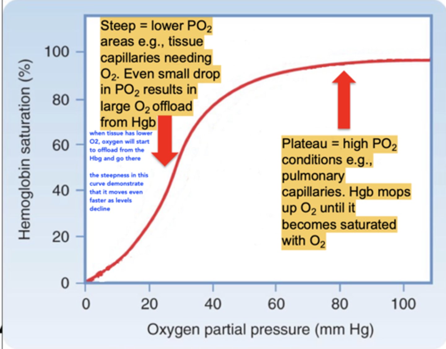
Oxygen-hemoglobin Dissociation Curve
when tissue has lower O2, oxygen will start to offload from the Hbg and go there
the steepness in this curve demonstrates that desaturation is even faster as levels decline
What causes a left shift in Oxygen-hemoglobin Dissociation Curve
Decreased P50 (increased affinity)
decreased temperature
decreased 2,3 DPG
increased (alkaline) pH
decreased PCO2
Normal destruction of RBCs
Spleen and marrow phagocytes (part of the RES) break down RBCs, releasing free hemoglobin
The released hemoglobin binds haptoglobin to facilitate access to degradative enzymes. Hgb- haptoglobin complexes increase and serum haptoglobin decreases in hemolytic anemia
Heme gets broken down to indirect bilirubin (unconjugated) which travels to the liver and become conjugated bilirubin to facilitate its removal as bile
Heme oxygenase opens porphyrin ring and releases free iron
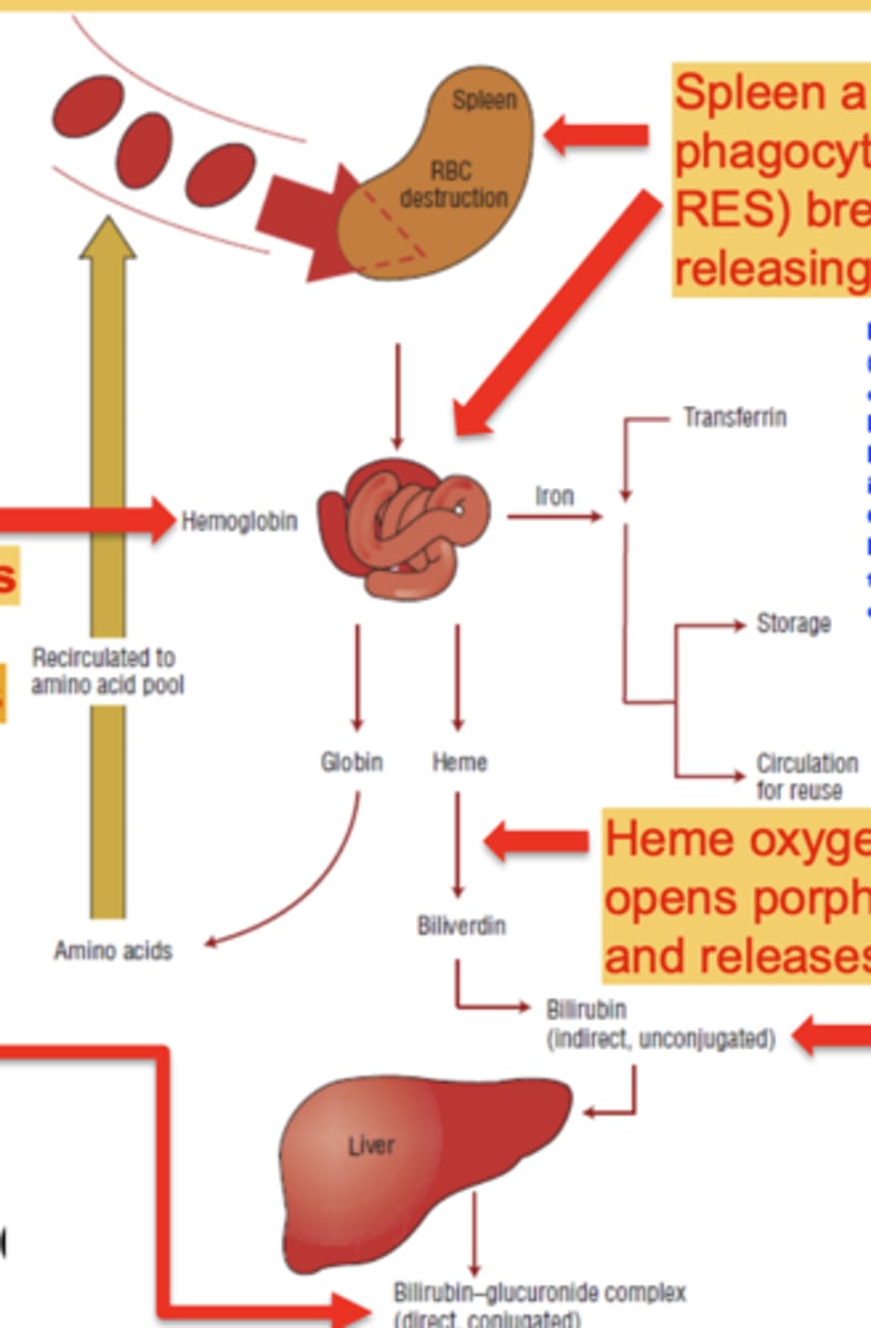
Direct (conjugated) bilirubin rises when...
bile excretion is obstructed
When do indirect (unconjugated) bilirubin levels rise?
in liver disease or excessive hemolysis
3 Different ways to classify anemia
- Morphology (size, shape and colour)
- Etiology (cause of the anemia)
- Pathophysiology
Morphology classification of anemia (Size)
What levels are used to classify this?
Characterized by Mean Corpuscular Volume
MCV = Hematocrit ÷ RBC count
Anemia with normocytic MCV can indicate which causes?
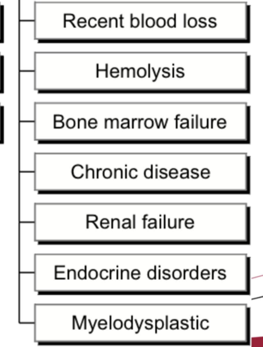
Normocytic: RBCs are at their normal size (80-100 fL)
Acute blood loss, anemia of chronic disease/chronic inflammation, hemolysis, chronic kidney disease
normolytic can be misleading especially when there Is a mix of small and large, so you would look at the RDW
What is Megaloblastic Macrocytic MCV anemia and What can cause it?
Megaloblastic - due to impaired DNA synthesis, which inhibits nuclear division. Production of abnormal immature cells called megaloblasts.
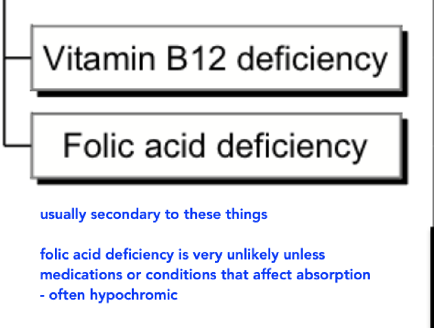
• E.g. Folate/Vitamin b12 deficiency, drug-induced
RBCs are larger than normal (>100 fL)
What is NON Megaloblastic Macrocytic MCV anemia and What can cause it?
Non-megaloblastic – usually as a result of more reticulocytes circulating (e.g. recovery from hemolytic or bone marrow insult, nutrient repletion, hematopoietic cell transplant) OR altered RBC membrane composition (e.g. hypothyroidism, liver dysfunction)
RBCs are larger than normal (>100 fL)
T/F: alcohol can cause megaloblastic AND nonmegaloblastic anemia
True
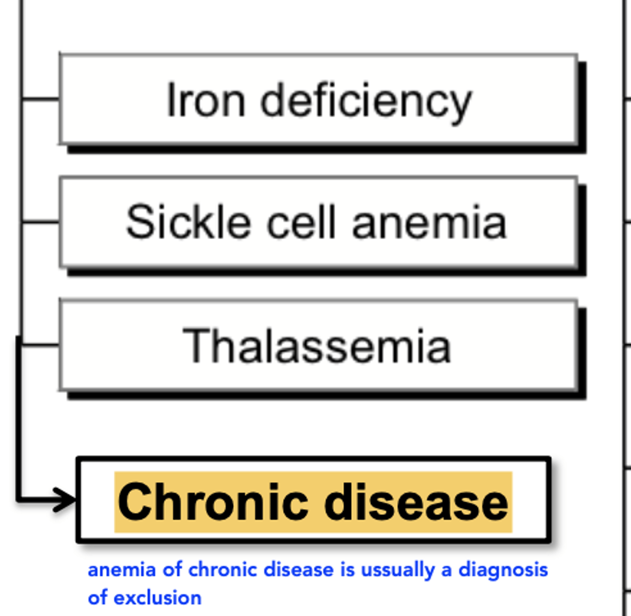
Anemia with microcytic MCV can indicate which causes?
• Often associated with impaired hemoglobin synthesis
• Examples: iron deficiency, thalassemic disorders
• Often also hypochromic
Anemia Characterized by Red Cell Distribution Width (RDW) and which anemias cause high RDW?
– A measure of variation in RBC size expressed as a percentage (SD of MCV÷MCV x 100)
– Normal RDW = between 11-16% variation
– High RDW = greater than 16% variation
• Vitamin, mineral or iron deficiencies, patients who received blood transfusions (increased variation between patient’s own and donor’s RBCs)
Normal shape of RBCs
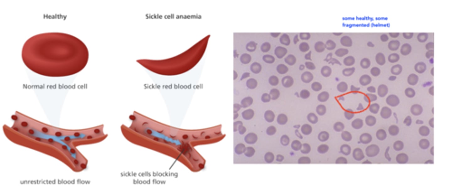
biconcave disks
able to deform to squeeze through capillaries
Abnormal shapes of RBCs
– Crescent-shaped and sticky (sickle cell anemia)
– Spherical, also called spherocyte (e.g., hemolytic anemia)
– Fragmented, also called schistocytes or “helmet” cells (e.g., thrombotic thrombocytopenic purpura) happens when RBC breaks
Morphology classification of anemia based on colour
Based on MCH and MCHC
– Normochromic: RBCs have normal colour (MCH and MCHC normal)
– Hypochromic: RBCs are paler than normal (MCH and MCHC low)
What cases of anemia would exhibit normochromic MCH and MCHC
Example: acute blood loss, EPO deficiency
What cases of anemia would exhibit hypochromic MCH and MCHC
Example: iron deficiency, chronic disease, thalassemia
Causes of macrocytic anemia
Causes of microcytic, hypochromic anemia
Causes of normocytic anemia
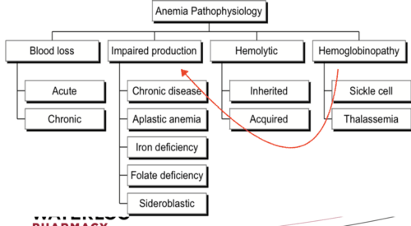
3 types of anemia pathophysiology that can be used in classification
1) inadequate RBC production
2) blood loss
3) RBC destruction
Inadequate production of normal RBCs
Deficient erythropoiesis due to:
deficient erythropoietin, iron, folate, vitamin B6/B12 and/or globin (thalassemia);
abnormally shaped RBCs (sickle, spherical, fragment); impaired bone marrow function; malignant bone marrow disorders
Causes of blood loss leading to anemia
GI bleeds, injuries, childbirth, surgery, tumours, GI cancer/polyps, GI ulcers, heavy periods
Causes of RBC destruction leading to anemia
Hemodialysis; hemolysis due to autoimmunity, infection, trauma, and other inherited RBC defects (sickle cell, thalassemias, enzyme deficiencies); drug- induced causes, prosthetic heart valves
Anemia classification by pathophysiology (Flowchart)
Initial laboratory investigations for anemia:
– Complete Blood Count (CBC)
• RBCs, WBCs, Hemoglobin (Hgb), Hematocrit (HCT), Platelets
– RBC indices (MCV, MCH, MCHC)
– Examination of stool sample for occult blood
– Reticulocyte index
What can reticulocyte index determine
can help determine if anemia is secondary to bone marrow problem
Reflects how quickly immature RBCs are being produced by the bone marrow and released into circulation
Reticulocytes Circulate in blood for how long before maturing to final RBC?
around 2 days
- 1% of RBCs being replaced daily by maturing retics
- Normally reticulocyte index is 0.5-2.5% (0.005-0.025)
If reticulocyte index is low when anemic...
– Anemia is due to impaired bone marrow function and bone marrow unable to compensate for anemia
– Iron deficiency, B12 deficiency, anemia of chronic disease, malnutrition, renal insufficiency, malignancy (either bone marrow is not getting the right signals, or its not responding)
If reticulocyte index is high when anemic...
– Bone marrow is functioning normally in response to
anemia; cause of anemia unlikely related to bone marrow
– Acute blood loss
– Hemolysis (eg. reticulocyte index > 2.5% is suggestive)
Peripheral blood smear
– Stained blood smears placed on a microscope slide
– Allows for morphological examination: RBC size, shape, colour
– Can assess for variations in cell size (anisocytosis) or variations in cell shape (poikilocytosis)
– These variations can help to assess functional status of bone marrow and defects in RBC production
anisocytosis
presence of red blood cells of unequal size
poikilocytosis
cells are irregularly shaped
What does serum iron measure
iron bound to transferrin (NOT including what's bount to hemoglobin)
- Diurnal property (higher when drawn in morning, lower when drawn in afternoon; consistency is key)
When is serum iron decreased
When there is:
• Infection, inflammation, anemia of chronic disease (more iron is redistributed to ferritin)
• Iron deficient anemia (less iron to bind to transferrin)
When is serum iron increased
When there is:
• Hemolytic anemia (RBCs break down and release iron into bloodstream)
• Iron overload (e.g. excessive iron supplementation or genetic condition hemochromatosis)
Total Iron-Binding Capacity (TIBC) and how it is measured
Measures the iron-binding capacity of transferrin
• Performed by adding an excess of iron to plasma to saturate all transferrin (normally only ~30% of available iron-binding sites are filled)
• Each transferrin can only carry 2 iron atoms
• Remaining excess unbound iron is removed
• Serum iron concentration then measured – this is the amount of iron bound to transferrin at 100% transferrin saturation
Total Iron-Binding Capacity or TIBC is higher than normal when:
– Body iron stores are low (transferrin production is
increased --> more binding sites)
– Oral contraceptive use
– Pregnancy
Transferrin protein production increased with variety of other proteins. Increased transferrin naturally means more sites to bind to iron
_____________ + low serum iron suggest IDA
High TIBC
Total Iron-Binding Capacity or TIBC is lower than normal when:
when there is decreased amount of transferrin itself:
– Infection
– Malignancy
– Inflammation
– Liver disease (transferrin synthesized by liver)
– Uremia
*Transferrin is a negative acute phase protein; production in the liver decreases under a state of inflammation, trauma or infection
% Transferrin SATuration or TSAT
• The percentage of iron-binding sites on transferrin that is bound to iron
• TSAT(%) = (serum iron ÷ TIBC) x 100
• Normally~20-50%
• In iron deficient anemia (IDA), TSAT typically 15% or lower
What is the best test for iron levels in the body
Serum ferritin
best test because it assesses a person's iron stores
- good overall assessment of how much in the body
- but may not be helpful in someone with RA or any chronic inflammation
Ferritin (where is it found)
• Ferritin is a measure of storage iron (in the form of iron-protein complexes) in the macrophages of the liver, spleen, bone marrow
• Good indicator of overall iron deficiency or iron overload
Low serum ferritin likely means:
- Iron deficiency anemia (IDA), because ferritin decreases only when truly iron deficient/low body iron stores
High serum ferritin indicates:
– Truly a higher body storage of iron OR
– Chronic infection or inflammation, regardless of true iron status
• Can increase by 3000% in response to infection
– Hence ferritin is of limited clinical utility in chronically ill patients
*Ferritin is a positive acute phase protein; it increases under a state of inflammation, trauma or infection
Folic acid (folate) deficiency
• Decreased level may indicate folate deficiency anemia, that may co-exist with B12 deficiency anemia
Folate in RBCs ________ volatile than folate in serum:
less
– Slow to decrease in acute process (e.g., drug-induced deficiency)
– Slow to increase with oral folate supplementation
– Less susceptible to rapid changes in diet or alcohol intake
– Serum folate more useful to assess acute folate deficiency
– RBC folate more useful to assess body folate storage
What lab values can tell us about folate deficiency
• Elevated MCV and low Hgb (macrocytic anemia suggesting B12 or folate deficiency) may prompt drawing a serum folate level
T/F: Generally, there is not much correlation between serum folate and RBC folate
false, there is usually good correlation
- In most patients, the addition of RBC folate testing does not add substantial amount of clinical information to serum folate results
- Serum folate is more useful for acute deficiency while RBC more useful for overall folate status
usually ordering a serum folate test is enough
T/F Low serum vitamin B12 level indicates deficiency
true
T/F Vitamin B12 deficiency may occur in conjunction with folate deficiency
true
If you suspect either folate or b12 deficiency, what bloodwork should you confirm with? WHY is this important
– Both folate and vitamin B12 should be drawn if either is suspected to be deficient
– Treatment of folic acid deficiency can resolve the macrocytic anemia (i.e. mask Vitamin B12 deficiency), but does not treat the underlying neuropathy caused by Vitamin B12 deficiency
SOO if you treat only the folate deficiency and mask the b12 deficiency, this can cause neuropathy and CNS damage
How is vitamin B12 absorbed?
- stomach acid breaks down the bond of B12 from the protein
- it then binds to haptocorrin which protects the B12 from being destroyed by stomach acid
- when this complex reaches the small intestine, pancreatic enzyme protease breaks down this bond so the cobalamin is free to bind to intrinsic factor made by stomach parietal cells
- the complex produced is what gets absorbed by the ileum
if not enough intrinsic factor: can't all be absorbed
if not enough enzymes: cant break it down to bind with intrinsic factor to form the complex
Where is B12 found
Meat, dairy and eggs
Schilling’s Urinary Excretion Test - What is it used for?
Used to diagnose vitamin B12 deficiency caused by lack of intrinsic factor which facilitates ileal absorption of B12
Schilling’s Urinary Excretion Test: STAGE 1
– An oral dose of radio-labeled B12 is given, followed by a large dose (1000 mcg IM) of non-labeled B12 one hour later to saturate tissue binding sites
If sufficient intrinsic factor is present, the labeled B12 that was absorbed from ileum would not bind to the saturated tissue binding sites, and then be excreted in the urine
– Normal result ≥8% of the labeled dose excreted in urine in 24 hours
Schilling’s Urinary Excretion Test: STAGE 2
– 5-7 days later, oral dose of radio-labeled B12 +
intrinsic factor are administered.
– If normal amount of labeled B12 is now found in urine, then intrinsic factor deficiency is confirmed as the cause of B12 deficiency anemia (=pernicious anemia)
– If urinary excretion of labeled B12 still low, proceed to stage 3
Schilling’s Urinary Excretion Test: STAGE 3
– Determines if B12 deficiency is due to B12
malabsorption in the ileum d/t bacterial overgrowth
– Tetracycline 250 mg QID x 10 days (to kill the bacteria), then oral dose of radio-labeled B12 is given
– Improved excretion indicates bacterial overgrowth is interfering with B12 absorption
*Note that in all stages, one hour after the radio-labelled B12 is given PO, a large dose (1000 mcg IM) of non- labeled B12 is given to saturate tissue binding sites
Schilling’s Urinary Excretion Test: STAGE 4 (if stage 3 is abnormal)
– Pancreatic enzymes given orally x 3 days, then oral
dose of radio-labeled B12 is given
– Improved urinary excretion of labeled B12 indicates malabsorption is due to pancreatic insufficiency
• Food bound B12 is released from the stomach and binds to a protein called haptocorrin that is produced in salivary glands
• The B12-haptocorrin complex then needs to be cleaved by pancreatic enyzmes in the ileum to release free B12, thus allowing for intrinsic factor-mediated absorption
______________ elevated in both Vit B12 and folate deficiency
Homocysteine
What levels are elevated with Vit B12 deficiency but NOT for folate deficiency?
Methylmalonic Acid (MMA) elevated in Vit B12 deficiency only
Coombs Test
• Anti-globulin test indicating that the hemolytic anemia is caused by an autoimmune response
Direct vs indirect Coombs test
Direct Coombs Test
– Detects self attacking antibodies bound to RBCs
– Positive finding usually indicates autoimmune- mediated hemolysis
Indirect Coombs Test
– Measures antibodies in the serum
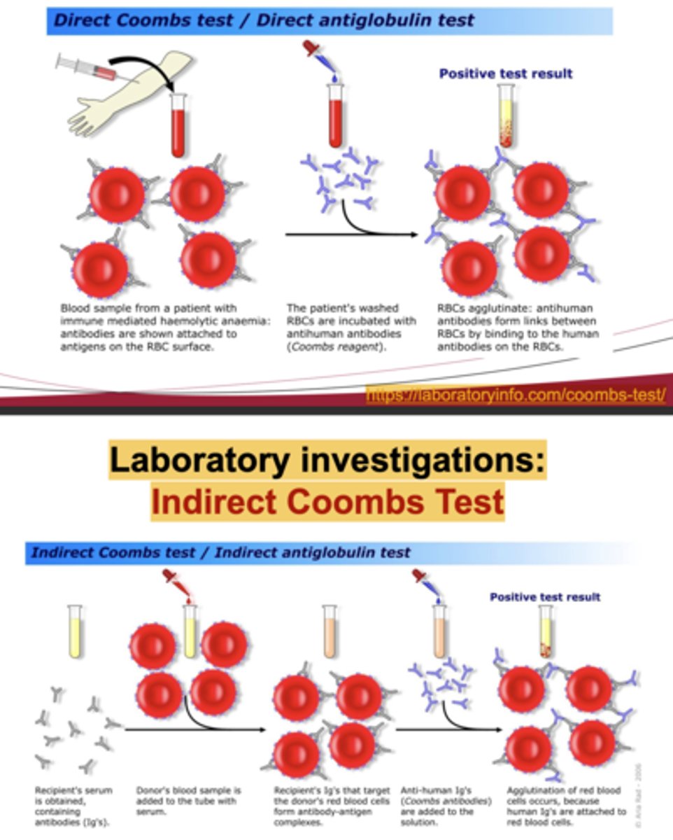
When could we use the indirect coombs test
if we were checking if persons blood is suitable to be donated - "will anything here react with the recipient?" also used in pregnancy testing
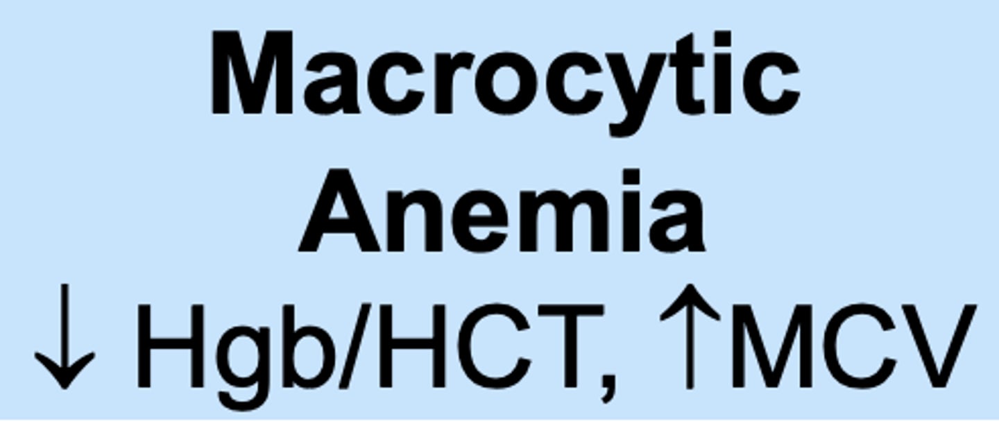
MCV and Hbg/HCT in macrocytic anemia
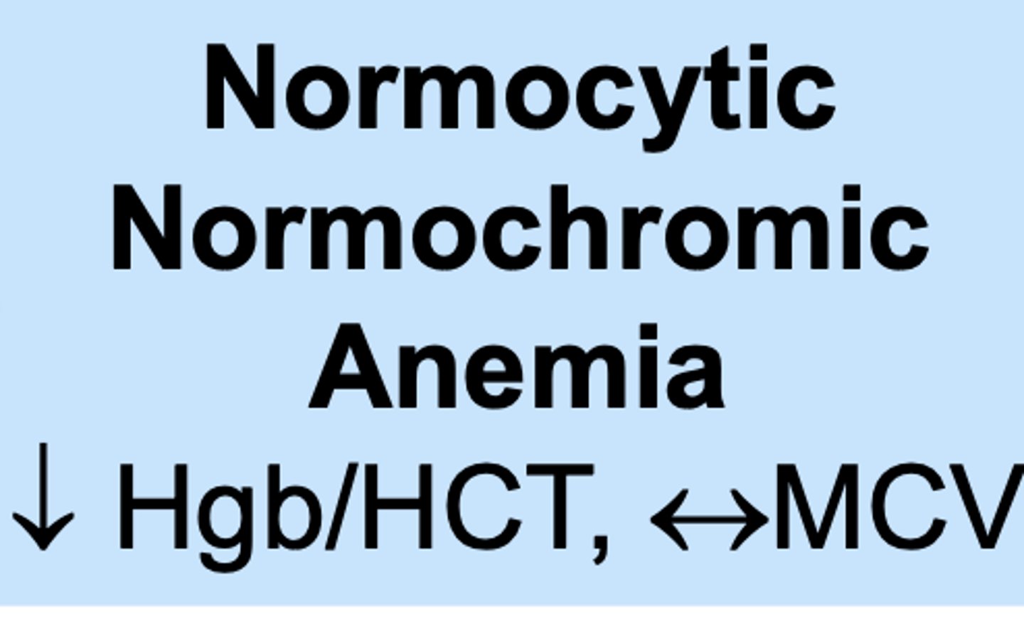
MCV and Hbg/HCT in normochromic/normocytic anemia
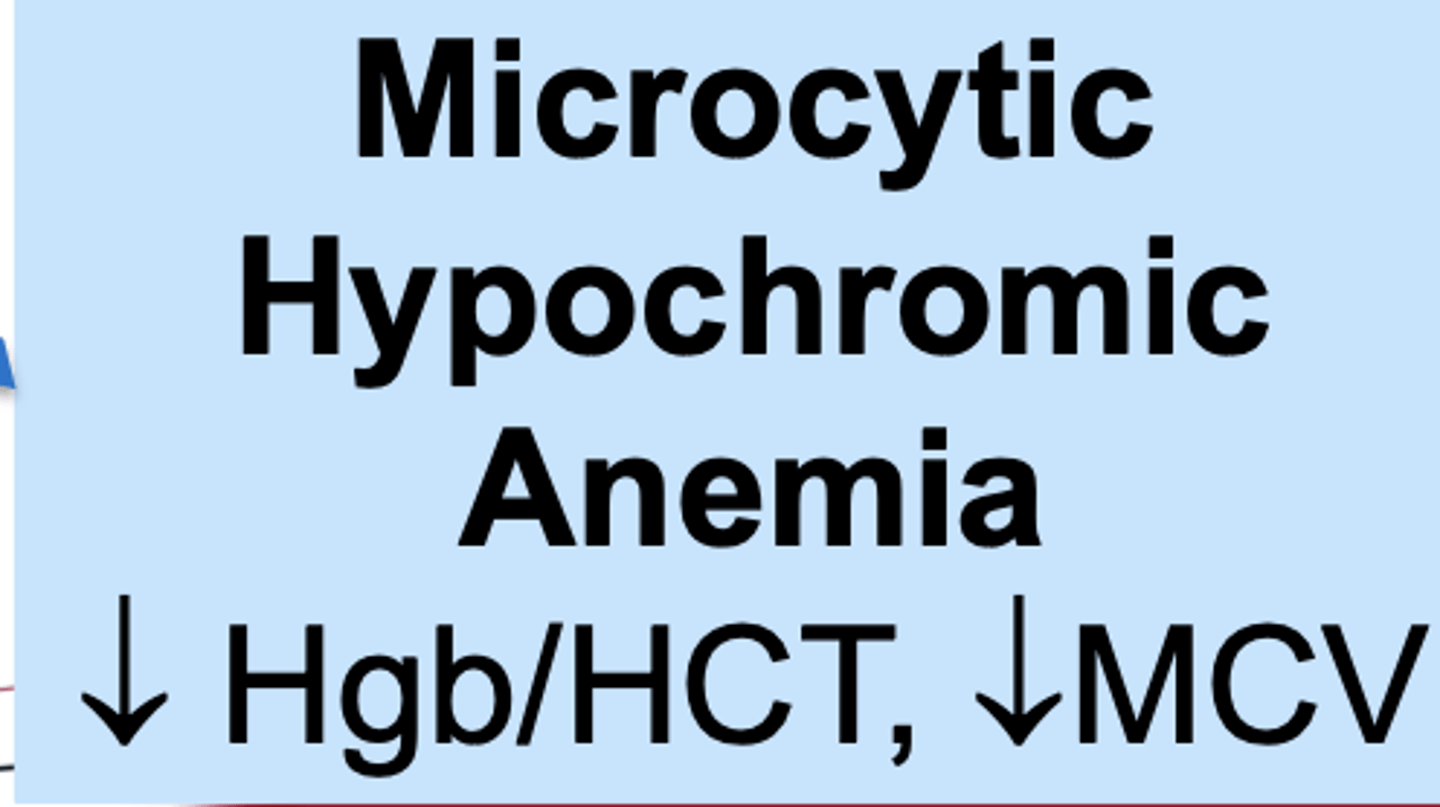
MCV and Hbg/HCT in microcytic hypochromic anemia
What tests would tell us if mactrocytic anemia is B12 caused
Low B12
Stage 2 Schilling's = pernicious anemia
high homocysteine / MMA
What tests would tell us if macrocytic anemia is folate caused
low serum folate
low RBC folate
high homocysteine
Other causes of macrocytic anemia, and what levels they are characterized by
Drug-induced (some)
Alcohol
Myelodysplastic
syndromes
Multiple Myeloma
Hypothyroidism
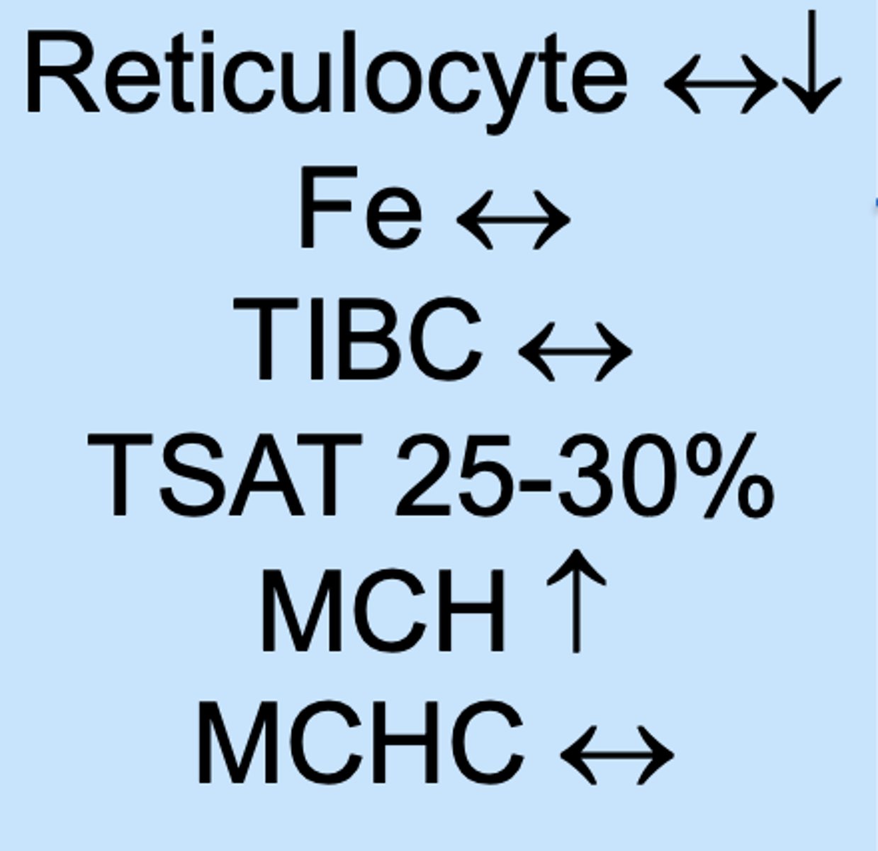
Normocytic normochromic anemia with high reticulocyte count indicattes...
acute blood loss