Cardiac Physiology
 Call Kai
 Learn
Learn Practice Test
 Spaced Repetition
 Match
Match Flashcards
 Knowt Play
1/476

There's no tags or description
Looks like no tags are added yet.
Name | Mastery | Learn | Test | Matching | Spaced | Call with Kai | Chat |
|---|
No analytics yet
Send a link to your students to track their progress
477 Terms
Why do humans have a cardiovascular system
Evolutionary consequence of multicellular organisms - cells inside body don't have access to environment for diffusion
Hemodynamics
the study of blood moving through the circulatory system, looking at the relationship between blood flow, blood pressure, and resistance to blood flow
Formula for blood flow
F = ΔP/R
F unit
Flow (mL/min)
ΔP
Pressure difference between two fixed points (mmHg)
R
Resistance to flow (mmHg/mL/min)
Which way does blood flow
From higher pressure to lower pressure - the pressure difference is the driving force for blood flow
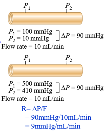
To have flow, the pressure difference....
Must overcome resistance to blood flow (ΔP > R)
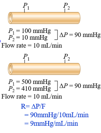
How can our bodies change the factors effecting blood flow
By changing resistance at a constant pressure in arterioles
Hydrostatic pressure
the pressure that blood exerts on the walls of the blood vessels
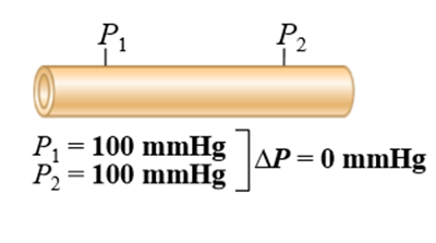
What happens if there is no pressure difference between two points (ΔP = 0)
Then the flow is 0mL/min
What factors determine resistance to blood flow
blood viscosity, vessel length, vessel diameter
How does viscosity affect resistance to blood flow
Higher hematocrit = more cells in blood = more viscous blood = greater resistance to flow
How does blood vessel length effect resistance
the longer the vessel, the greater the resistance. Blood comes into contact with the vessel walls more. creating more friction
How does blood vessel diameter effect resistance
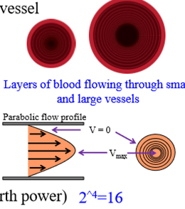
In a smaller diameter vessel, a greater proportion of blood is in contact with the wall = more friction = more resistance
- Larger diameter - blood forms concentric layers, and the inner layers do not touch the vessels
What is the use of Poiseuille's equation
Displays the relationship between viscosity, vessel radius, and vessel length for how they effect resistance
Poiseuille's equation
R = 8 L η/ π r^4
- R = resistance to blood flow
- L = vessel length
- n = blood viscsity
- r = radius
In Poiseuille's equation, resistance is proportional to:
Increased vessel length, increased viscosity
In Poiseuille's equation, resistance is indirectly proportional to:
Radius
Which factor has the greatest impact on resistance
Radius (as is it raised to the power of 4)
In which way does the human body increase resistance
Constricting and relaxing blood vessel muscle
What are the functions of the cardiovascular system
- Deliver oxygen and nutrients, and remove metabolic waste products
- Fast chemical signaling to cells by circulating hormones or neutrotransmitters
- Thermoregulation (transfer heat from muscles to the core, or lose heat through the skin)
- Mediation of inflammatory and host defense responses against invading organisms
Components of the cardiovascular system
Heart, blood vessels, blood
Types of blood vessels
arteries, arterioles, capillaries, venules, veins
Arterioles
Small branching vessels with high resistance
Capillaries
Transport of blood between small arteries and veins - exchange of materials between blood and body cells
Which way does blood flow in arteries
Away from the heart
Which does blood flow in veins
Towards the heart
Used of closed circulatory system
Greater pressures to be generated when the heart contracts
How many chambers does the heart have
4 - 2 atria, 2 ventricles
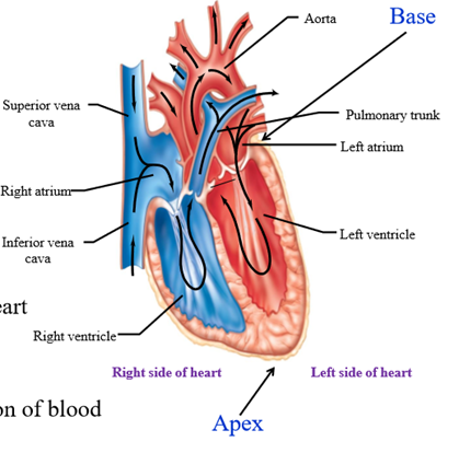
Atria
Thin walled, low pressure chambers that receive returning blood, and move blood into the ventricles
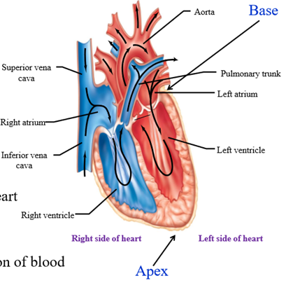
Ventricles
Thick walled, high pressure chambers that are responsible for the forward propulsion of blood when they contract
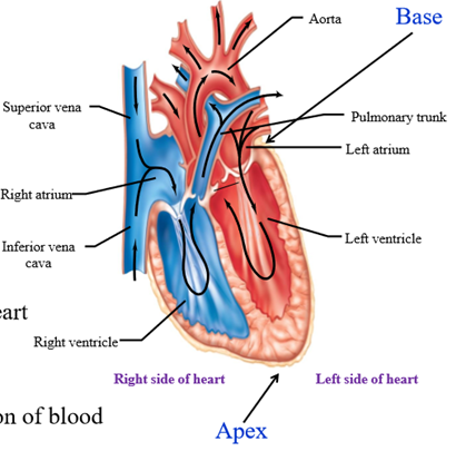
Base of the heart
Where the blood vessels attach
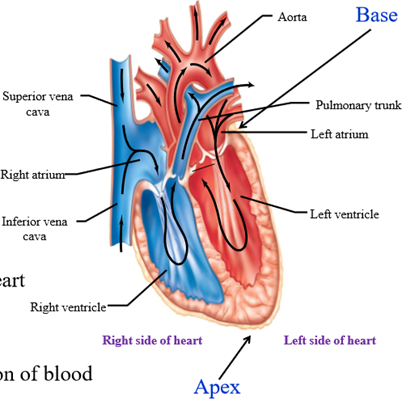
Septa
Divides the sides of the heart, making sure that low oxygenated and high oxygenated blood are seperate
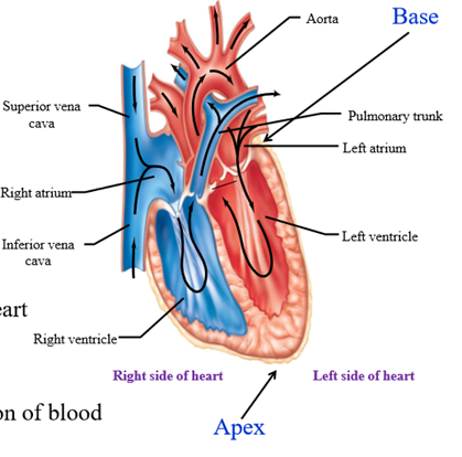
Where does the left side of the heart pump blood to
The systemic circuit
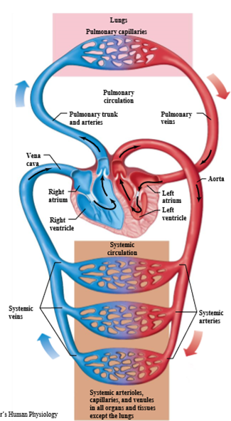
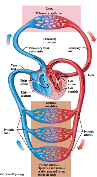
Where does the right side of the heart pump blood to
The pulmonary circuit
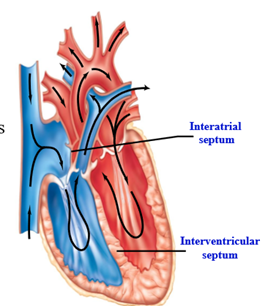
Interatrial septum
Wall separating the atria
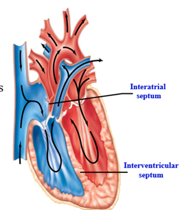
Interventricular septum
Wall separating the ventricles
Pulmonary circuit
Carries blood to and from the gas exchange surfaces of the lungs. Blood enters lungs poorly oxygenated and leaves highly oxygenated
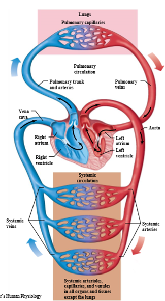
Systemic circuit
Transports blood to and from the rest of the bodies. Oxygen leaves the left side of the heart oxygenated, and leaves tissues deoxygenated
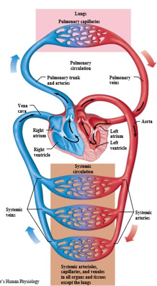
Left side of the heart
Receives blood from the pulmonary circulation and pumps it to systemic circulation
Right side of the heart
Receives blood from the systemic circulation and pumps it to the pulmonary circulation
Order of blood flow
Moves from pulmonary circuit to the heart, and then the systemic circuit before returning to the heart
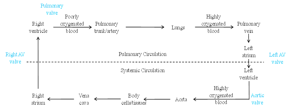
Blood flow cycle
1. Right ventricle (deoxygenated blood)
2. Pulmonary trunk/artery
3. Lungs (oxygenated here)
4. Pulmonary vein
5. Left atrium
6. Left ventricle
7. Aortic valve
8. Aorta
9. Body cells/tissues (deoxygenated)
10. Vena cava
11. Right atrium (and begin again)
Arteries
Carry blood away from the heart - mostly oxygenated blood except pulmonary trunk/artery
Veins
Carry blood to the heart - mostly deoxygenated blood except the pulmonary veins
Series flow
Blood passes through the pulmonary and systemic circuits in sequence
Parallel flow
Flow of blood to organs - each organ is supplied by a different artery, allowing for independent regulation
Which organ is an exception to parallel flow
The liver - receives blood in series
When the right ventricle contacts, where does blood flow
Blood is pumped the lungs
When the left ventricle contracts, where does blood flow
Blood pumped to the body
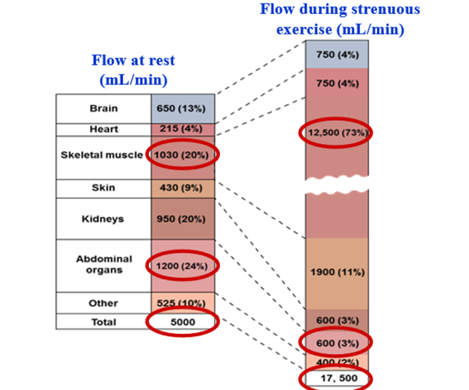
Distribution of blood flow during excersice
Cardiovascular system increases the rate of blood flow, and alters the distribution of blood flow to the muscles (instead of abdominal organs)
Pericardium
Fibrous sac surrounding the heart and the roots of great blood vessels leading into and out of the heart
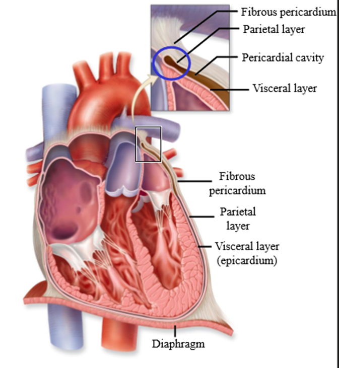
Functions of the pericardium
- Stabilizes the heart in the thoracic cavity
- Provides protection to the heart by physically surrounding it
- Reduces friction as the heart beats by secreting the pericardial fluid
- Limits overfilling of the heart chambers, preventing sudden distention (stretching)
Structures of the pericardium
Fibrous pericardium, parietal pericardium, visceral pericardium
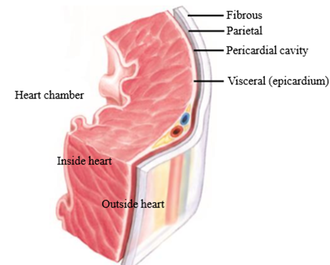
Fibrous pericardium
Outer layer of the pericardial sac - provides protection for the heart and stabilizes the heart in the thoracic cavity, prevents rapid overfilling of the heart
Parietal pericardium
Connected to the fibrous pericardium and attaches to it. Secretes pericardial fluid into the pericardial cavity to decrease friction during heartbeats
Pericardial cavity
Open area separating the parietal pericardium from the visceral pericardium - filled with pericardial fluid
Visceral pericardium
Innermost layer of the pericardial sac
- AKA the epicardium when it comes into contact with the heart muscle
Serous layer
Layer of cells that secrete fluid - made up by parietal + visceral pericardium
Pericarditis
inflammation of the pericardium caused by virus or bacteria - causes blood vessels to become leaky
Cardiac tamponade
acute compression of the heart caused by pericardial fluid accumulation - heart chambers cannot fill with adequate amount of blood
How to treat cardiac tamponade
Drain pericardial fluid, then treat the cause
Why is the muscle wall of the left ventricle thicker than the right ventricle
Allows the left ventricle to generate higher pressures, allowing blood to pump around the systemic circulatory system
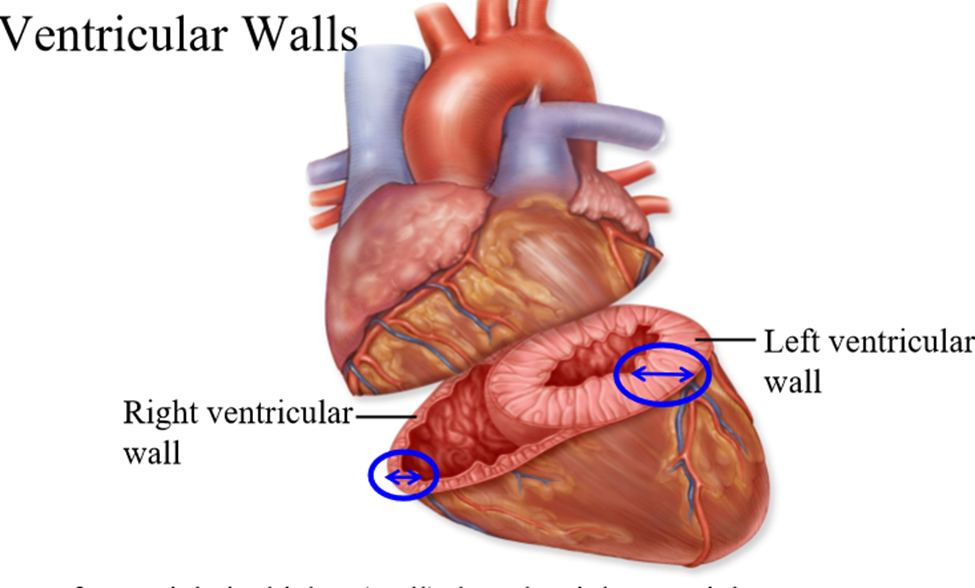
Why is the right ventricle muscle wall not as thcik
Blood only needs to pump from the right ventricle to the lungs (pulmonary system)
Layers of the heart wall
epicardium, myocardium, endocardium
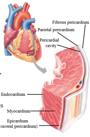
epicardium (visceral pericardium)
Layer outside heart muscle, covering the surface of the heart and connected to the myocardium.
Epicardium function
Protective layer for the heart
Myocardium
Muscular wall of the heart underneath the epicardium
Role of the myocardium
Contains myocytes (muscle cells) that contract when the heart beats
Endocardium
Innermost layer of the heart wall, lines the cavities/heart valves
Function of the endocardium
Made of endothelium - provides a smooth surface for blood to flow oveer
Which side of the heart has a thicker myocardium
Left side
Myocytes
Cardiac muscle cells (Y shaped)
Myocyte connection
Connected longitudinally - allows for greater connectivity (Y shaped cell allows for connection to 2 other myocytes)
Myocyte features
- Striated/striped with actin and myosin
- Single, centrally located nucleus
- Rich in mitochondria (ATP for muscles to contract)
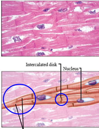
How are adjacent myocytes held together
Intercalated discs
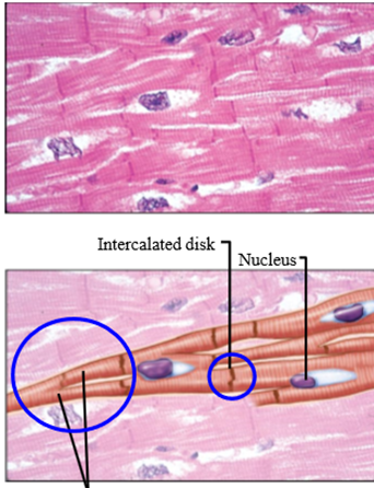
2 specialized intercellular junctions at intercalated disk
Desmosomes, gap junctions
Desmosomes
Adhering junctions that hold cells together in tissues subject to considerable mechanical stress/stretching
- Mechanically couples one heart cell to another
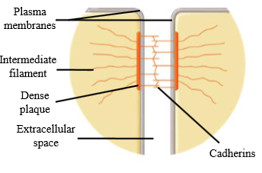
Proteins involved in desmosomes
Cadherins, plaques, intermediate filaments
Cadherins
Attach from one cell to cadherins of other cells
What do cadherins attach to
Dense plaques, which attach to intermediate filaments that are in the cell
Gap junctions in intercalated disks
Communicating junctions that electrically couple heart cells via ions
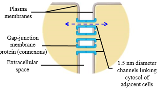
Use of gap junctions in heart cells
Spread action potential
Proteins in gap junctions
Connexons
Heart muscle arrangement
Spiral arrangement around heart chamber (c.f biceps - straight line)
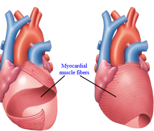
Why is heart muscle arranged in a spiral
Helps squeeze blood into arteries when ventricles contract
How many heart valves are there
4 - 2 AV valves + 2 Semilunar valves
Bicuspid valve
valve between the left atrium and the left ventricle.

Tricuspid valve
valve between the right atrium and the right ventricle
Semilunar (arterial) valves
found between the ventricles and the arteries into which the ventricles pump their blood
Aortic valve
Semilunar valve separating the left ventricle and the aorta
Pulmonary valve
Semilunar valve separating the right ventricle and the pulmonary trunk
What are the valves made of
Collagen covered by endothelium
Valve rings
Cartilage that valves attach to
Blood flow through the valves
· Poorly oxygenated blood returns to right atrium.
· Right AV valvue - blood flows to the atrium
· Blood moved from right ventricle to pulmonary trunk via pulmonary valve
· Oxygenated blood returns to the left atrium - blood pushed through tricuspid valve to left ventricle
· Blood pushed from left ventricle to aorta via aortic valve
What is the function of the heart valves
Ensure unidirectional blood flow to the heart, preventing blood flowing backwards
How do valves open and close
Valves open/close passively due to difference in pressure
Is energy expended to open valves
No
How does a heart valve open
When the pressure is greater behind the valve, the valve opens
