Vertebrate Zoology Exam 2 - Amniotes
1/59
Earn XP
Description and Tags
Name | Mastery | Learn | Test | Matching | Spaced |
|---|
No study sessions yet.
60 Terms
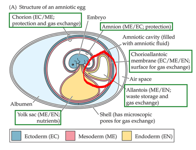
Yolk
Energy supply
In yolk sac (extraembryonic membrane, outside of embryo’s body)
Yolk sac is made of mesoderm and endoderm
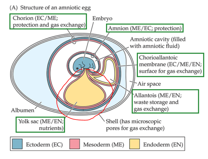
Amnion
Forms folds of ectoderm + mesoderm
Formed by mesoderm on the outside and ectoderm on the inside
Amniotic cavity, surrounds to protect embryo from mechanical shocks, similar to mammals’ amniotic fluid (cushions embryo)

Chorion
Forms folds of ectoderm + mesoderm
Chorion has mesoderm on inside (pink layer)
Encloses other extraembryonic membranes + contacts inside surface of eggshell
Also works with allantois to form membrane for gas exchange (chorioallantoic membrane)
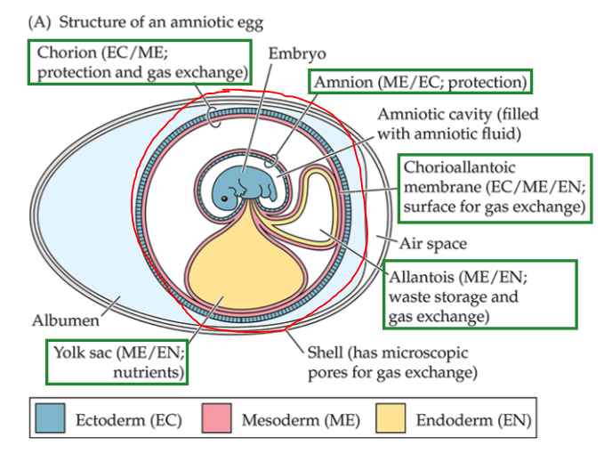
Chorioallantoic Membrane
Chorion + allantois work together to make a surface for gas exchange

Allantois
Inside = endoderm, outside = mesoderm
2 Functions
Stores nitrogenous wastes
Expands to contact the chorion to form + vascularize the chorioallantoic membrane
What was the ancestral amniote egg likely like?
Flexible, leathery eggshell
The Composition of Waterproof Skin
Multilayered keratinous epidermis
Thick dermis
Hydrophobic lipids in skin limit water loss
Keratin
Forms scales, hair, feathers, nails, beaks, and horns
(*Scales, hair, and feathers are homologous structures)
Alpha Keratin
Contained in keratinized skin of extant amniotes
Beta Keratin
Second, harder type of keratin not found in mammals but found in extant sauropsids
Metanephric Kidney
Derived character of amniotes
Drained by a duct called the ureter
Responsibilities:
Maintains homeostasis of body fluids
Regulates extracellular fluid volume, osmolality, ion concentrations, and pH of blood
Gets rid of nitrogenous wastes
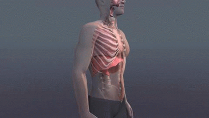
Costal Ventilation
Moving air into and out of lungs using movements of the ribs
A character of amniotes based on phylogenetic inference and anatomy of fossils
Derived Features of Amniotes + Functional Significance
Lateral flange of the pterygoid bone - jaw adductor muscle division for diff. modes of processing food
Second cervical vertebra modified into an axis - greater mobility of head on neck, allow schanges in feeding + social behavior
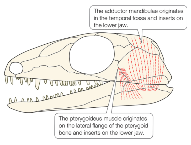
Astragalus bone and mesotarsal joint - change in ankle allows more limb-based (rather than axial-based) locomotion
More than one sacral vertebra (base of spine) - reflects increasing use of hindlimbs in limb-based locomotion
Amniotic egg - no need to return to water for reproduction
Waterproof skin - limits water loss and greater independence from water
Penis - internal fertilization, essential for amniote eggs
Metanephric kidney - improved water retention
Costal aspiration - higher metabolic rates and loss of CO2 via lungs rather than skin
Trachea with cartilaginous rings - longer neck, changing feeding + social behavior
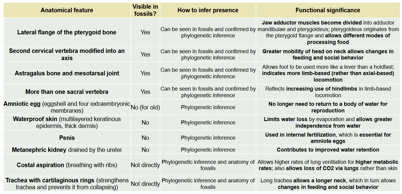
Function of: Lateral flange of the pterygoid bone
Lateral flange is the origin for the division of jaw adductor muscles into adductor mandibulae and pterygoideus muscle (jaw-closing muscle)
Plays many roles in the feeding systems of amniotes
Function of: Second cervical vertebra modified into an axis
Greater mobility of head on neck
Allows changes in feeding and social behavior
Function of: Astragalus bone and mesotarsal joint
3 small proximal tarsal bones fuse to form the astragalus
Mesotarsal joint is the plane of bending
Allows foot to be used like lever than holdfast
Indicates more limb-based locomotion (role of the hindlimbs)
Function of: More than one sacral vertebra (base of spine)
Reflects increasing use of hindlimbs in limb-based locomotion
Function of: Amniotic egg
Eggshell and four extraembryonic membranes (yolk, amnion, chorion, & allantois) allows independence from water for reproduction
Function of: Waterproof skin
Limits water loss by evaporation
Allows greater independence from water
Function of: Penis
Used in internal fertilization, which is essential for amniote eggs
Function of: Metanephric kidney
Contributes to improved water retention
Function of: Costal aspiration
Breathing with ribs
Higher metabolic rates
Allows loss/release of CO2 via lungs rather than skin
Function of: Trachea with cartilaginous rings
Longer trachea = longer neck
Changes in feeding and social behavior
What did the ankle of basal tetrapods look like? How did it function?
No distinct ankle joint (mesotarsal joint)
Axial muscles power locomotion
Feet functioning mainly as pivot points around which the hindlimb rotates
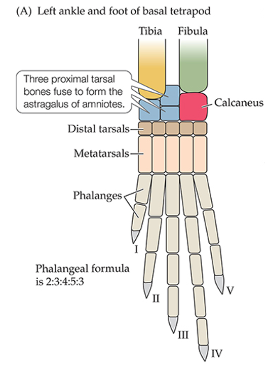
What is the ancestral conflict between locomotion and respiration? (i.e. what 2 things are “required” if both must occur simultaneously?)
Locomotion - bending the trunk unilaterally from side to side for locomotion
Respiration - compressing the rib cage bilaterally to ventilate the lungs
Can’t run and ventilate lungs at same time
How do Synapsids overcome the conflict between locomotion and respiration?
Spine moves up-down (dorsoventral flexion), avoids conflict with lung ventilation and actually assists lung movement
Why are some lizards (basal amniotes) limited to short bursts of activity?
They retain ancestral modes of locomotion and ventilation, can only rely on short dashes
As ATP and creatine phosphate are used up, muscles switch to anaerobic metabolism
If dash is too long, can no longer use phosphates OR anaerobic metabolism, they need oxygen in order to be replenished
Adductor
Adds/in/TOWARD body
Abductor
Away from body
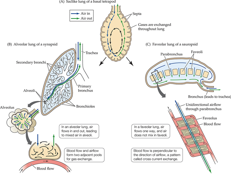
What type of airflow do Synapsids have?
Bidirectional/tidal airflow (in-and-out)
Diaphragm
Muscularization of the postpulmonary septum
In extant mammals
Helps move lungs without conflicting with locomotion
Postpulmonary Septum
Sheet of connective tissue in the coelom that separates lungs from digestive organs and forms separate pleural (lungs) and peritoneal (abdominal) cavities

Synapsid VS. Diapsid Temporal Fenestrae
Synapsid - ONE temporal fenestra on each side of head (lower only)
Diapsid - TWO temporal fenestra pairs (lower and upper)
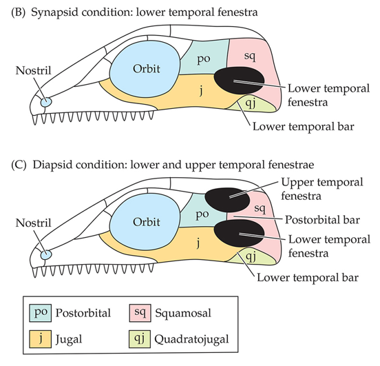
Skull hole (temporal fenestrae) functions
Stronger bite forces
Compared to reptiliomorph, simple jaw-closing muscles beneath skull roof
Synapsid VS. Sauropsid Lungs
Synapsid - alveolar, tidal ventilation (compliant)
Sauropsid - faveolar, undirectional ventilation (rigid, noncompliant)
Gastralia
“Free-floating” bones in the ventral abdominal wall (basal amniotes)
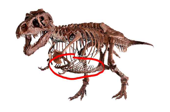
Mobile gastralia
Muscle on pelvis and inserts on the gastralia
Contracts, pulling gastralia, increasing volume of abdominal and pulmonary cavities and produces inhalation
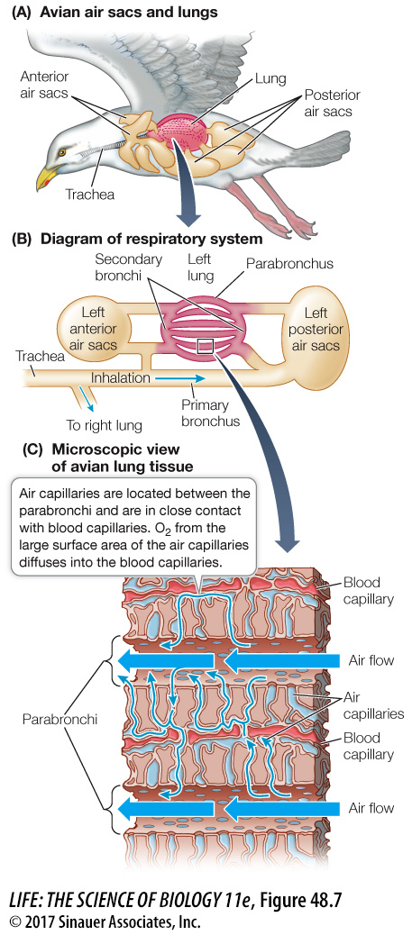
Air sacs
Located in bones anterior and posterior to lungs
Retained in birds (and some crocodylians)
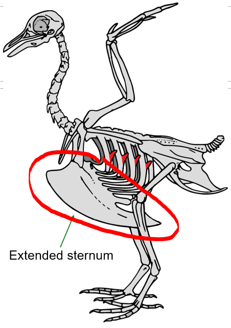
Extended Sternum
Replaces gastralia in birds as a method of ventilating the lungs
Moves, pivots, and helps drive air into air sacs
Immobile lungs
In some derived sauropsids (like pterosaurs)
Only ventral portions are compliant
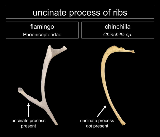
Uncinate processes
Muscles attached to ribs to facilitate movement of ribs and sternum for respiration
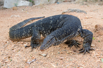
Gular Pumping
Monitor Lizards
Ventilation by drawing air into gular region through the nares (expands and contrats entire throat)
(Image: Asian Water Monitor)
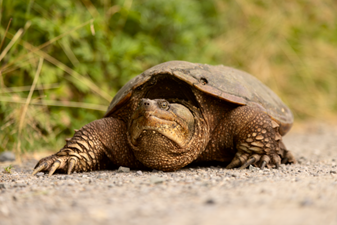
Muscular Sling
Turtles
Can’t move ribs or ventral surfaces of bodies (bc of shell) to change volume of lungs, instead uses a sling created by abdominal muscles
Pelvic Ventilation
Crocodylomorphs
Derived character
Rotate pubic bones, increasing volume of abdominal cavity for lungs to expand
Hepatic piston
Extant crocodylians
Moves liver to help ventilate lungs
Describe avian respiration.
“Two- breath” model - always at least one breath in each reservoir
Parabronchial lungs
Unidirectional flow, cross-current exchange system (air and blood pass in opposite directions)
Thin capillaries for easier oxygen diffusion
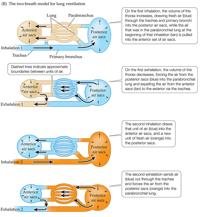
Air Capillaries
Gas-exchange structures of bird lungs, millions of interconnected small tubules radiating from the parabronchial lungs
Downside of alveolar lung:
On exhale, there is a moment where we are not getting oxygen
Why is high blood pressure bad?
Can damage the delicate, thin lung tissue
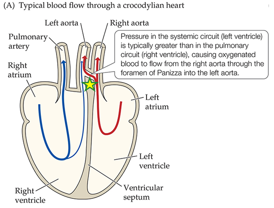
Why did amniotes evolve separate pressure systems for circulation? What are those 2 systems?
Prevent high blood pressure causing damage to the delicate lung
Systemic (body) pressure is ALWAYS higher than pulmonary (lung) pressure because of lungs’ thin tissue
What direction do arteries flow blood?
Away from heart
What direction do veins flow blood?
Toward heart
What 2 lineages independently reduced to a single systemic arch? Which arch is retained in each lineage?
Arch 4 develops into the systemic aorta
Mammals - retains left arch
Birds - retains right arch
Which 2 lineages have a ventricular septum in their hearts?
Mammals and birds
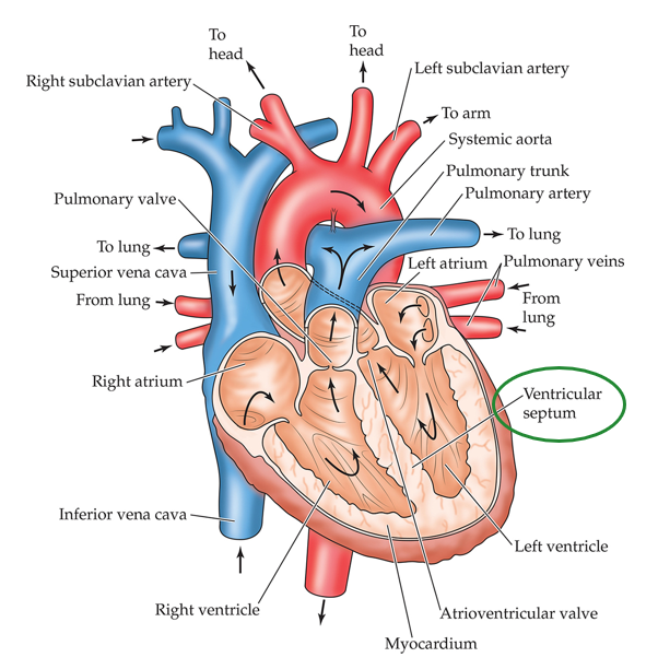
Ventricular septum
A permanent septum separating the ventricle into systemic and pulmonary sides
Allows different blood pressures in each circuit
What is the order of blood flow in hearts with a permanent septum?
(Mammals and bird hearts)
STARTS in the Right (pulmonary), deoxygenated blood
Inferior vena cava
Right atrium
Right ventricle
To pulmonary artery, going to the lungs
Now to Left (systemic), oxygenated blood
From lung, to left atrium
Left ventricle
To rest of body through the systemic aorta
What is the order of blood flow in hearts without a ventricular septum?
(Turtles and lepidosaurs)
Right (Pulmonary), deoxygenated
Starts in right atrium
Flows into cavum venosum
Over the muscular ridge to cavum pulmonale
To pulmonary artery
Left (Systemic), oxygenated
Left atrium
Cavum arteriosum
Leaves through aortas
What groups can perform shunting of blood?
Turtles, Lepidosaurs, & Crocodylians
How does a right to left shunt benefit lizards, turtles, and crocodylians?
A pulmonary to systemic shunt allows a higher rate of blood flow and allows for quicker warming of the body
Foramen of Panizza
Hole allowing flow to both aortas from the left side of Crocodylian heart