RADIOLOGICAL ANATOMY OF THE UPPER EXTREMITY
1/285
There's no tags or description
Looks like no tags are added yet.
Name | Mastery | Learn | Test | Matching | Spaced | Call with Kai |
|---|
No analytics yet
Send a link to your students to track their progress
286 Terms
How is the shoulder or pectoral girdle defined?
The shoulder or pectoral girdle is defined as the polyarticular complex extending from the base of neck to inferior edge of major pectoralis muscle.
CONNECTS THE UPPER LIMB TO THE AXIAL SKELETON
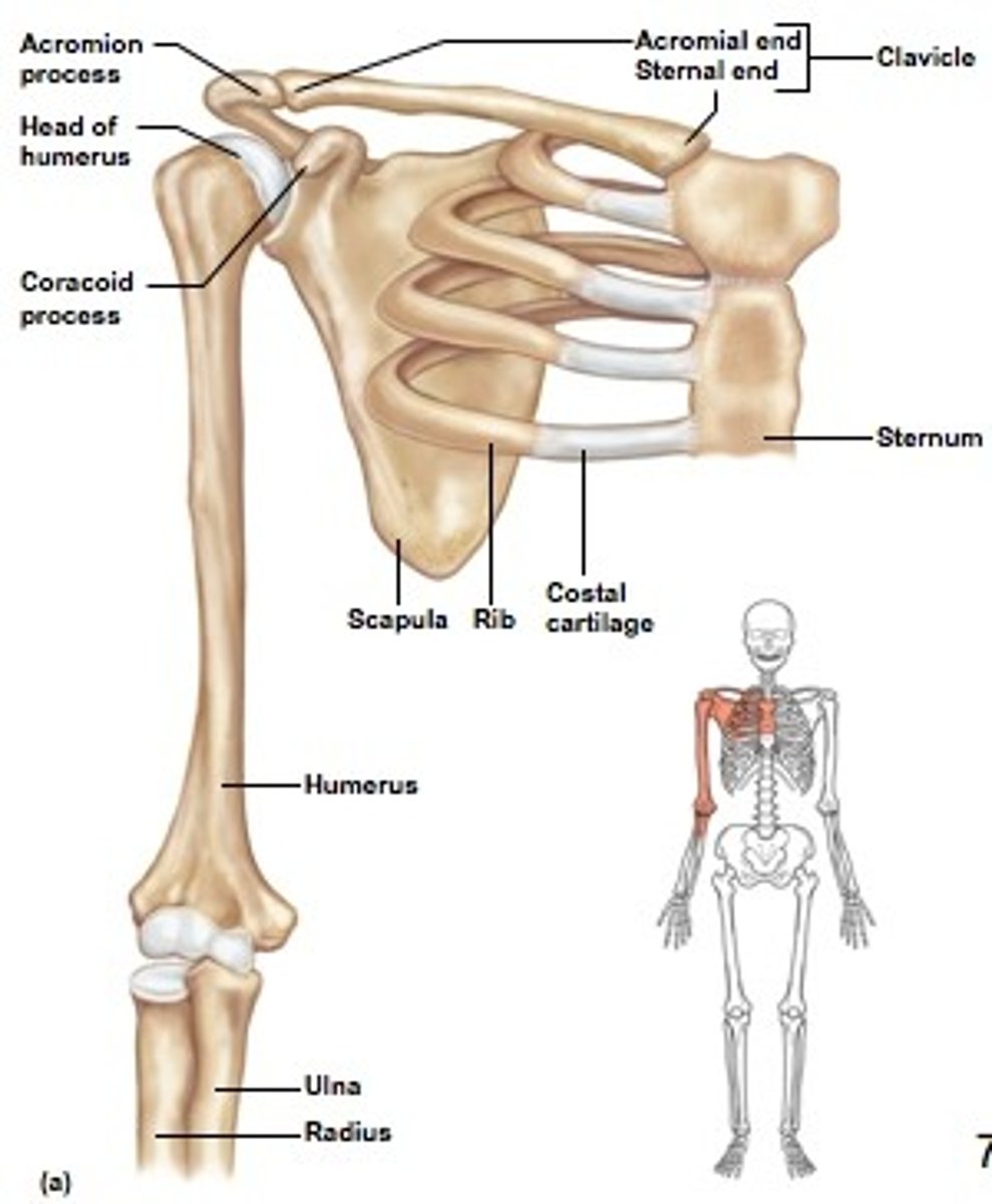
What bones are found in the shoulder or pectoral girdle region?
1) scapula
2) clavicle (collarbone)
3) sternum
4) humerus
5) rib cage.
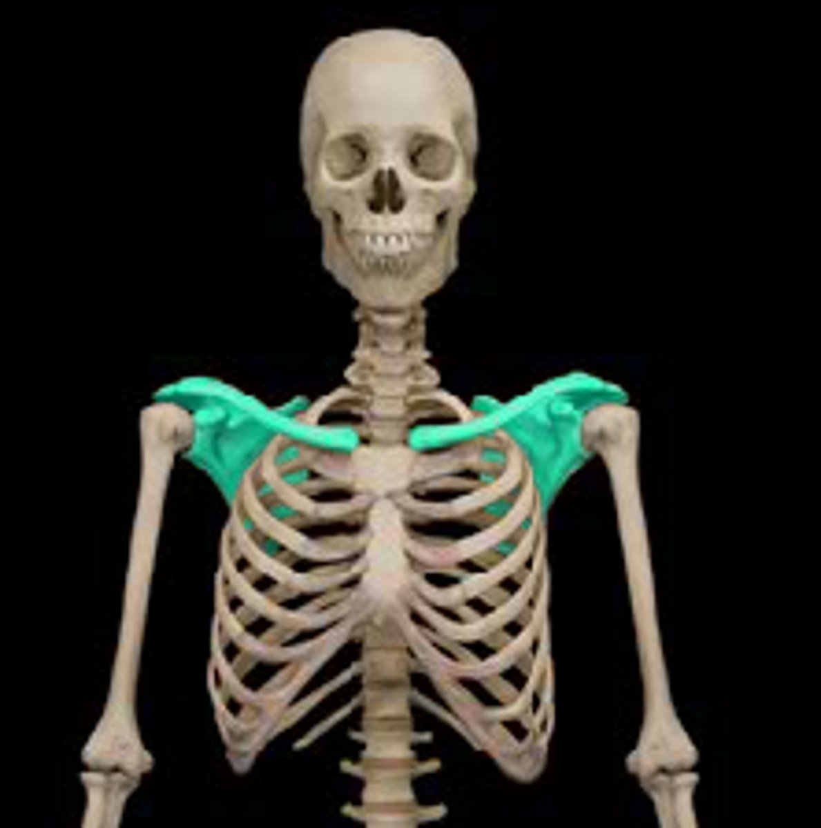
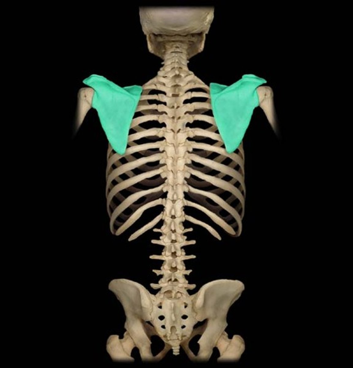
What is the general shape and placement of the scapula?
The scapula is a flat, roughly triangular bone located on the posterolateral aspect of the thoracic cage (behind, next to thoracic cage), forming the posterior limit of the shoulder girdle.
* sub scapular fossa faces thoracic cage
What are the features of the anterior and posterior compartments of the scapula?
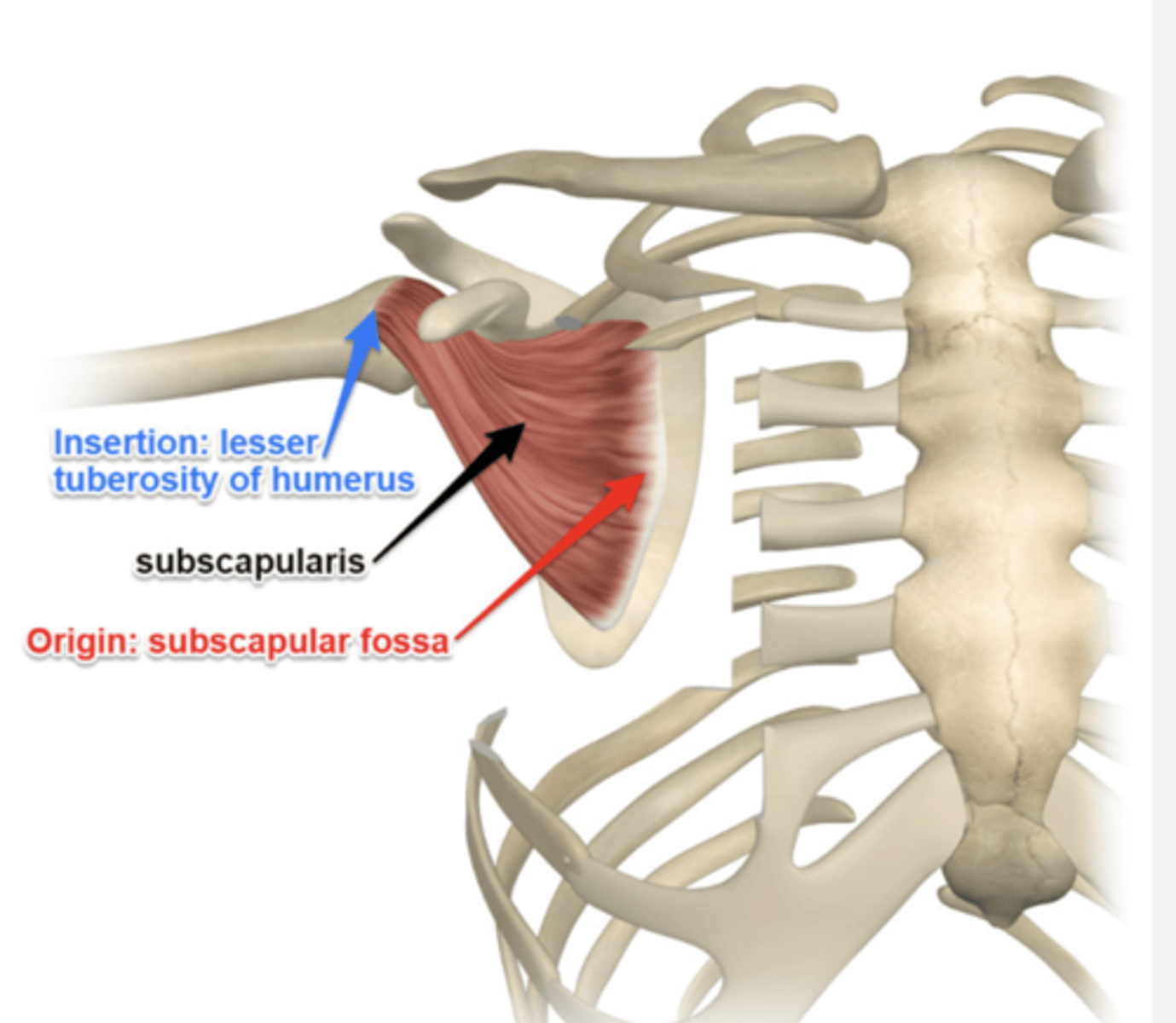
1) anterior (costal) component the subscapular fossa
facing the thoracic cage for subscapularis muscle attachment
2) posterior component divided by the spine into the infraspinous and supraspinous fossae.
Describe the spine of the scapula and its continuation.
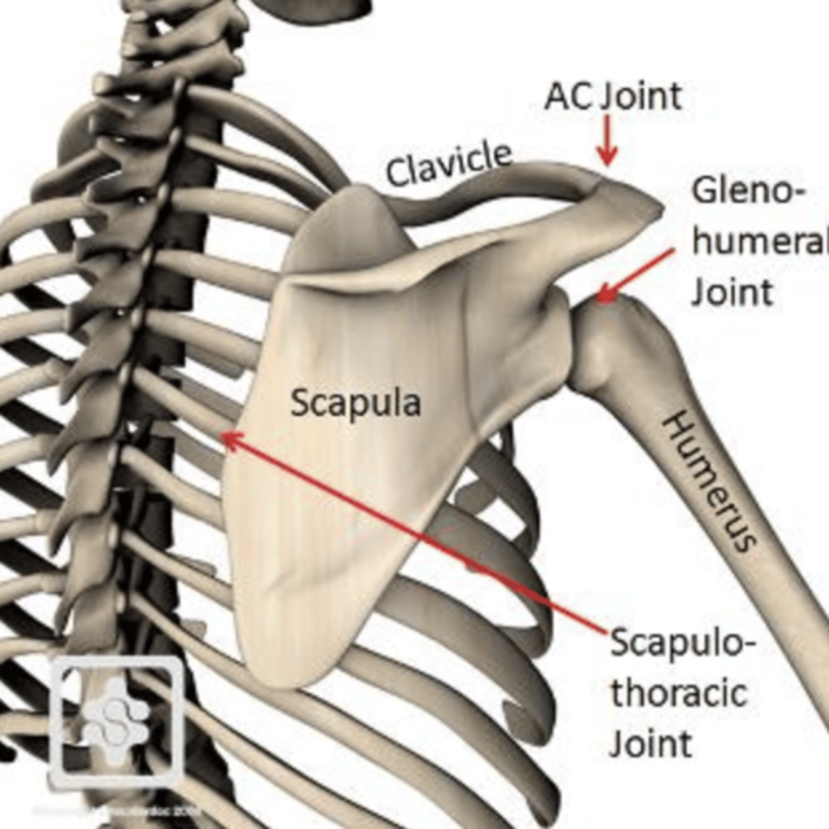
The spine of the scapula starts at the medial border and continues laterally as the acromion, which articulates with the clavicle's acromial end.
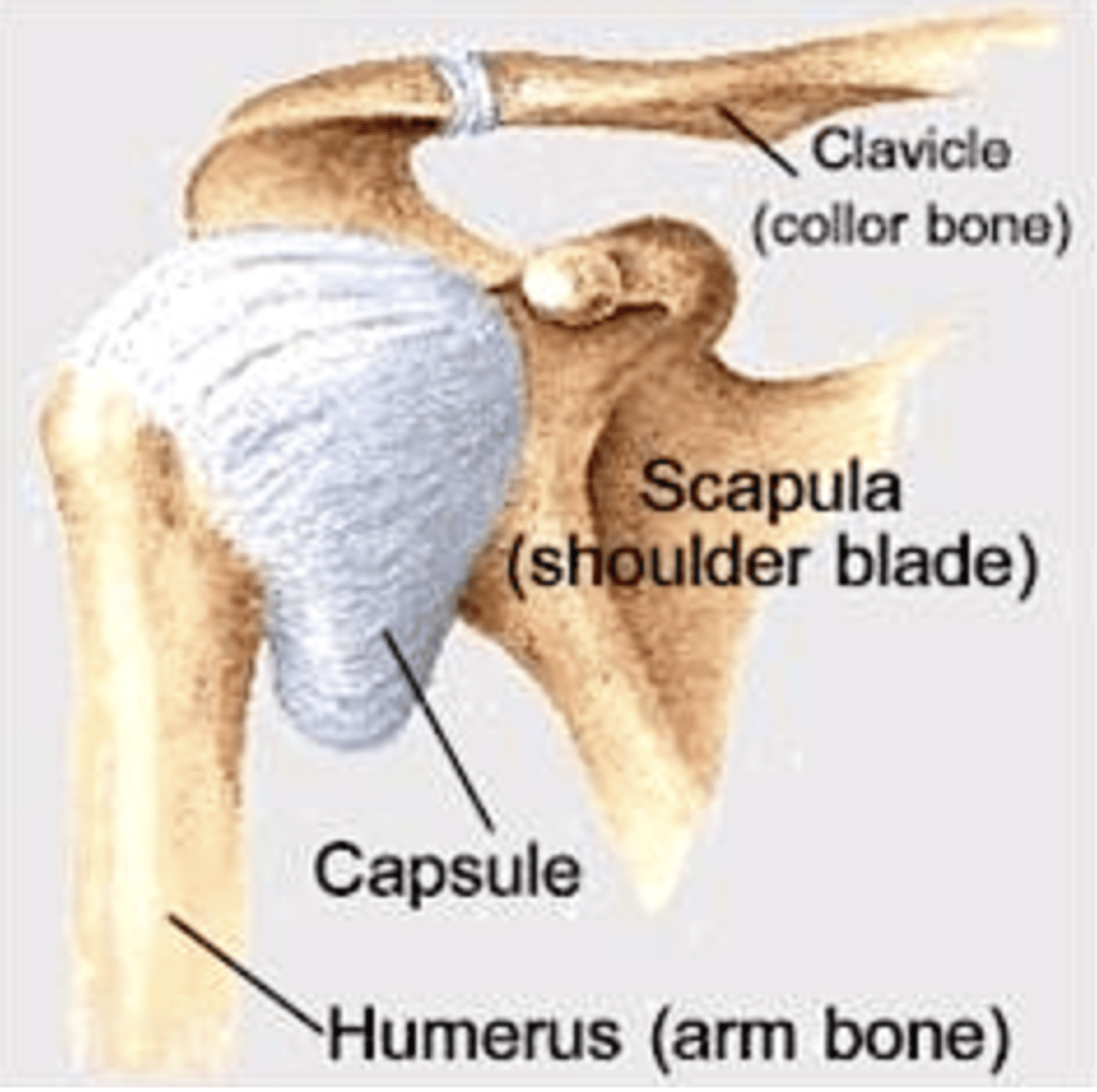
What is the role of the glenoid cavity?
The glenoid cavity is a concave, oval fossa that articulates with the head of the humerus at the glenohumeral joint, directed anterolaterally and slightly superiorly.
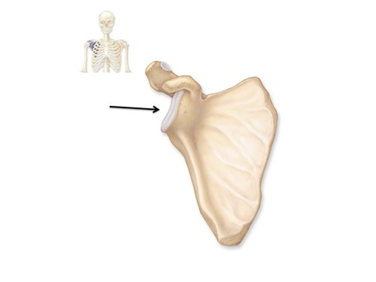
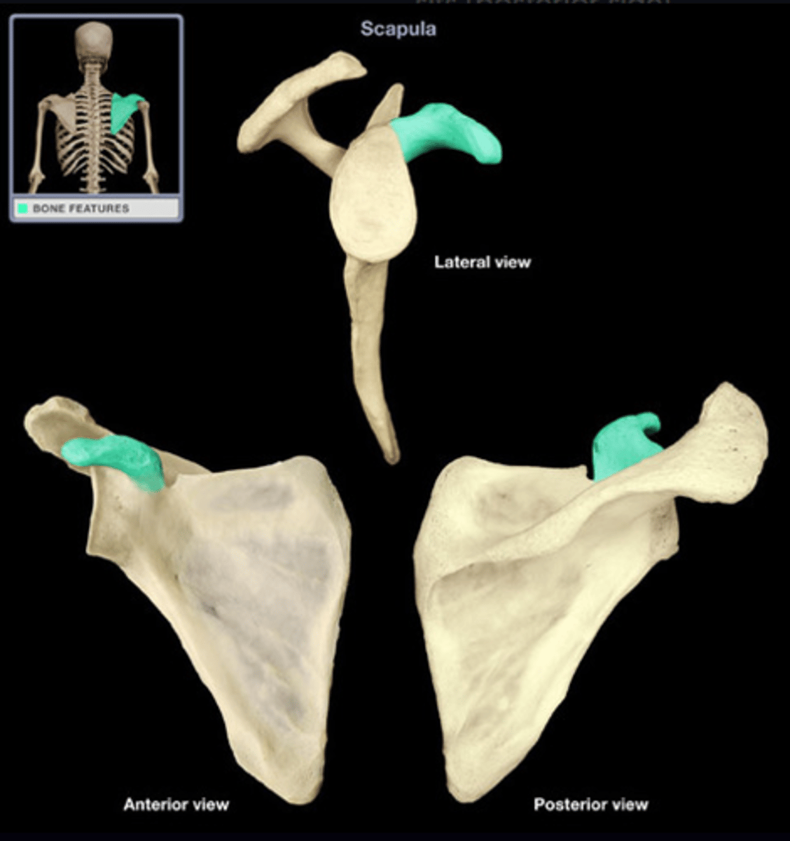
What is the significance of the coracoid process?
The coracoid process is a beak-like projection superior to the glenoid cavity, projecting anterolaterally and involved in muscle attachment.
How is the scapula positioned in AP radiological studies?
In AP radiological projections, the scapula overlaps with the ribs and thoracic cage, requiring the arm to be raised to decrease overlap.
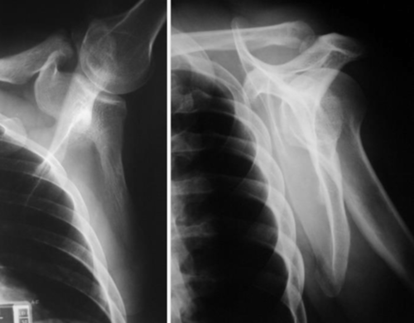
What is the purpose of the scapular Y view (lateral scapula shoulder) in radiology?
The scapular Y view is orthogonal (perpendicular) to the AP shoulder view, used to assess suspected dislocations and fractures of the scapula by showing the coracoid and acromion process in profile.
RECALCO... good for
1) suspected dislocations/ fractures
2) evaluation of coracoid process and acromion
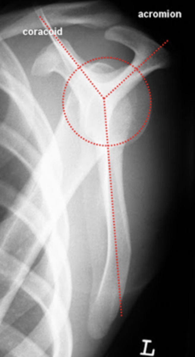
Be able to differentiate between the AP and Y lateral view
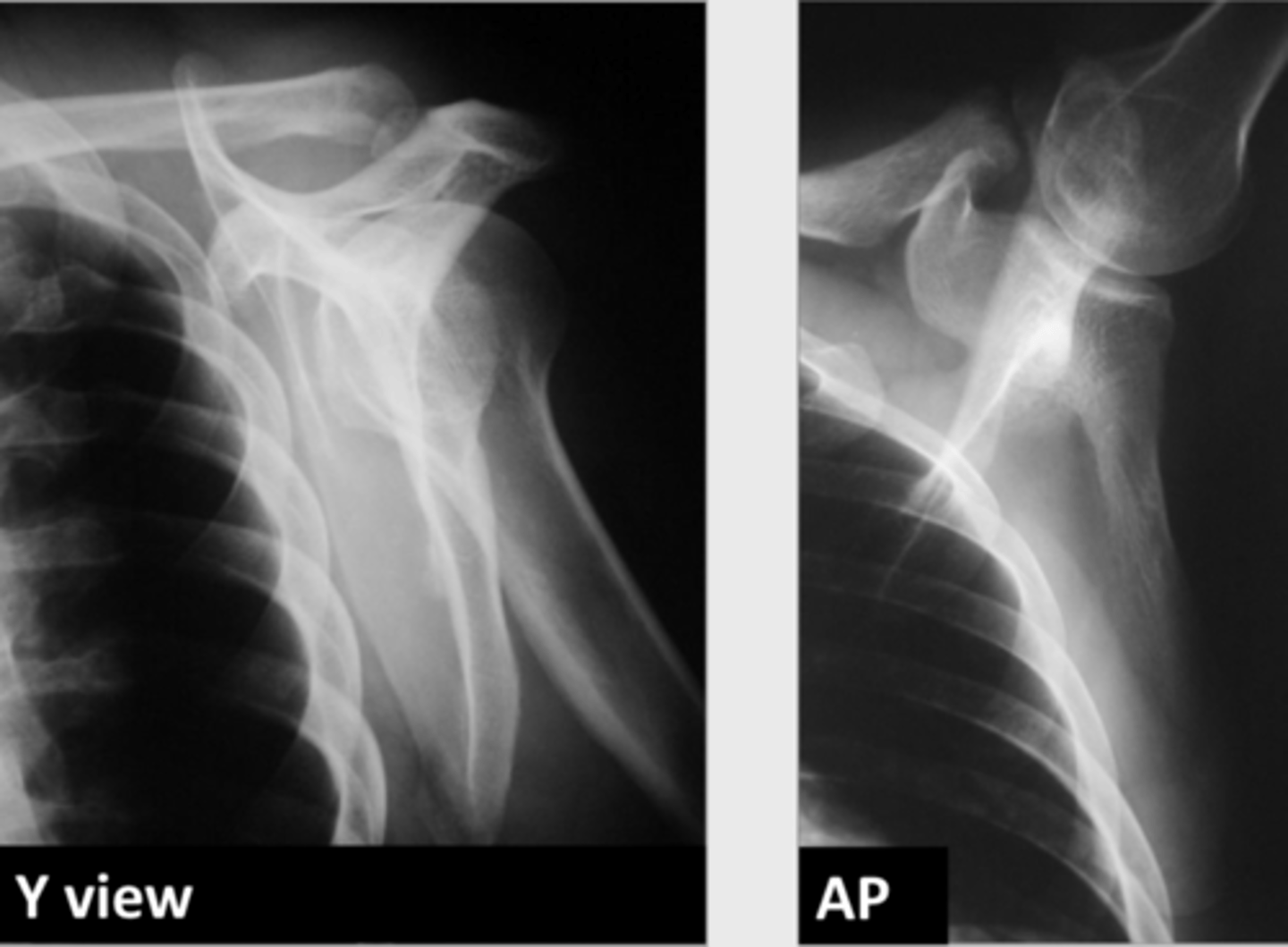
1) acromial process with the clavicle
2) the upper area of the glenoid with the origin of the coracoid
3) the gleno-humeral joint
4) the inferior part of the scapula (4).
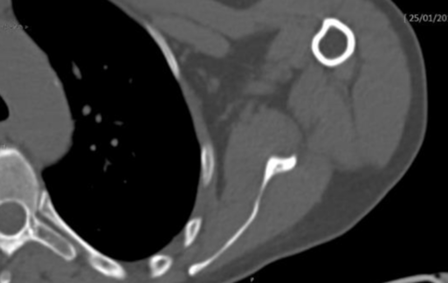
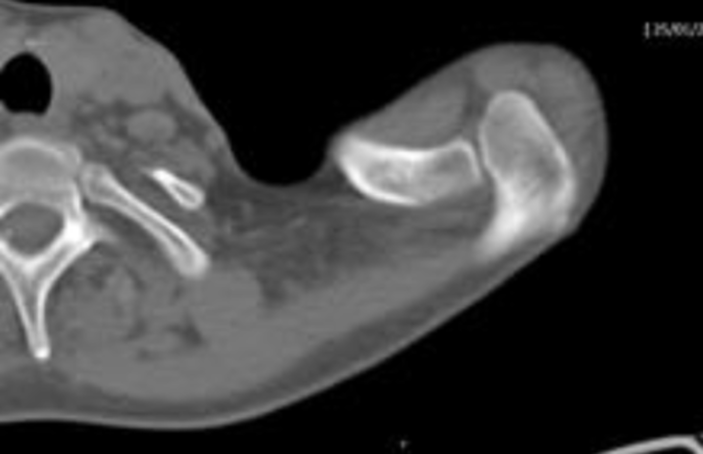
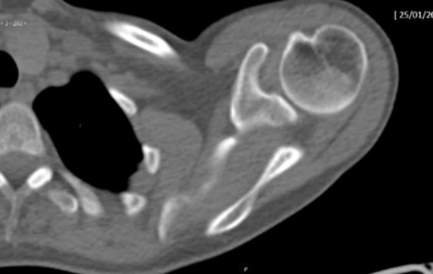
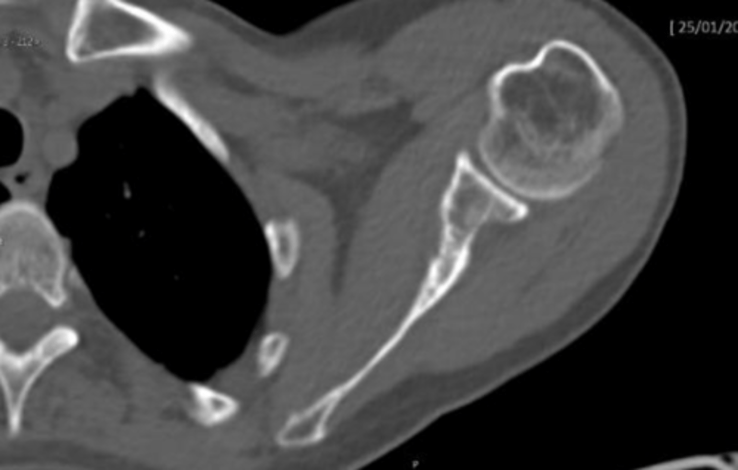
Compare the utility of MRI to CT in studying the scapula.
MRI can study the scapula but provides images that are not as anatomically detailed as those obtained from CT imaging.
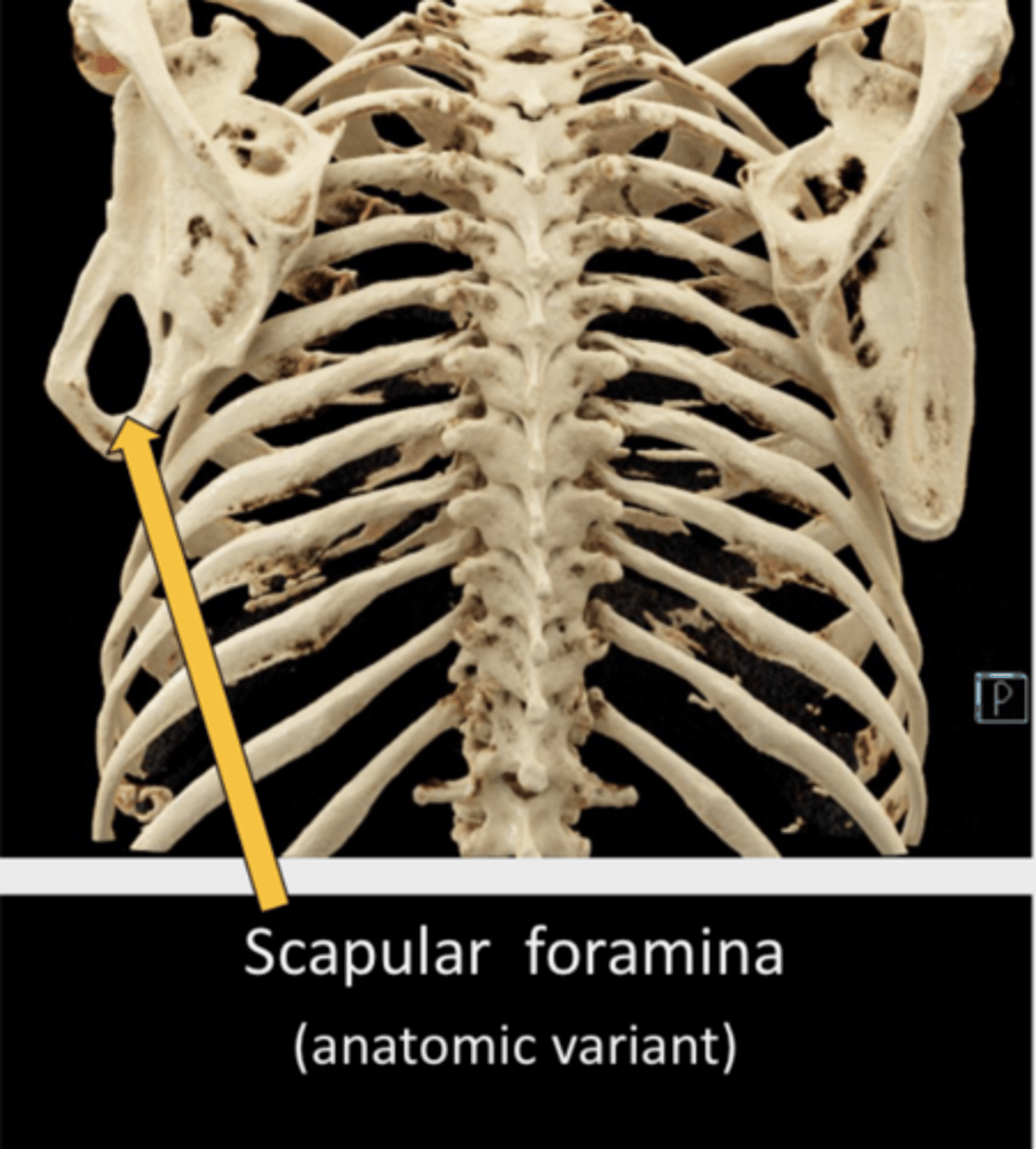
Describe an anatomic variant of the scapula
scapular foramina
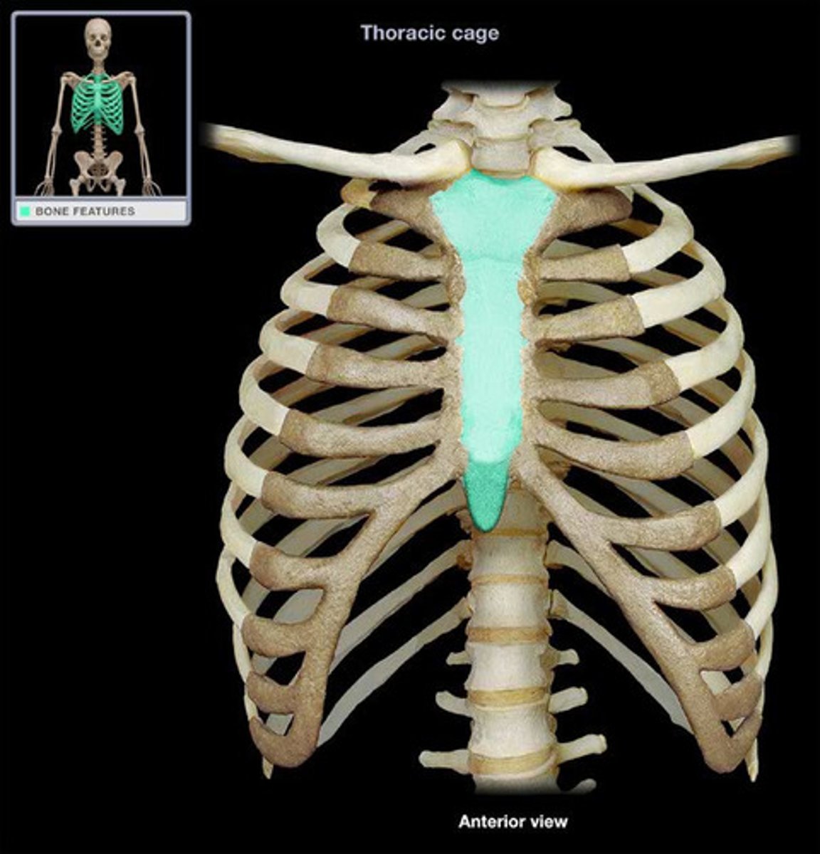
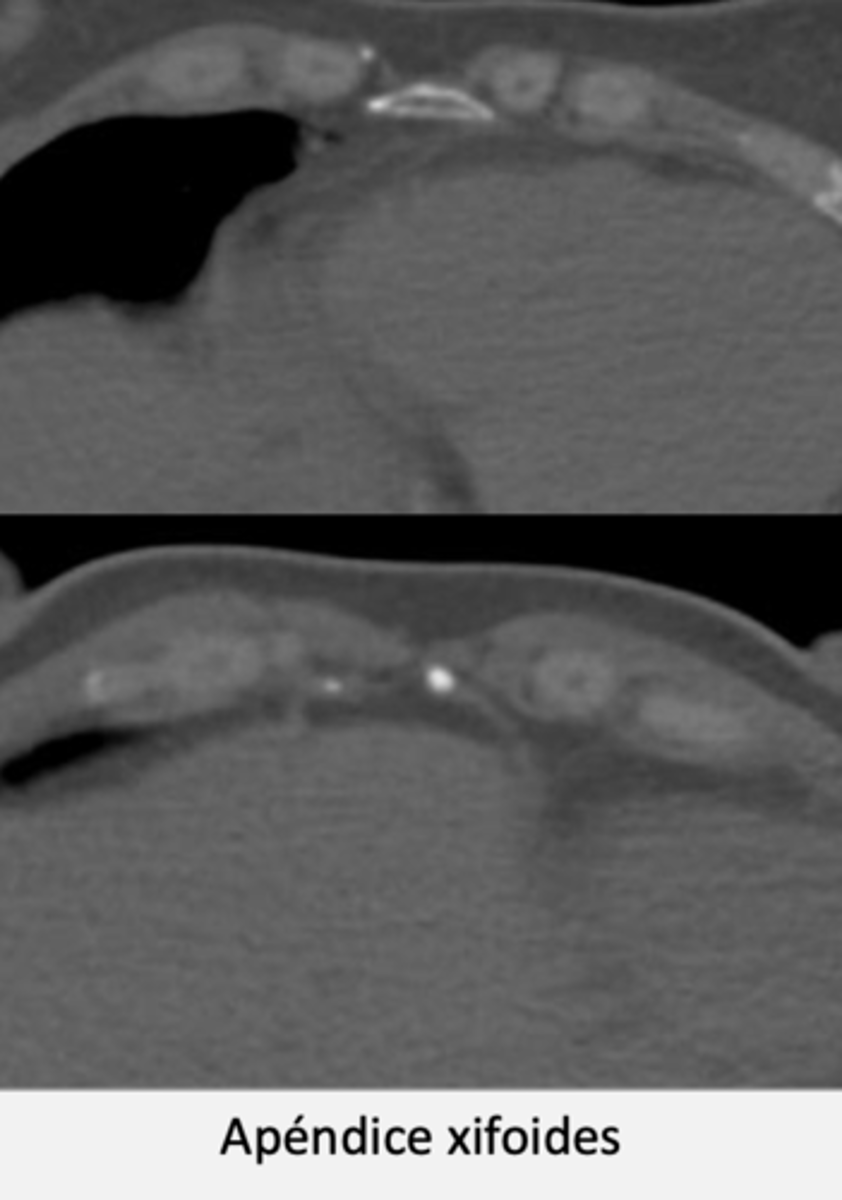
What is the sternum's role in the body?
The sternum is a long, flat bone in the central chest area, connecting to the ribs via cartilage to form the anterior part of the thorax, protecting thoracic organs from trauma.
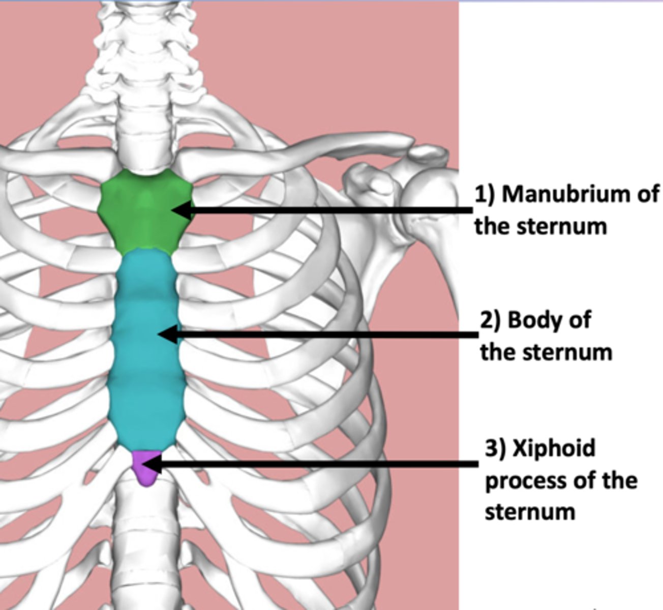
What are the three regions of the sternum?
1) the manubrium
2) the body,
3) xiphoid process.
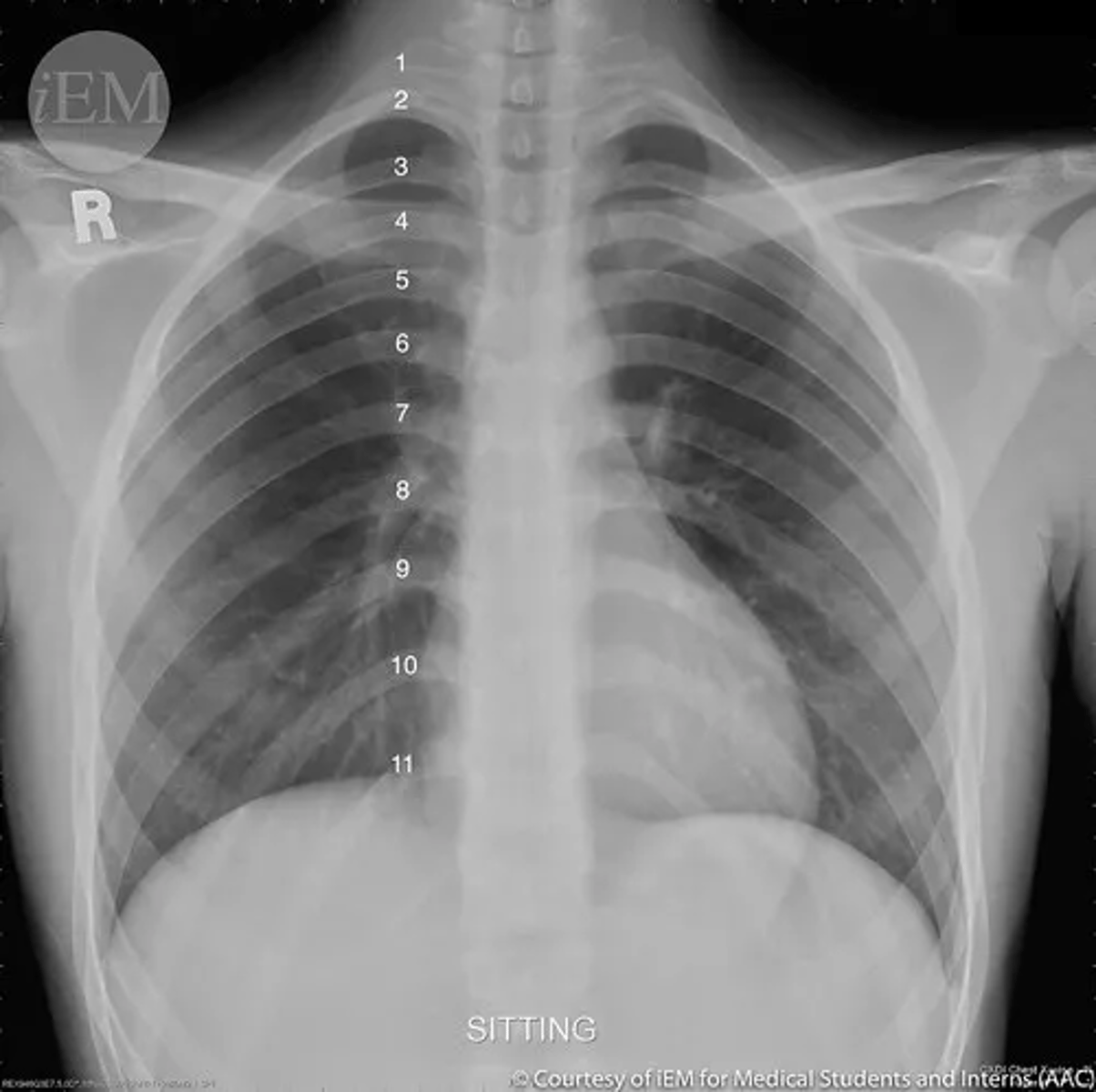
Which radiological view is preferred for studying the sternum and why?
The lateral view is preferred for studying the sternum because it allows for the appreciation of its shape and alignment.
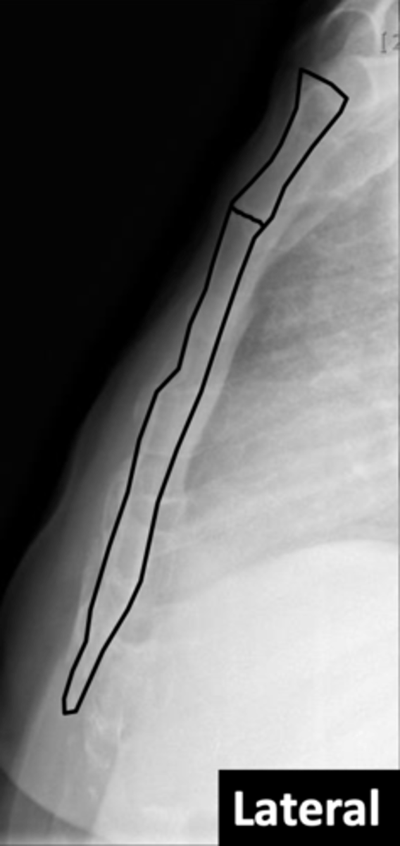
Why is an AP projection not useful for the sternum?
An AP projection is not useful for the sternum because it is superimposed with the vertebral spine and mediastinum, making differentiation of its parts difficult.
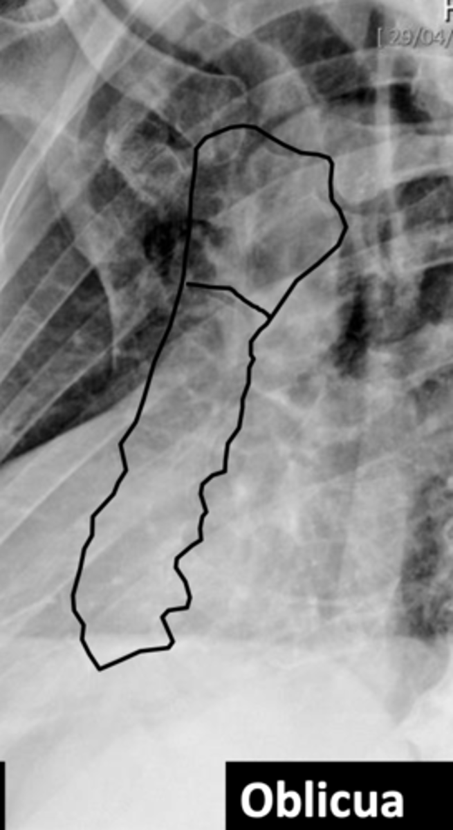
What alternative projection is used to study the sternum and what does it show?
A left anterior oblique projection is used to better differentiate the sternum's portions (manubrium, body, and xiphoid process) by avoiding overlap with the spine and mediastinum.
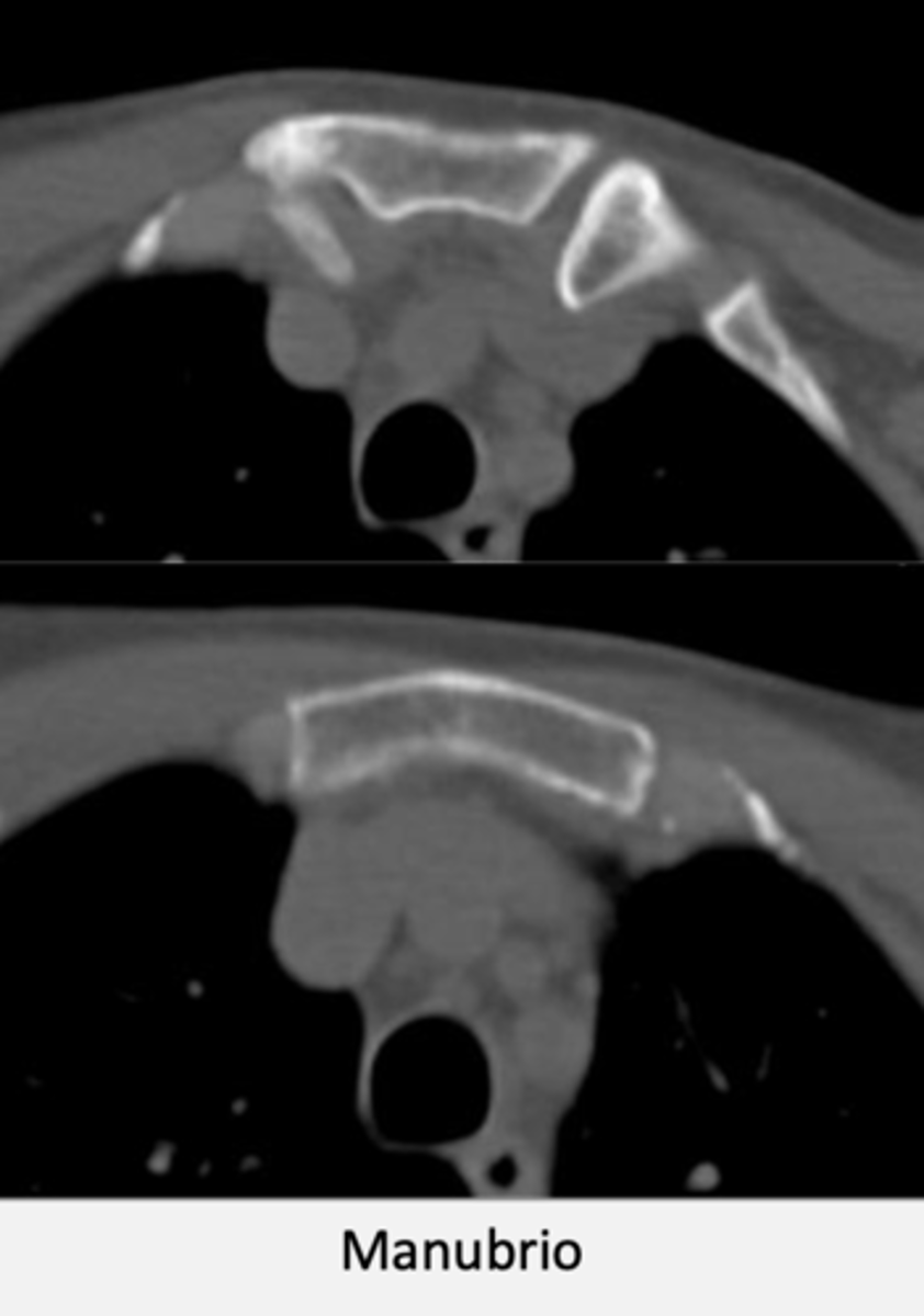
CT manubrium
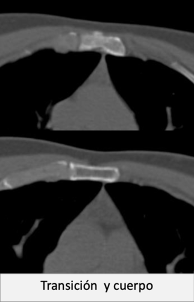
CT sternal body
CT xiphoid process
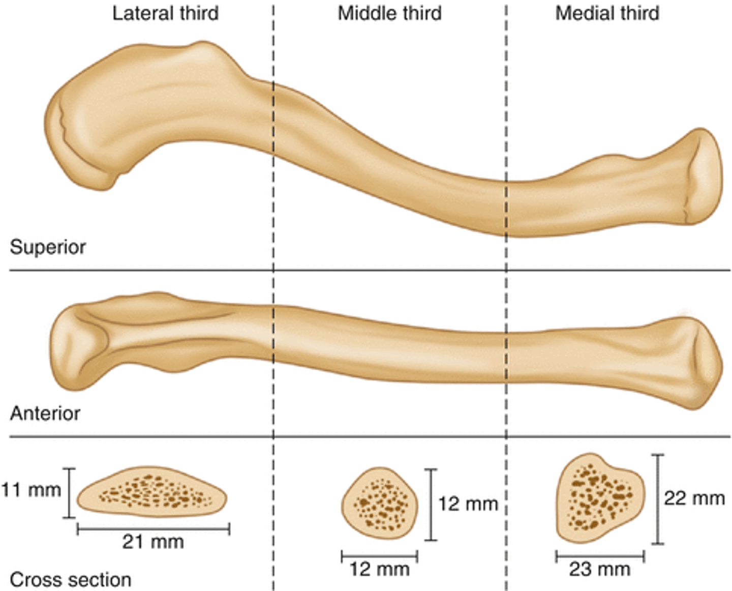
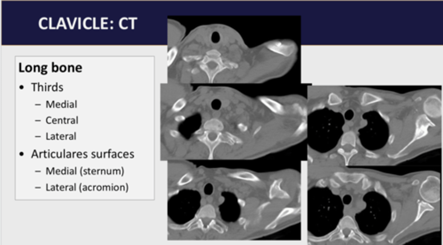
How is the clavicle structurally characterized?
The clavicle is a long, superficial S-shaped bone, subdivided into three portions: medial (proximal), central, and lateral (distal).
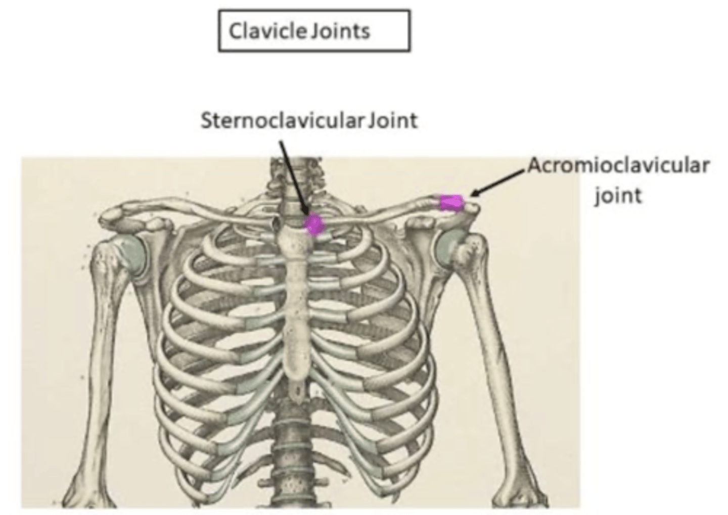
What are the two articulations of the clavicle?
The clavicle articulates at two planar (2 planes) diarthrosis joints:
1) sternoclavicular joint
2) acromioclavicular joint
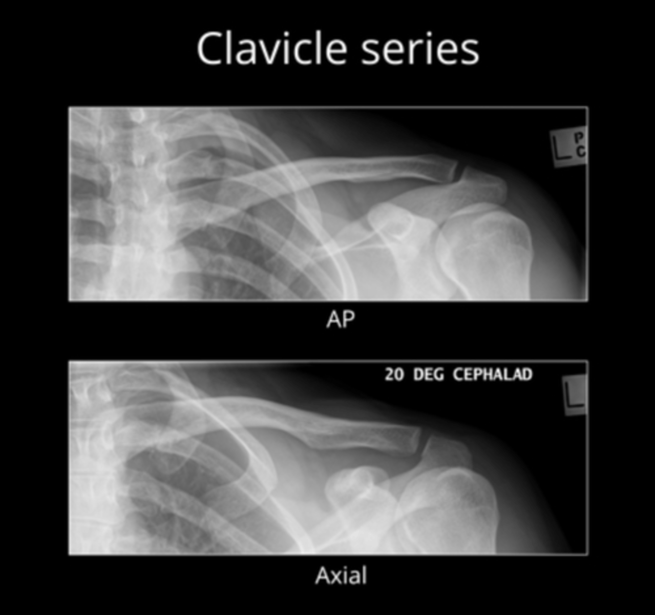
What is the standard radiological study for the clavicle?
The standard radiological study for the clavicle is usually an AP (anteroposterior) image.
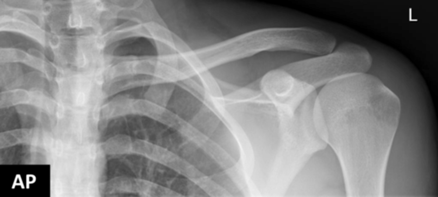
How is a complementary radiological projection of the clavicle obtained?
A complementary projection is obtained by obliquing the incidence of the X-ray beam to achieve a partially infero-superior vision.
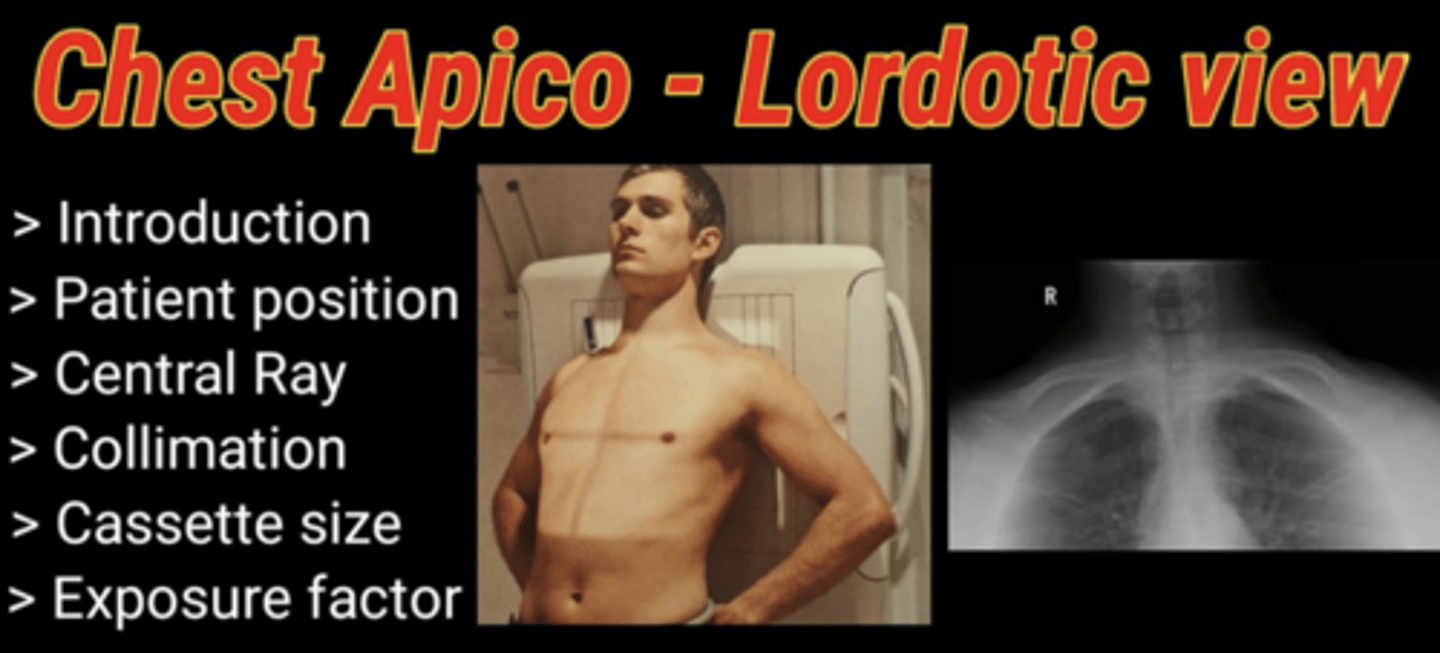
Why might an apical or lordotic view be used in chest x-rays involving the clavicle?
An apical or lordotic view may provide greater detail of the lung apices, as the clavicles can obscure subtle lesions when superimposed over the lungs in standard views.
This radiographic technique is used to move the clavicles out of the way, reducing overlap with upper parts of lungs in the image. Provides greater detail and visibility of lung apices, allow for detection of subtle lesions or abnormalities that might be obscured by clavicles in standard chest X-ray views.
Clavicle CT scan
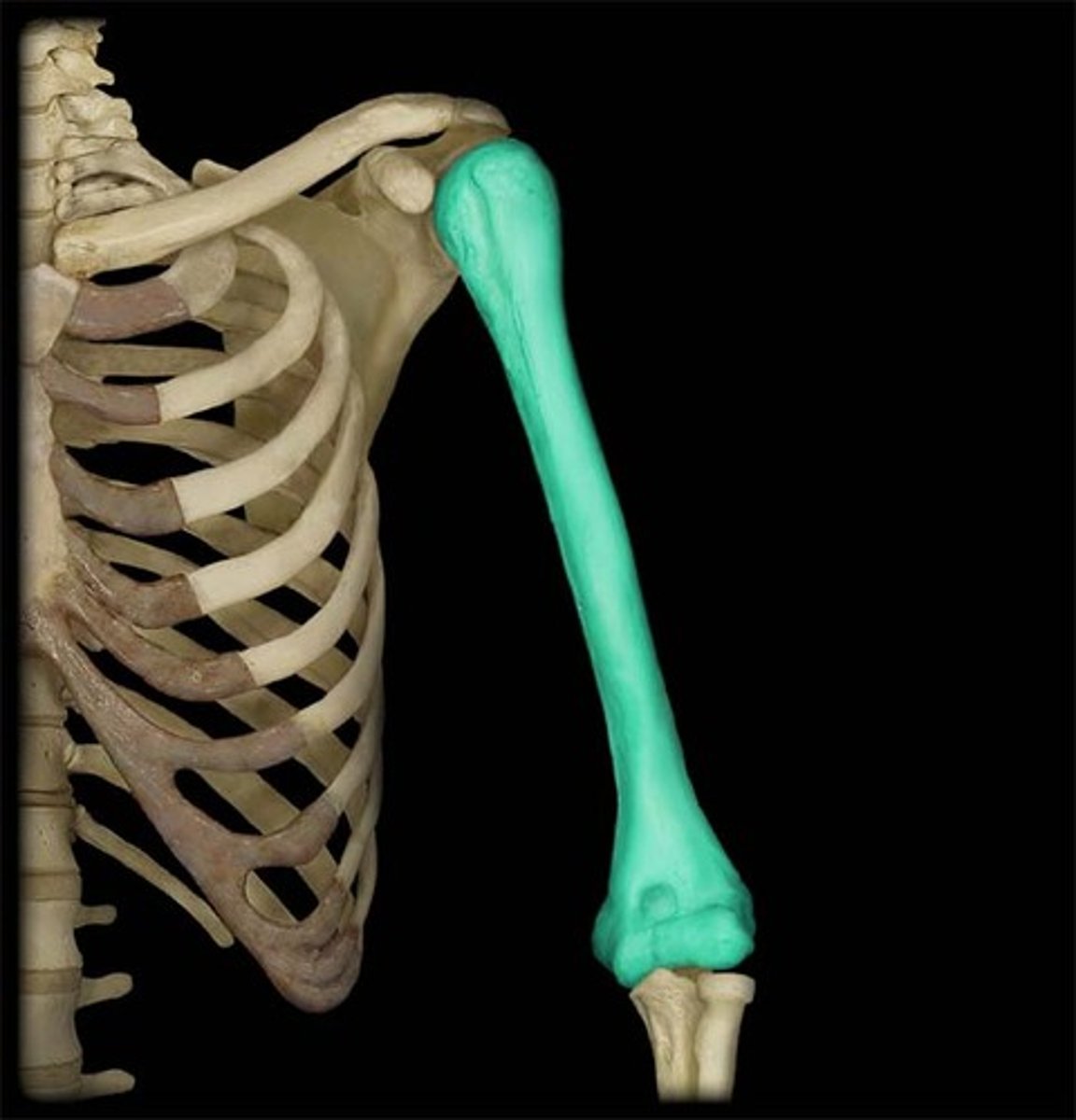
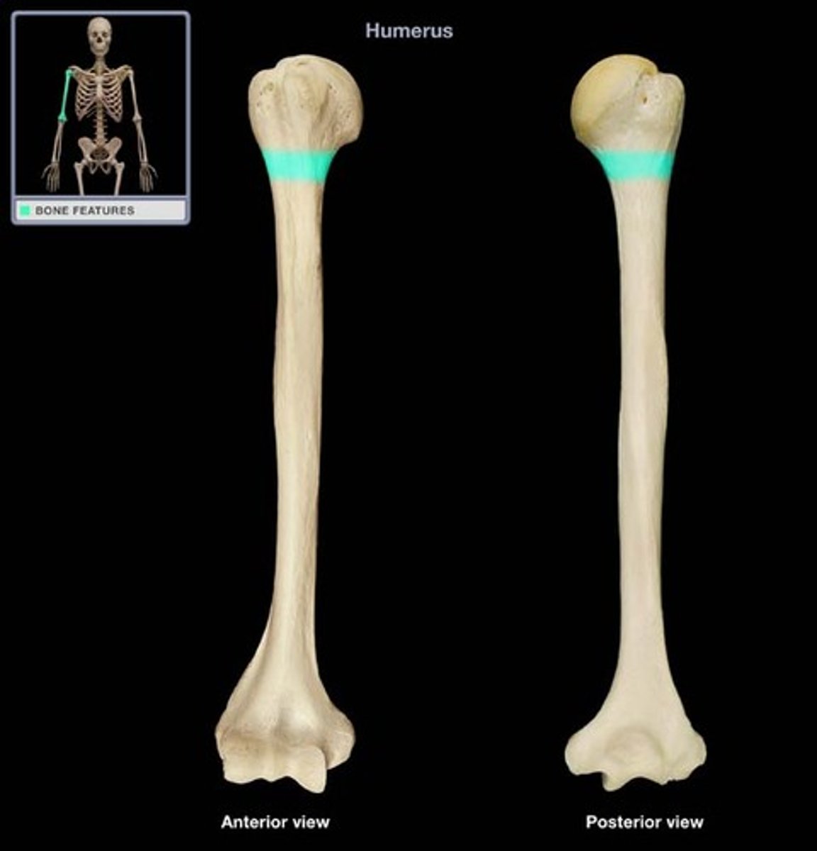
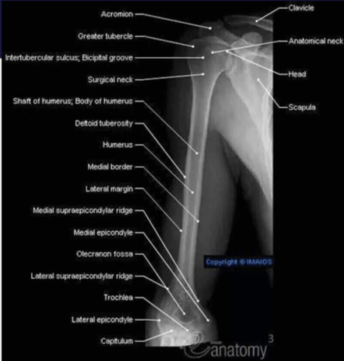
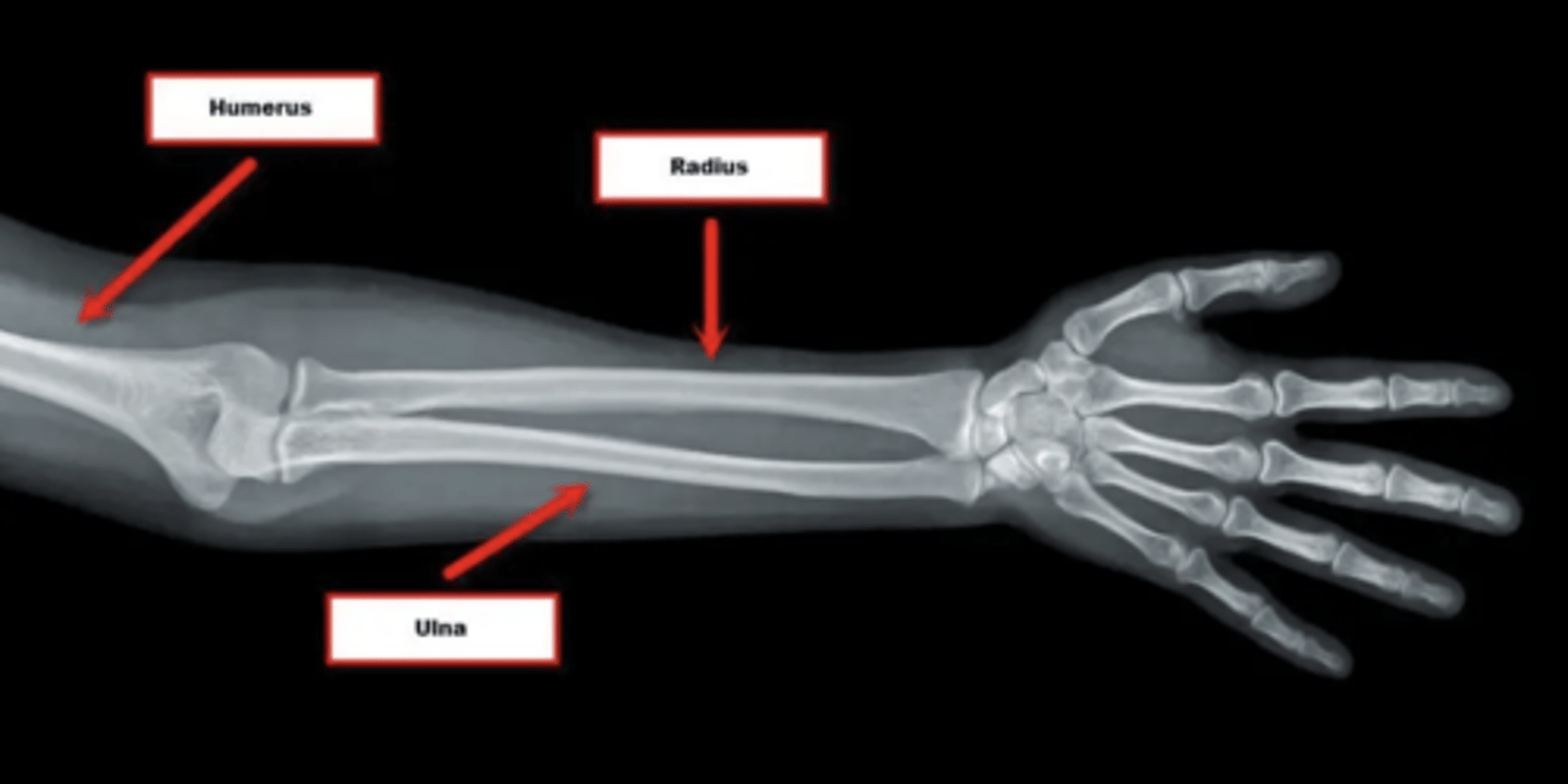
What bones does the humerus connect?
The humerus connects the scapula and the two bones of the forearm (radius and ulna).
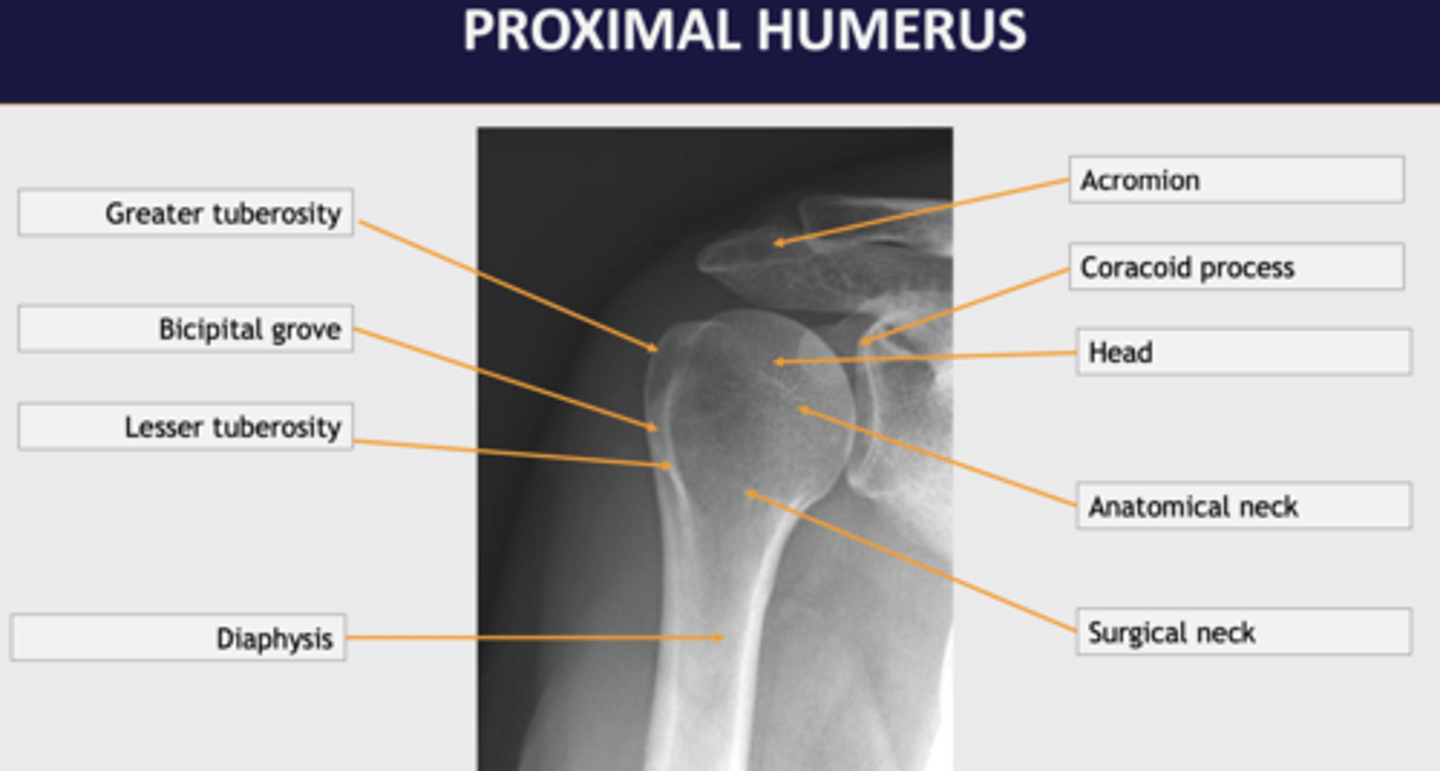
Describe the proximal end of the humerus.
The proximal end of the humerus consists of a rounded head, a narrow anatomical neck, and two short processes known as the greater and lesser tubercles, with the head articulating with the glenoid fossa of the scapula.
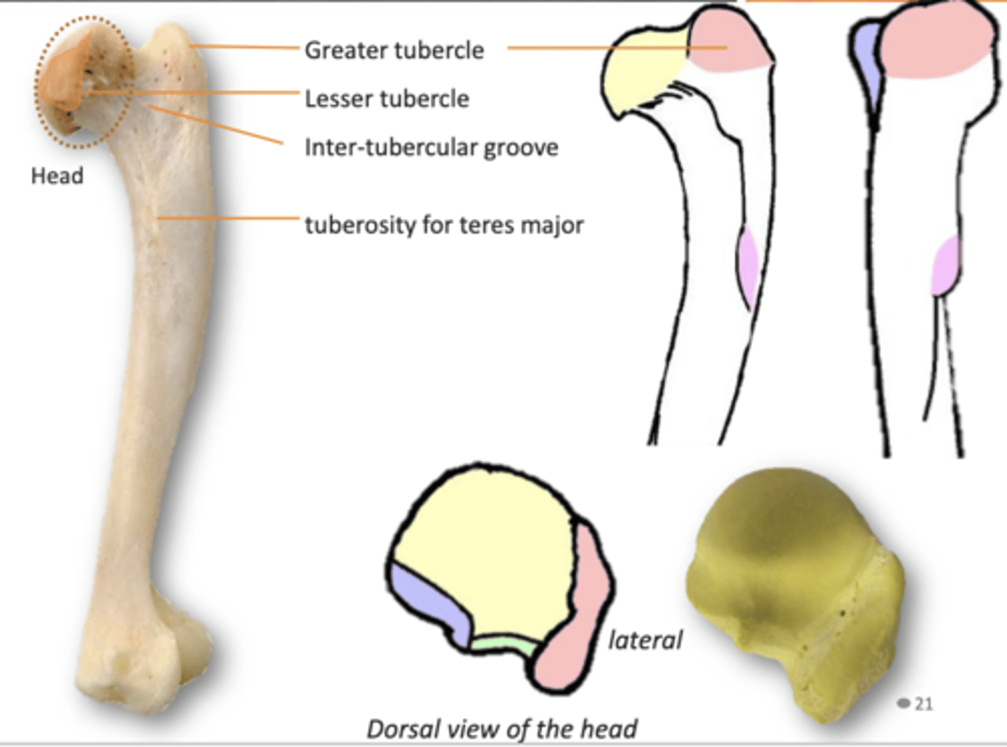
What is the significance of the humerus's anatomical neck?
The anatomical neck is a slight narrowing below the articular surface of the head, where the shoulder joint capsule is attached.
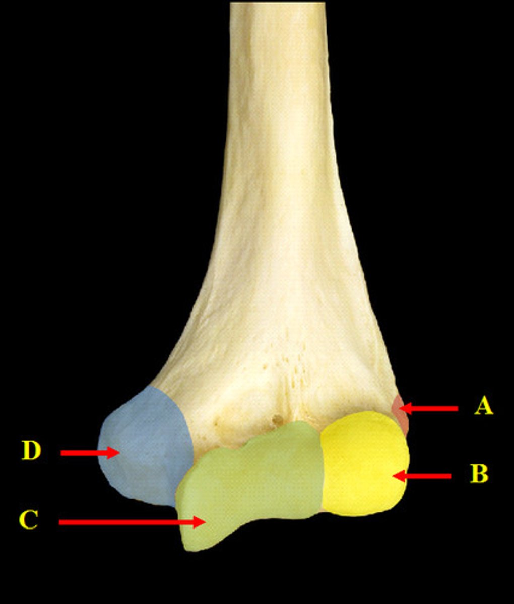
Where does the supraspinatus muscle attach on the humerus?
Attaches to the upper facet of the greater tubercle of the humerus.

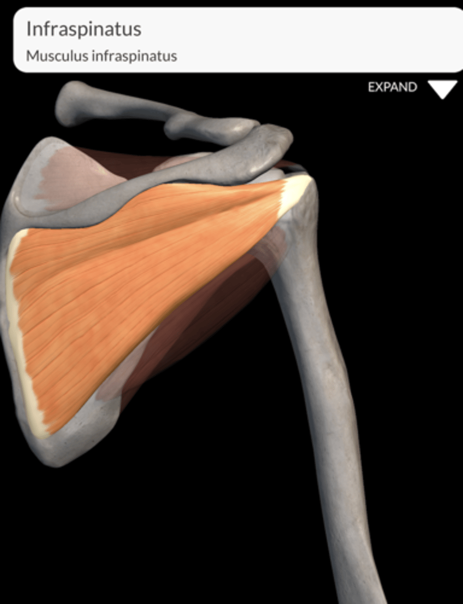
Where does the infraspinatus muscle attach on the humerus?
Attaches to the middle facet of the greater tubercle of the humerus.
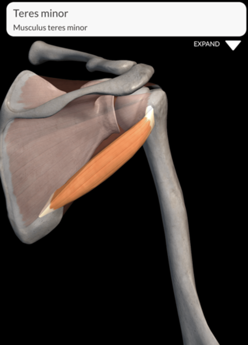
Where does the teres Minor muscle attach on the humerus?
Attaches to the lower facet of the greater tubercle of the humerus.
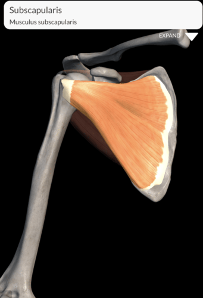
Where does the subscapularis muscle muscle attach on the humerus?
Attaches to the lesser tubercle of the humerus.
What is located in the intertubercular sulcus of the humerus?
The long tendon of the biceps brachii and a branch of the ascending circumflex humeral artery are located within the intertubercular sulcus.
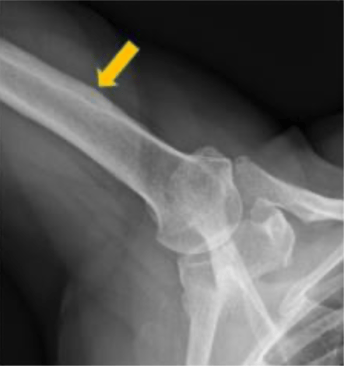
Why is the area just inferior to the tubercles referred to as the surgical neck of the humerus?
It's called the surgical neck due to its tendency to fracture.
Describe the diaphysis of the humerus.
The shaft or diaphysis of the humerus is cylindrical in its upper portion and more triangular distally, with a lateral cortical irregularity called the deltoid tuberosity for the deltoid muscle insertion.
What constitutes the distal end of the humerus?
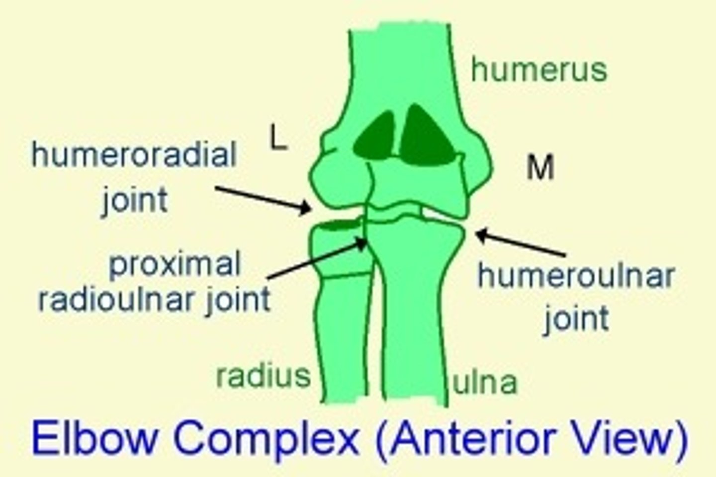
The distal end consists of an articular part with a modified condyle (medial trochlea and lateral capitellum) that articulates with the ulna and radius, and a non-articular part with medial and lateral epicondyles and three fossae (olecranon, coronoid, and radial fossae).
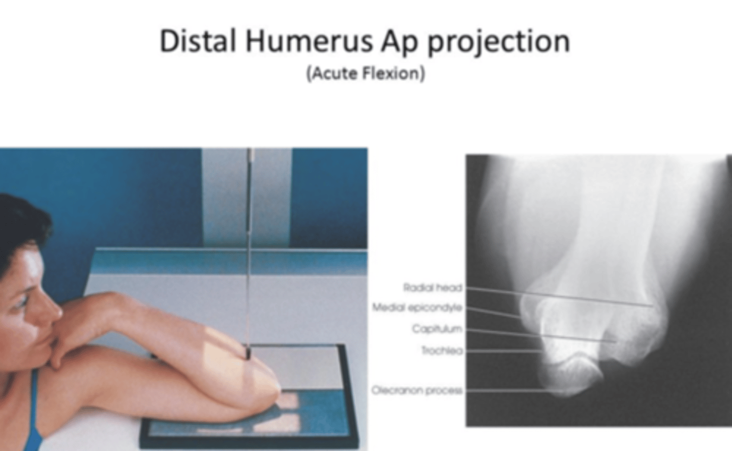
What are the key radiological projections for studying the humerus?
1) AP and lateral: general assessment
2) axial projection: shoulder, detailed views of the proximal region (head).
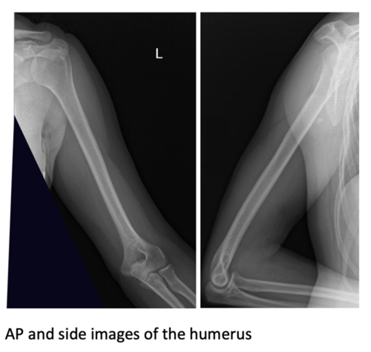
Radiology of humerus (full)
Radiology of humerus (proximal)
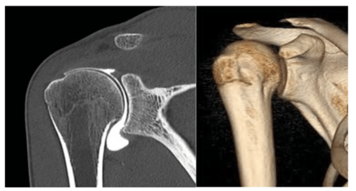
CT is often used in cases of fractures, to determine the number of fragments, dis- placements, angulations... 2D and 3D im- ages can be obtained. Coronal 2D image of CT arthrography (intraarticular con- trast) and 3D shoulder VR.
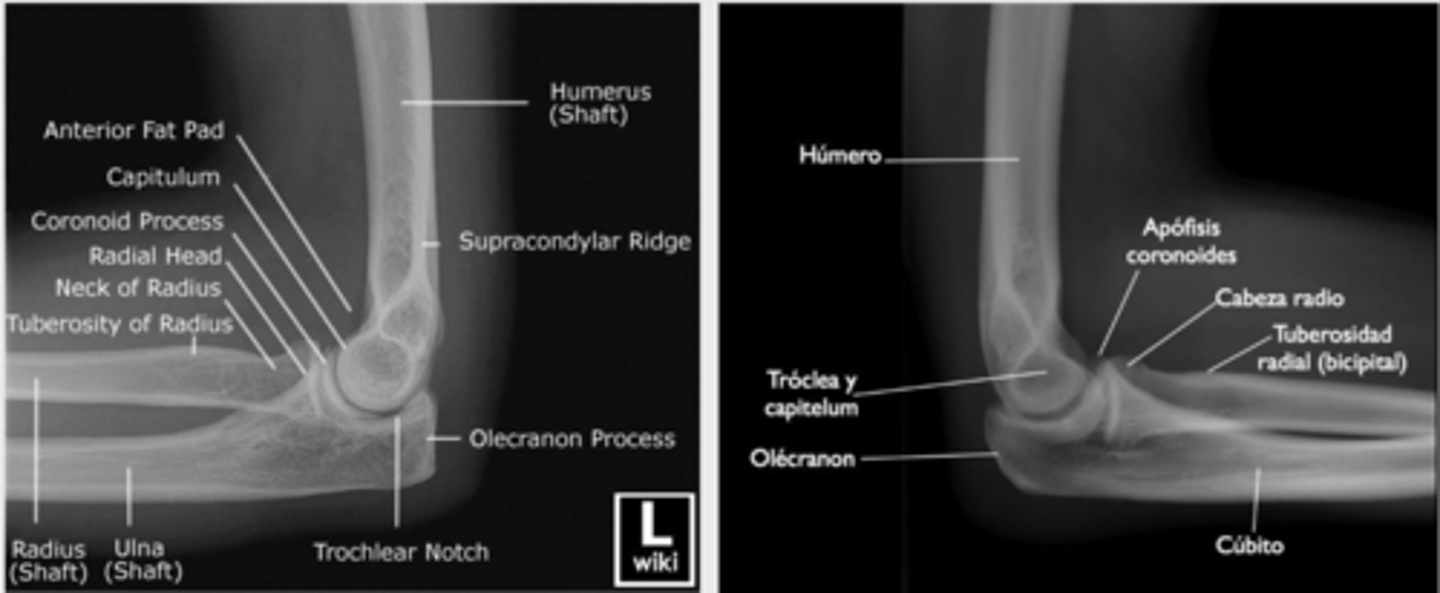
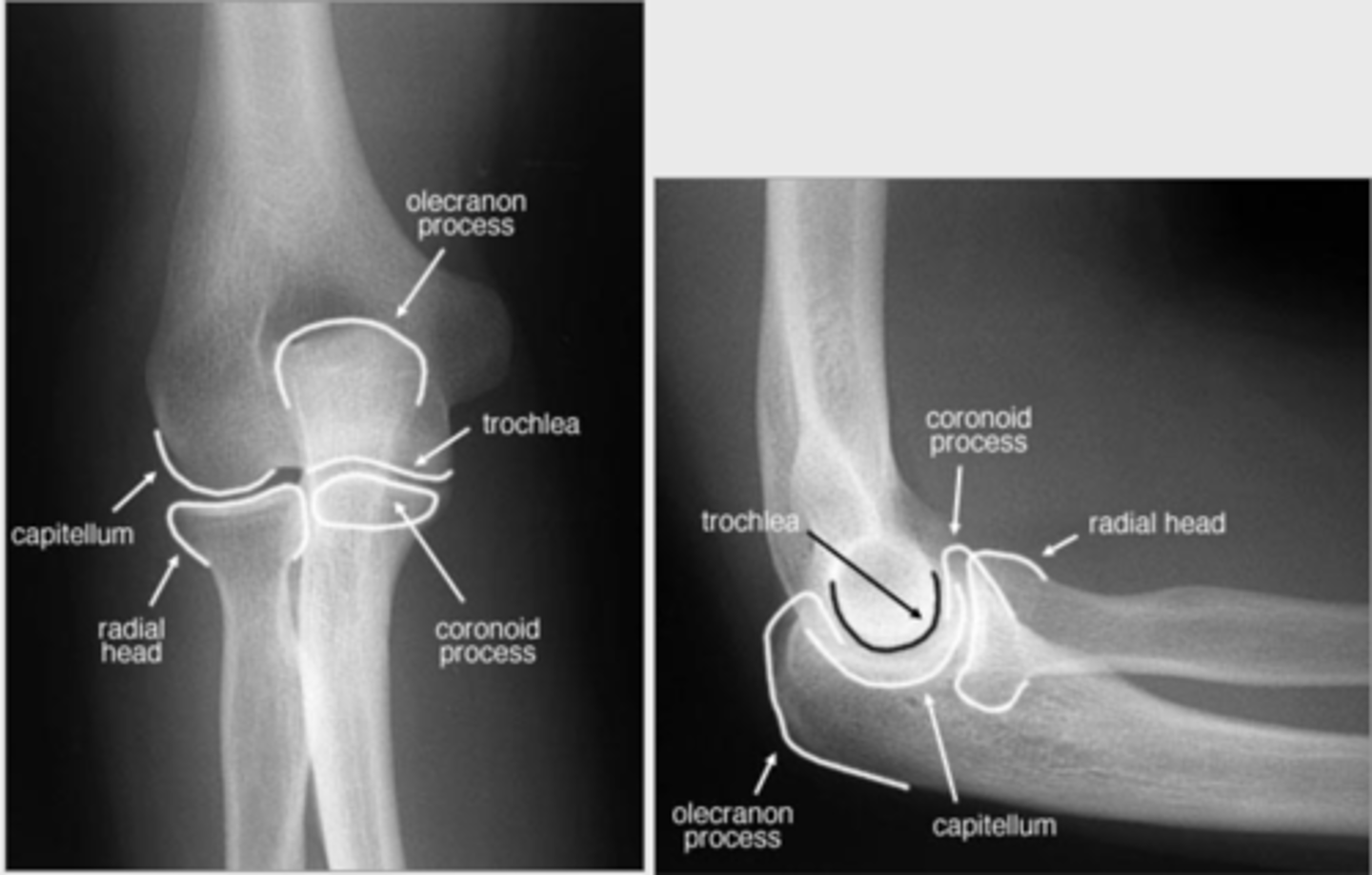
What radiographic projections are used to study the elbow?
The elbow is studied using AP (anteroposterior) and lateral projections.
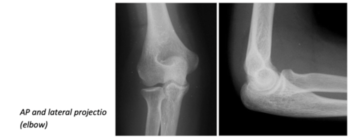
What structures are observed in the anterior view of the elbow?
In the anterior view, the capitulum (medial) and the trochlea (lateral) are observed, with the epitrochlear process (or medial epicondyle) being the most prominent.
How is the extension of the elbow limited?
The extension of the elbow is limited by the olecranon morphology, which fits into the olecranon fossa of the humerus, preventing larger extension.
What is noted about the articular surface of the radius in relation to the humeral condyle?
The articular surface of the radius is flat and confronted with the round-shaped humeral condyle, allowing wide mobility in both flexo-extension and rotation.
How are the elbow structures recognized in X-ray projections?
In lateral projection, the central axis of the radius aligns with the center of the region, with the radius being somewhat anterior to the ulna.
What happens in the AP projection performed during extension for the elbow?
The olecranon superimposes the humerus in the AP projection performed in extension.
"Superimposes" means to place or lay one thing over another, typically so that both are still evident.
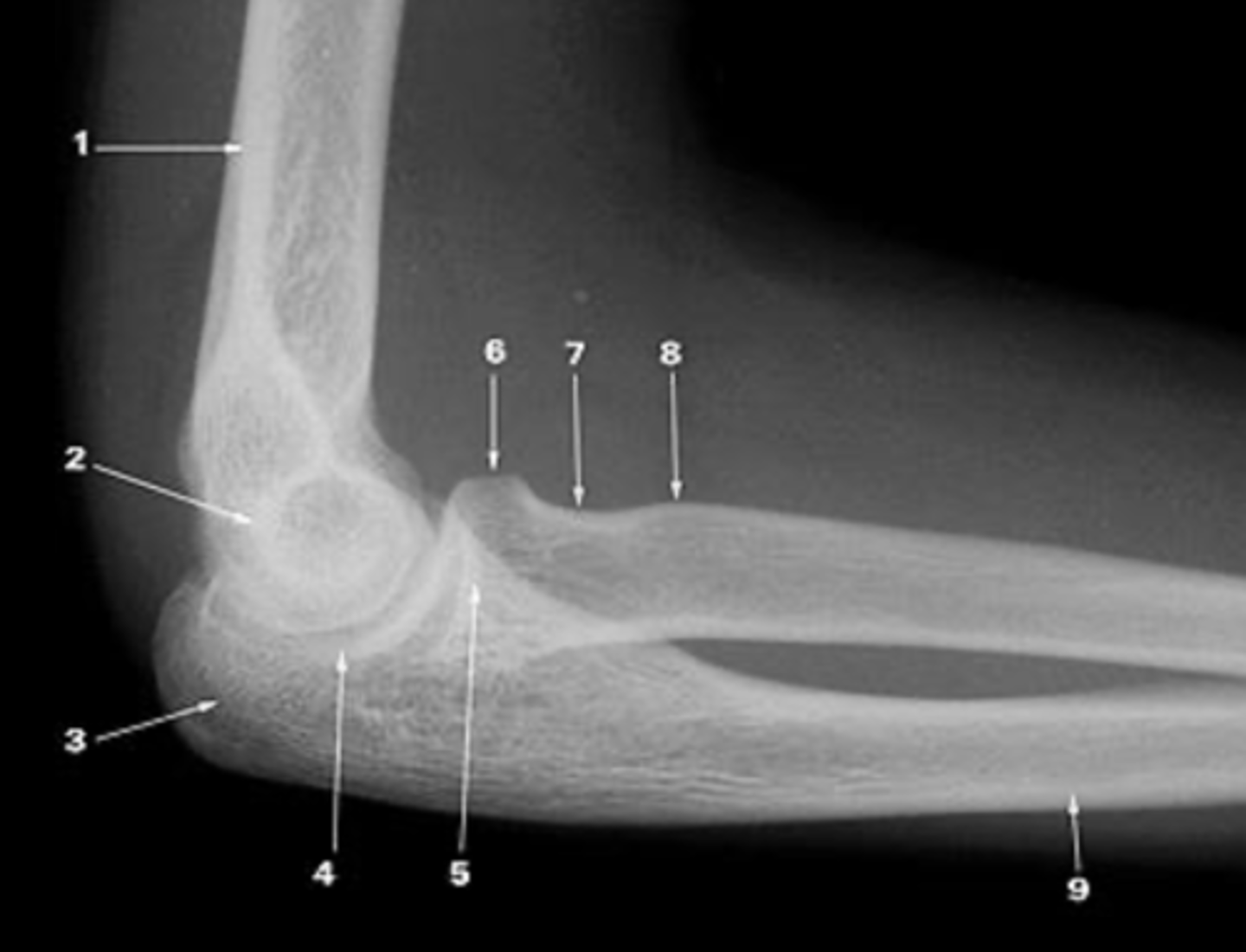
AP elbo x-ray
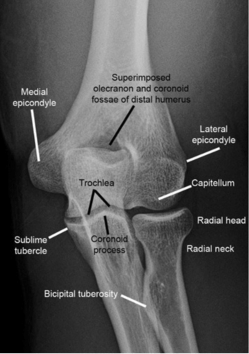
1. Lateral cortical of the humerus

2. Medial cortical of the humerus
3. Olecranon fossa

4. Medial epicondyle (Epitrochlea)

5. Lateral epicondyle

6. Capitellum (Condyle)

7. Olecranon

8. Trochlea

9. Coronoid process

10. Radio-ulnar joint

11. Head of the radius

12. Neck of the radius

13. Bicipital tuberosity

14. Ulna

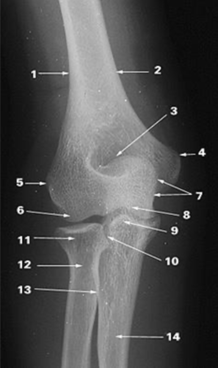
Elbow X-ray AP and lateral
1. Cortical humerus

2. Trochlea

3. Olecranon

4. Joint surface

5. Coronoid process of ulna
6. Head of radius
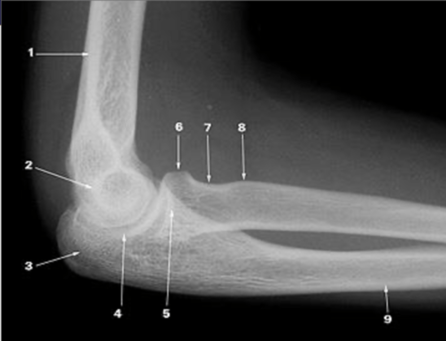
7. Neck of radius

8. Biccipital tuberosity

9. Ulna

What additional structures are found in the elbow's fossae?
The fossae of the elbow contain synovial joint fluid and fat pads.
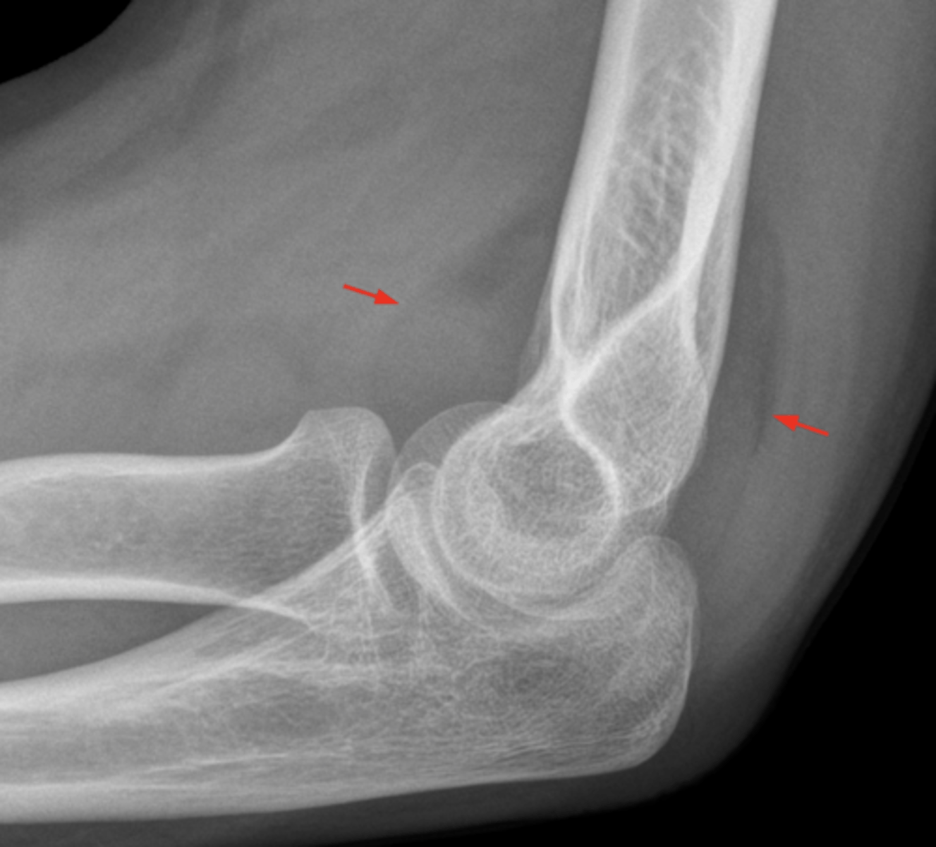
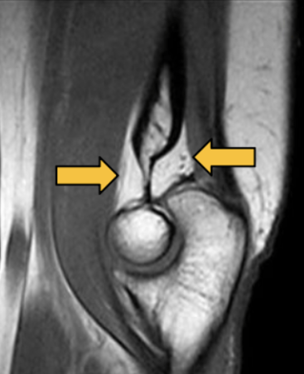
What is depicted in a sagittal MRI image of the elbow?
A sagittal MRI image of the elbow shows the coronoid and olecranon fossae, pointed and with fat inside.
What is the significance of the medial and lateral epicondyle processes?
The medial and lateral epicondyle processes serve as insertion points for different forearm muscle elements.
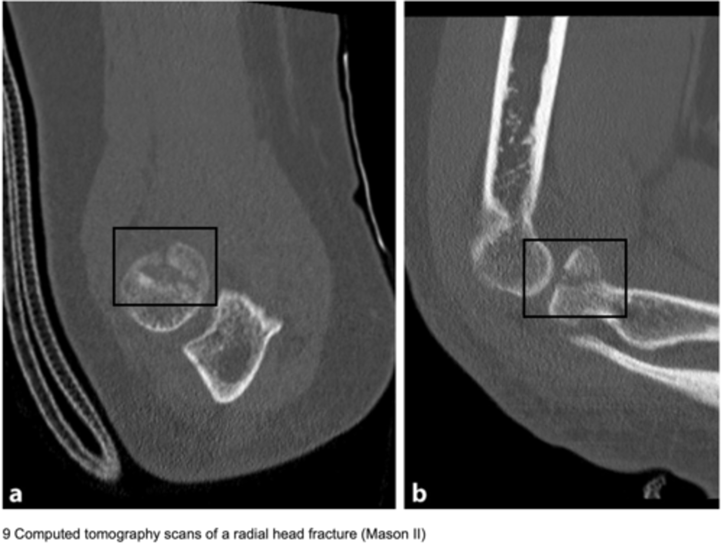
Which imaging technique is most used for detailed assessment of the elbow, especially for pathologies?
CT (Computed Tomography) is the most used technique for a detailed assessment of the elbow's radiological anatomy and the study of pathologies, mainly traumatic.
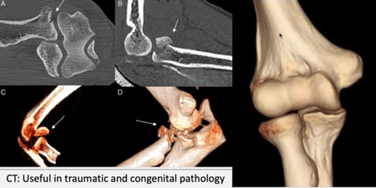
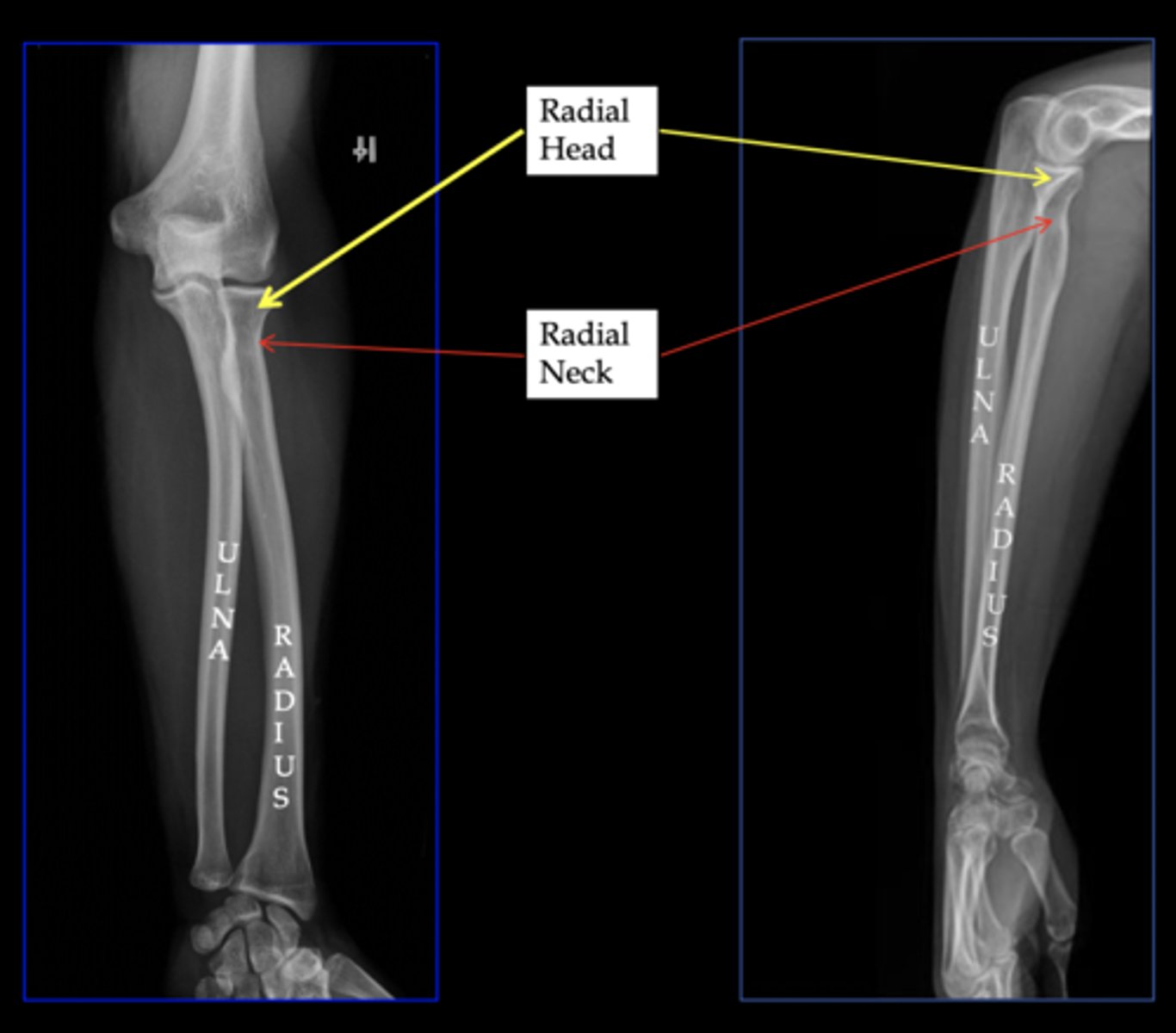
What can CT images reveal about the elbow?
CT images of the elbow can reveal conditions such as a fracture of the radius head.
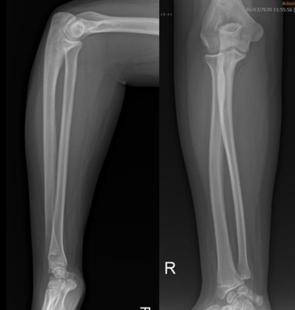
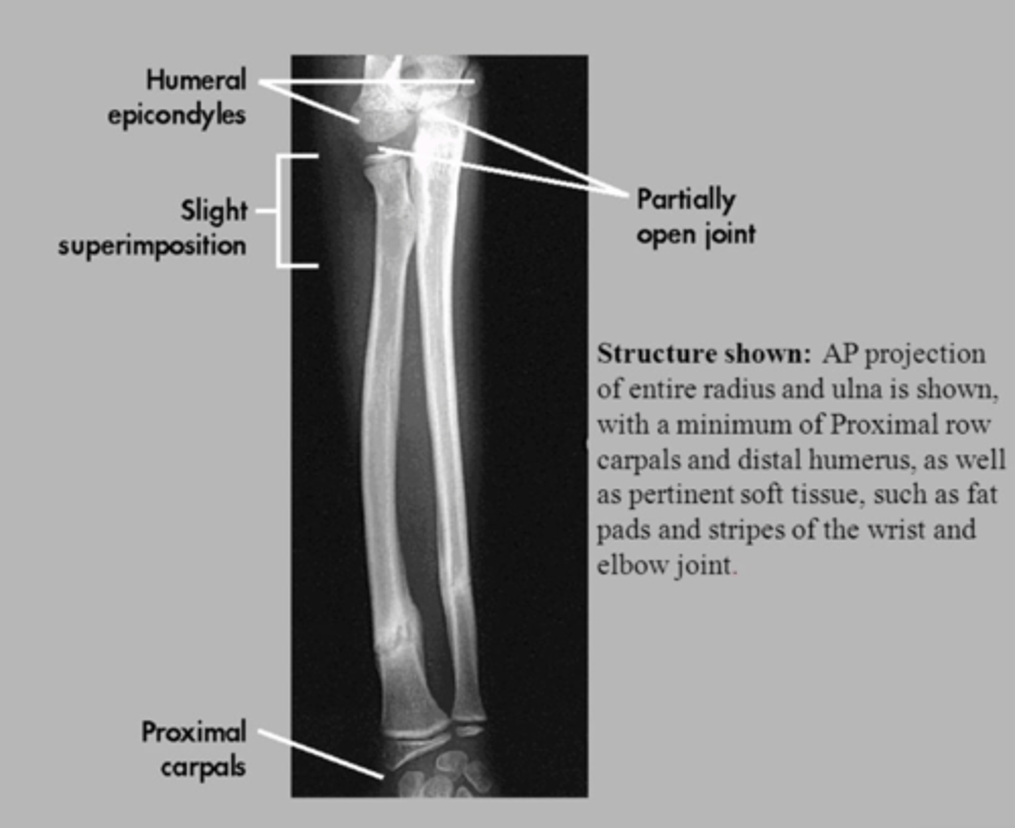
What radiographic projections are used for studying the forearm?
The forearm is studied using AP (anteroposterior) and lateral projections.
Describe the shape and curvature of the radius.
The body of the radius is prismoid in form, narrower above than below, and slightly curved, being convex lateralward.
How does the radius appear in lateral vs AP projections?
lateral projection: relatively straight
AP projection: presents discrete external convexity
Describe the ulna's morphology in lateral vs AP projections.
lateral projection: curved morphology
AP projection: relatively straight.
Compare the proximal and distal regions of the radius and ulna in terms of size.
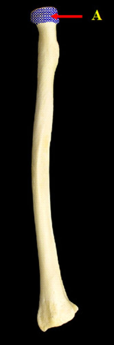
The head of the radius is relatively small compared to its distal epiphysis, while the ulna's proximal region is larger than its distal region.
Which carpal bones mark the central axis of the wrist?
The lunate and capitate bones mark the central axis of the wrist.
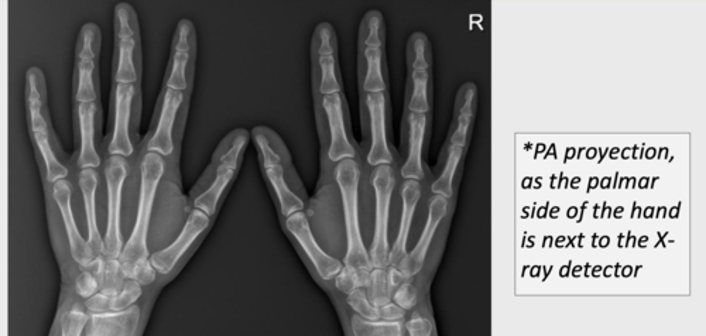
How is the radiological study of the wrist conducted?
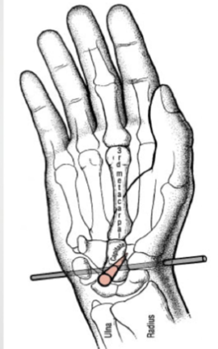
The wrist is studied radiologically with PA/AP and lateral projections, including specific projections for certain bones like the scaphoid (oblique projection) and pisiform, as well as assessments of stability in various positions.
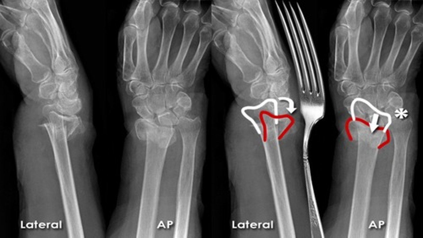
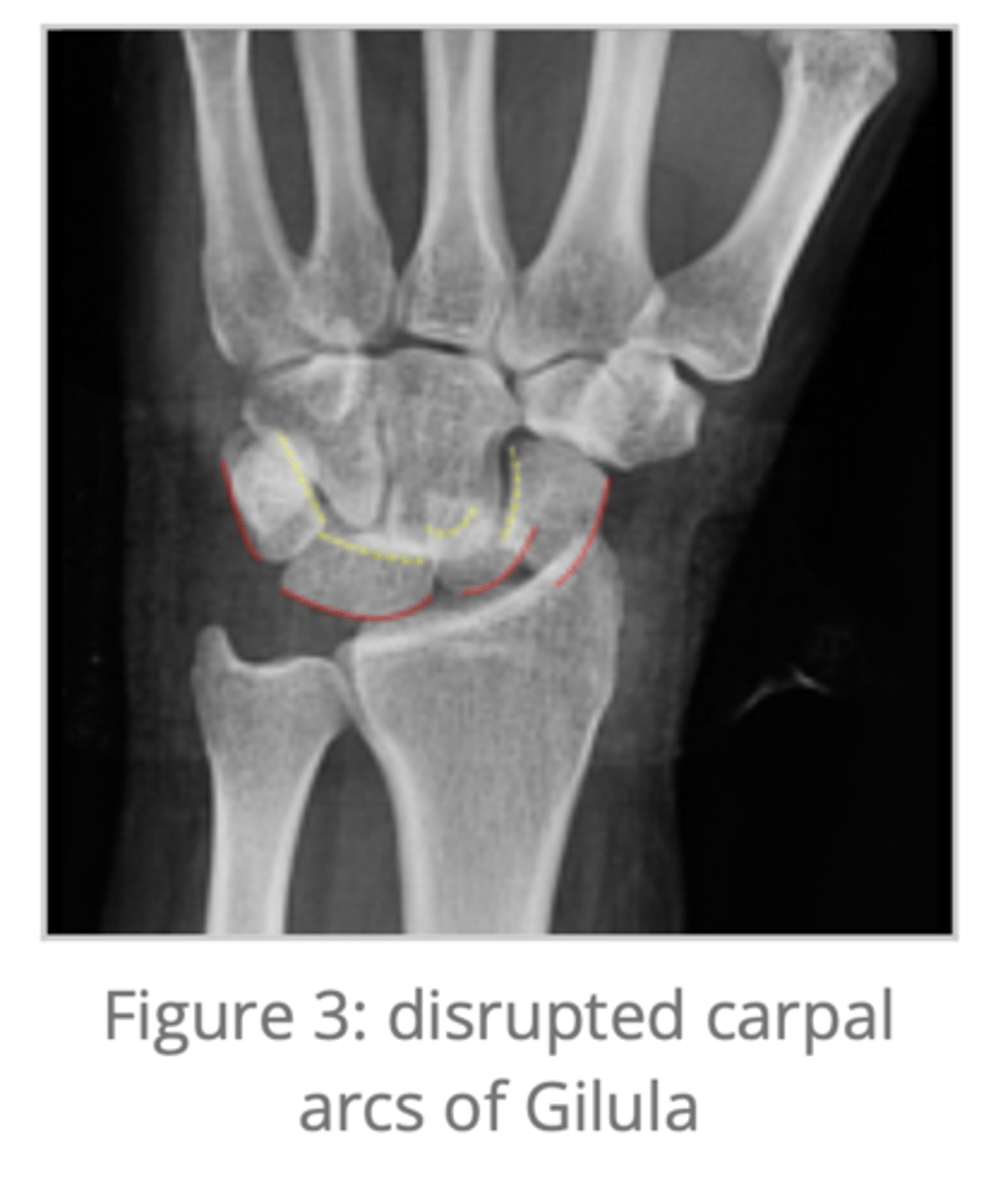
What are Gilula arcs or lines?
Gilula arcs or lines are three represented curved lines determining carpal alignment in the wrist, essential for identifying normal alignment or suspecting pathology.
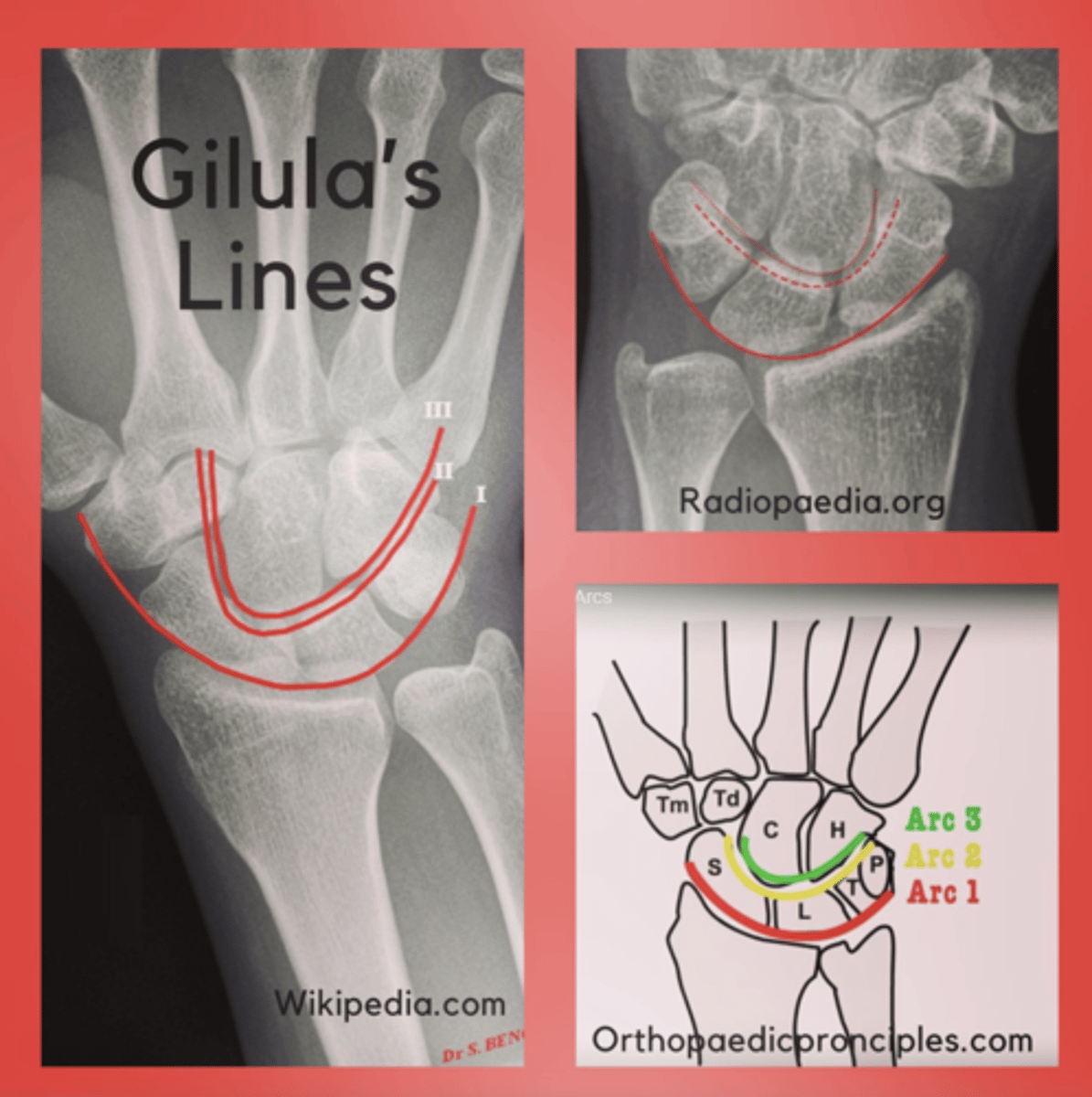
What does a disruption in Gilula arcs indicate?
Any loss of continuity in Gilula arcs should raise suspicion of wrist pathology.
Why is the scaphoid bone frequently fractured?
The lines of force during movement pass through the scaphoid bone due to its position relative to Gilula's arcs II and III, making it the most commonly fractured carpal bone.
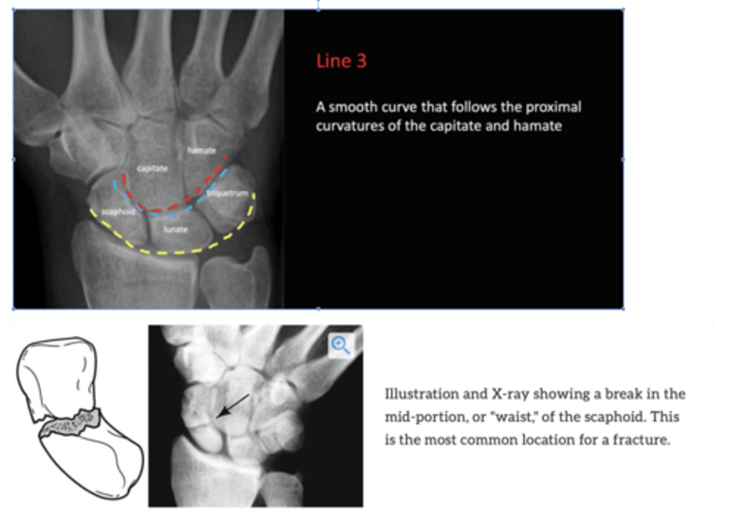
what can lead to carpal dysfunction?
Disruption of Gilula arcs involves significant carpal dysfunction
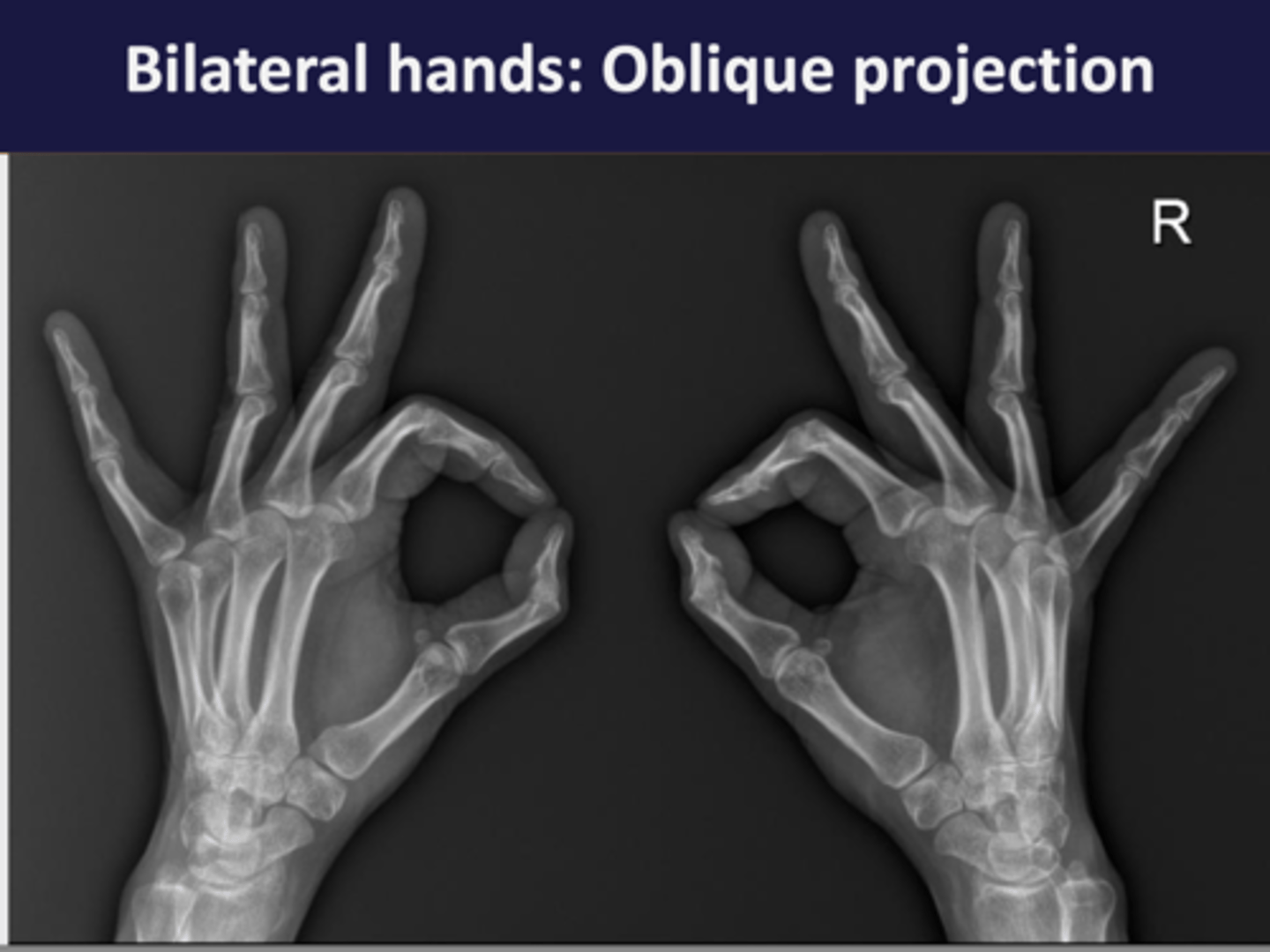
What radiographic projections are used to study the hand?
The hand is studied with AP (anteroposterior) and oblique projections to individually appreciate the different bone and joint elements.

PA projection
if thumb is looking out its AP
Oblique projection
Describe the first metacarpal's unique characteristics.
The first metacarpal is shorter and thicker, with a saddle-shaped articular surface at its base for articulation with the trapezium and is positioned at a right angle to the other four metacarpals, facilitating thumb flexion and extension towards the palm.
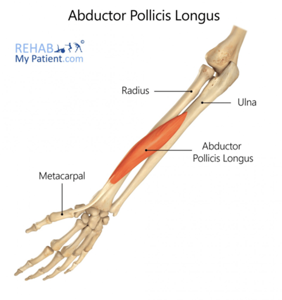
Where is the abductor pollicis longus muscle tendon inserted in the hand?
The tendon of the abductor pollicis longus muscle inserts on the palmar and lateral side of the base of the first metacarpal.
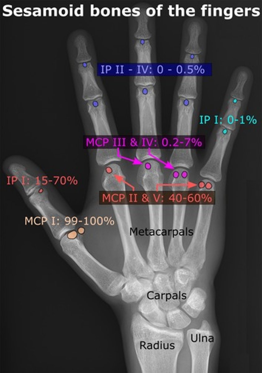
What is noted about sesamoid bones in the hand?
A sesamoid bone is typically present adjacent to the head of the first metacarpal, but sesamoids may also appear adjacent to the heads of other metacarpal bones.
How are fingers radiologically studied?
Fingers can be studied within hand projections, but individual AP and lateral X-rays are typically used for specific studies, as certain lesions may not be detected with the oblique view.
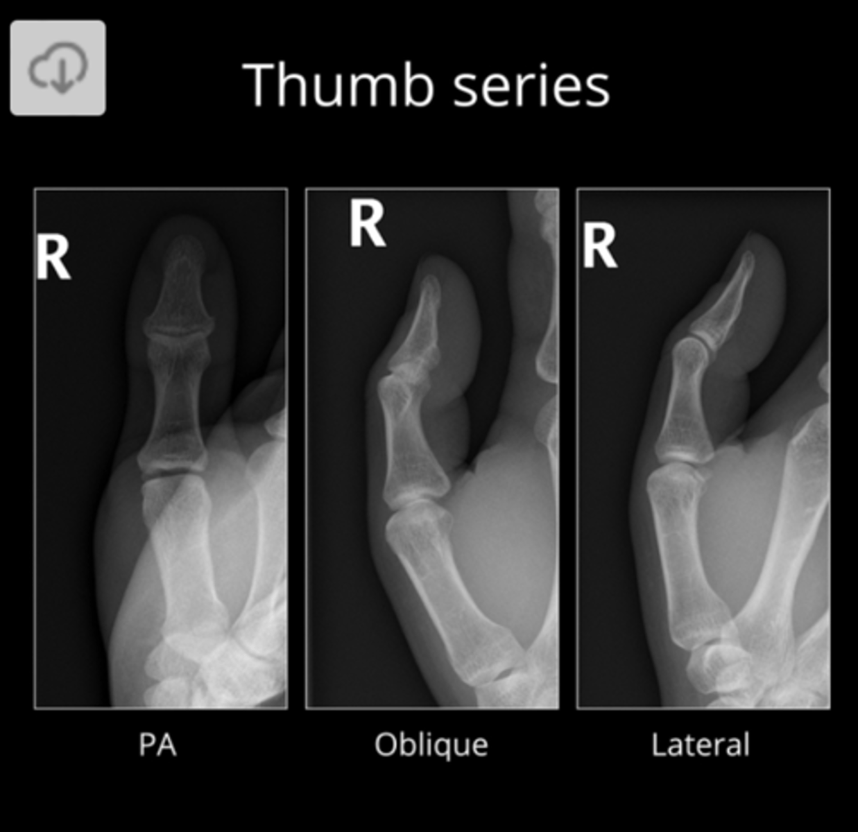
What is unique about radiographic projections of the first finger (thumb)?
The first finger or thumb requires specific positioning (tilted) for its radiographic projections due to its different orientation from the rest of the fingers.