comparative microanatomy of the urinary systems
 Call Kai
 Learn
Learn Practice Test
 Spaced Repetition
 Match
Match Flashcards
 Knowt Play
1/19

There's no tags or description
Looks like no tags are added yet.
Name | Mastery | Learn | Test | Matching | Spaced | Call with Kai | Chat |
|---|
No analytics yet
Send a link to your students to track their progress
20 Terms
regions of the kidney
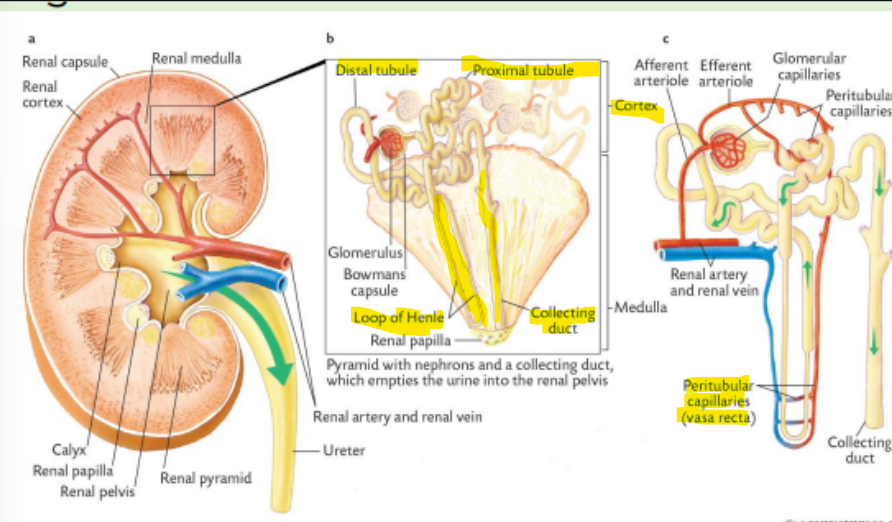
Cortex
Pars convolute (proximal tubule): Renal corpuscles, proximal and distal tubules, peritubular capillaries, and portions of collecting tubules
Pars radiata: Collecting tubules/ducts and portions of the loop of Henle
Medulla
Collecting tubules, thick and thin segments of the loops of henle and the vasa recta (peritubular capillaries)
renal nephrons
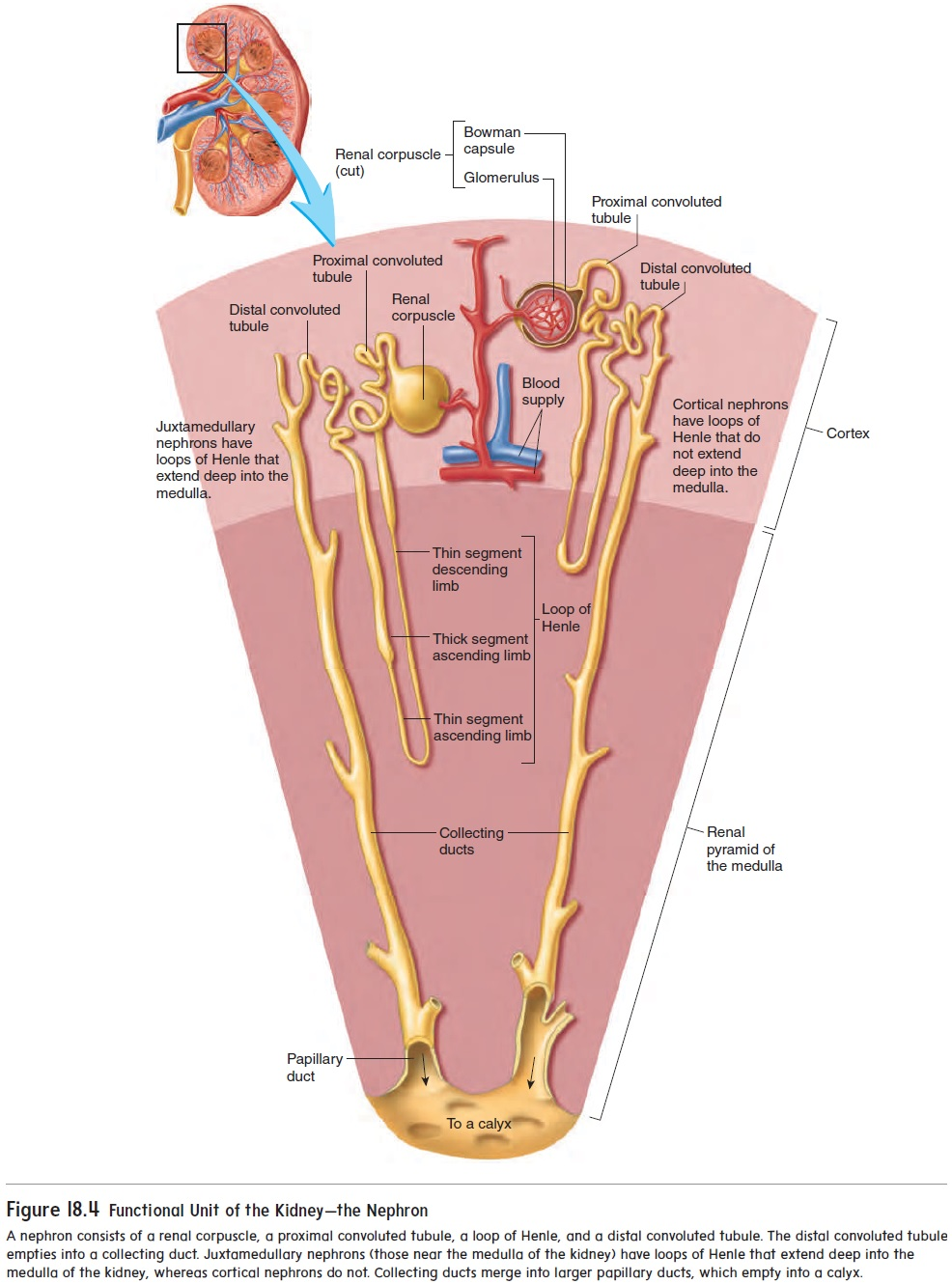
The functional units within the kidney are known as nephrons
Each nephron consists of a blood supply and a specialized network of ducts called a tubule
Vascular parts of the nephron include afferent arteriole, glomerulus, efferent arteriole and peritubular capillaries
Tubular parts include bowman’s capsule, proximal tubule, loop of henle, distal tubule and collecting duct
renal corpuscle
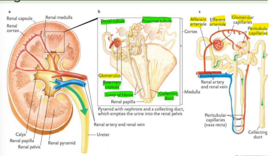
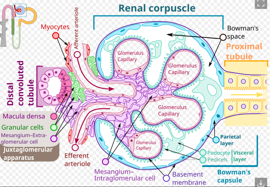
The renal corpuscle: Glomerulus, Bowman’s capsule/renal capsule, Bowman’s space
The glomerulus and its epithelial covering together will make the renal corpuscle
Once the ultrafiltrate is formed, the fluid enters Bowman’s space (the space between the capillary tuft and Bowman’s capsule)
Renal or Bowman’s capsule has two parts: parietal (squamous epithelial cells) and visceral layer (podocytes)
Blood enters the convulusions called glomerular tuft via the afferent arteriole at the vascular pole and leave via efferent arteriole
These two vessels are immediately adjacent to one another at the vascular pole (VP), which is at the opposite end of the glomerulus from the urinary pole (UP)
They can contract or dilate, changing the amount of blood that flows into the glomerular tuft
glomerulus
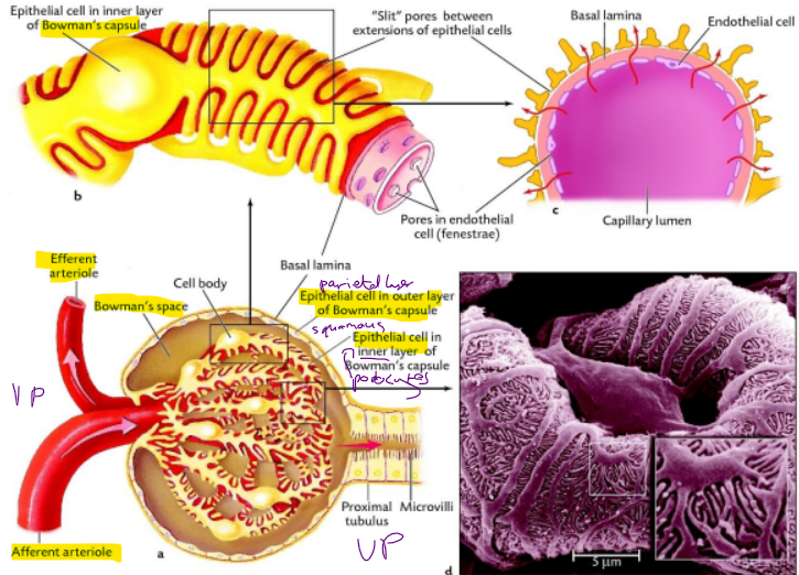
Each nephron begins with a blind expansion that is invaginated by a cluster of capillaries known as glomerulus
The glomerulus is a complex web of capillaries derived from the afferent arteriole
Glomeruli can be located in the cortex of the kidney or the corticomedullary junction
The glomerulus filters blood to produce a fluid, called filtrate
The major structures of the glomerular filtration barrier include: endothelium/pores, glomerular basement membrane (basal lamina), and podocytes
The glomerular endothelial cells are fenestrated
Another important feature of the endothelial cell is the glycocalyx, which covers the luminal surface of the cell - for a strong and functional vascular endothelium
Filtering out negatively charged molecules, leukocytes, red blood cells, platelets and larger proteins
The glomerular basement membrane is a product of the fusion of the basement membrane produced by endothelial cells and podocytes
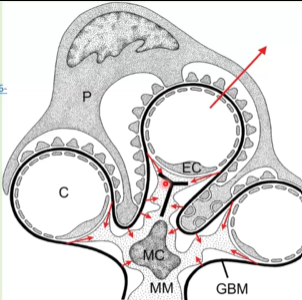
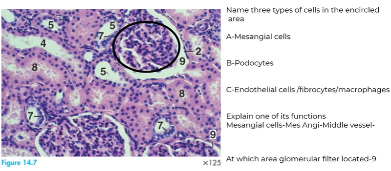
glomerulus-mesengial cells
Extraglomerular mesangial cells (not in pic):
(Juxtaglomerular apparatus)
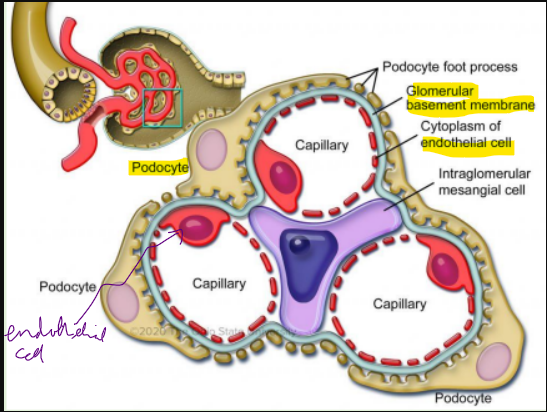
Intraglomerular mesangial cells (MC in pic) for:
Phagocytosis
Vasoconstriction
Histologically, there are 2 to 3 mesangial cell nuclei beneath each capillary loop
Collectively, the mesangial cells and the glomerular extracellular matrix that they produce constitute the mesangium
they’re basically immune cells
specialized, smooth muscle-like cells in the kidneys that support the structure of the glomerulus and help regulate blood filtration.
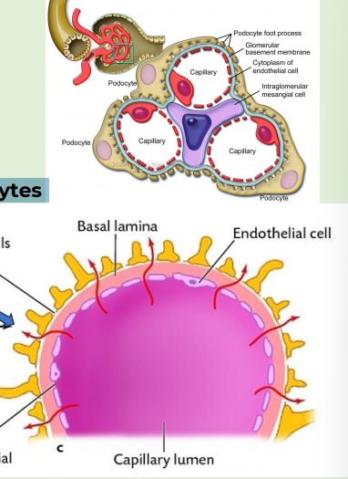
podocytes
Podocytes are specialised epithelial cells
Located on the “abluminal” surface of the capillary loop
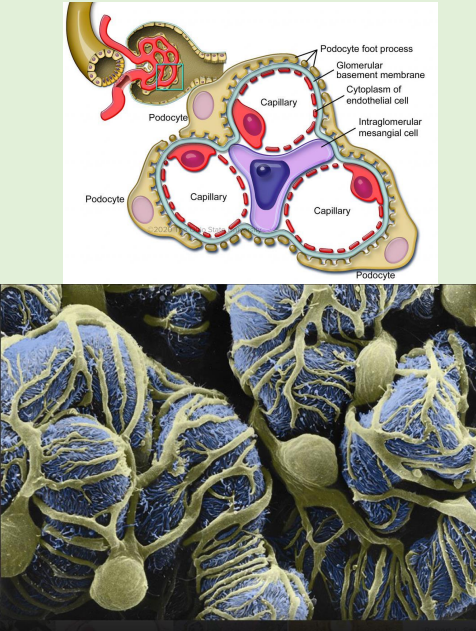
Podocytes have numerous processes, like tentacles, that surround the capillaries
Foot processes (FP) of podocytes (epithelial cell in inner layer of Bowman’s capsule) devoid of organelles except microtubules/ microfilaments
Podocyte FPs comprise a functioning slit diaphragm (SD) in between, a meshwork of proteins actively participating in podocyte signaling
visceral layer of bowman’s capsule
Support glomerular capillaries and are part of the glomerular filtration barrier
The filtration barrier: endothelial cells/pores, basal lamina, podocytes-slit pores (slit diaphragm) between podocyte foot processes
Podocytes respond to hormones and growth factors that are present in the circulation
Podocytes work with mesangial cells to support the structure and function of the glomerulus
the yellow wiggles around the pink circles are the slits
urinary system
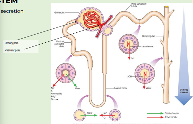
Filtration, resorption, secretion and concentration
so the glomerulus is the ultumate filtrate but now its part of the rest of the nephron
we have the
proximal convoluted tubule
loop henle - descending and ascending
distal convoluted tubule - v close to glomerulus
then collecting duct
many glomeruli give to collecting duct (many glomeruli for one collecting duct)
then papillary duct
then papillary
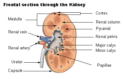
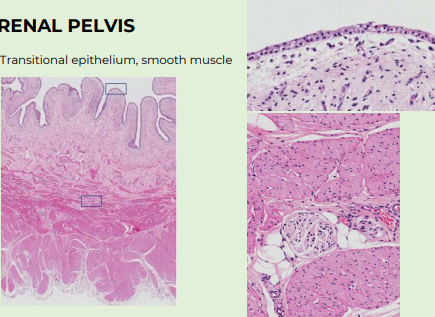
renal pelvis (major calyces converge to form the renal pelvis-large funnel shaped cavity in central part of kidney)
urinary system
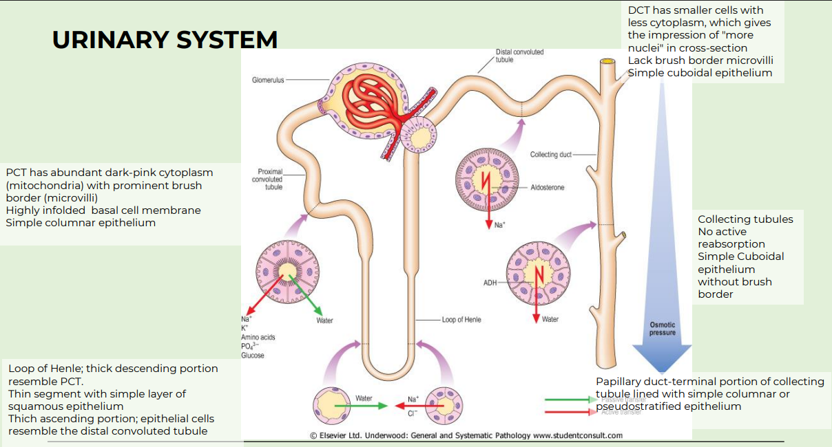
PCT
abundant dark pink cytoplasm-mitochondria
prominant brush border (microvilli) for increased SA
simple cuboidal epithelium
Loop of henle
thick descending portion resembling PCT -simple cuboidal
thick ascending portion resemble distal convoluted tubule - simple cuboidal
thin segments are squamous epithelium
DCT
Smaller cells with less cytoplasm-giving impession of more nuclei
lack brush border microvilli
simple cuboidal epithelium
Collecting tubule/duct
no active reabsorption
simple cuboidal epithelium without brush border
ADH influence
urinary system
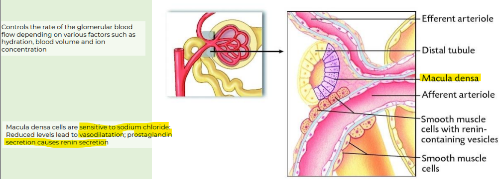
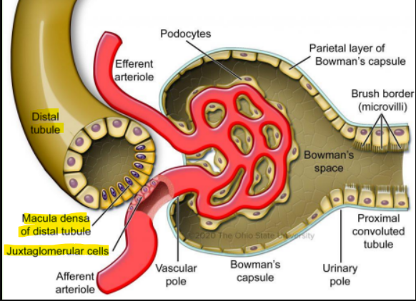
Lying just outside Bowman’s capsule and the glomerulus is the juxtaglomerular apparatus (JGA)
JGA includes macula densa, juxtaglomerular cells, mesangial cells
juxta = next to
macula densa
Macula densa(like dense black structures)
short segment of the distal convoluted tubule that is next to the afferent and efferent arterioles
the macula densa cells sense sodium chloride concentration
in the lumen and stimulates the juxtaglomerular cells to synthesize and release renin into the bloodstream
Juxtaglomerular cells
are within the interstial tissue between the macula densa and the vascular pole
This is a modified, smooth muscle cell lining the afferent arteriole that can contract or relax in response to the macula densa
histology
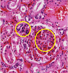
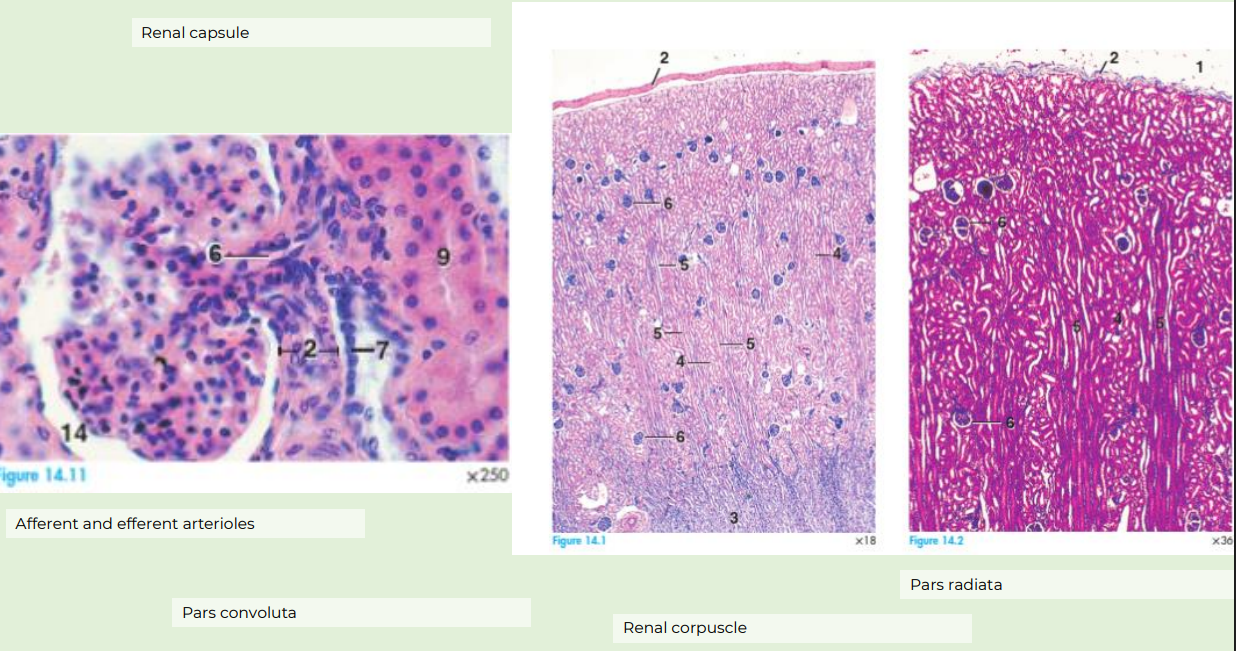
white space on 14 is bowman’s space, 14 points to the superficial cell layer i.e. bowmans capsule
14 - bowmans capsule
2- afferent arteriole
7 - macula densa
6- renal corpuscle
in second pic u see renal corpuscles (the blue dots scattered) ….check. radiata/tubules giving striated appearance (right pic shows straight vertical lines running towards centre are the straight tubules and collecting duct towards medulla)
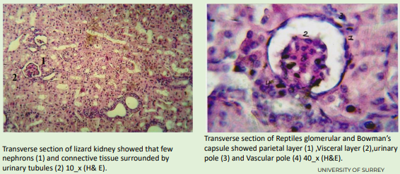
reptile kidney
Unusual glomerular appearance and renal portal system
urinary pole = bowmans space
reptile kidney lack loop of henle
fish kidney
Unusual glomerular appearance and renal system
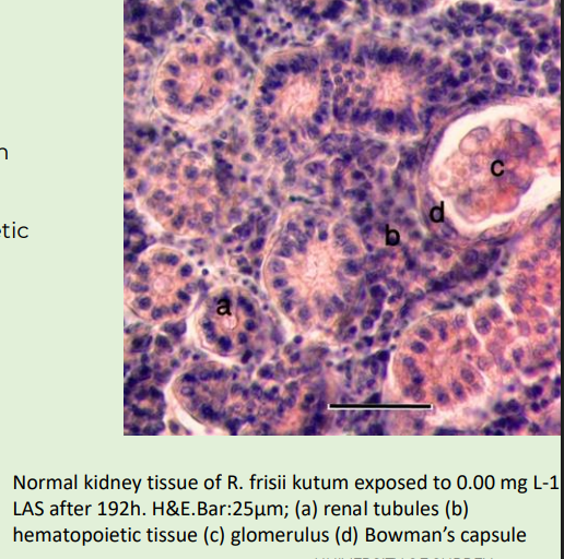
Interstitial cells have endocrine and haematopoietic function
No loops of Henle
Fresh water fish – few, small glomeruli
bird kidney
Cortical tissue dominates
There are two types of nephrons (most nephrons do not contain a loop of Henle) (reptillian and mammalian)
Cortical (reptilian) type is plenty and lacks loop of Henle (located entirely in the cortex)
Medullary type is mammalian type and less numerous and It has loop of Henle
16-bowmans space
11-proximal convoluted tubule
5-erythrocytes
7- mesengial cells
4- distal convoluted tubule
they have both two types of nephrons - mammalian (loop of henle) and reptilian
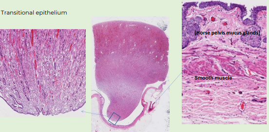
renal pelvis- transitional epithelium
Transitional epithelium, smooth muscle
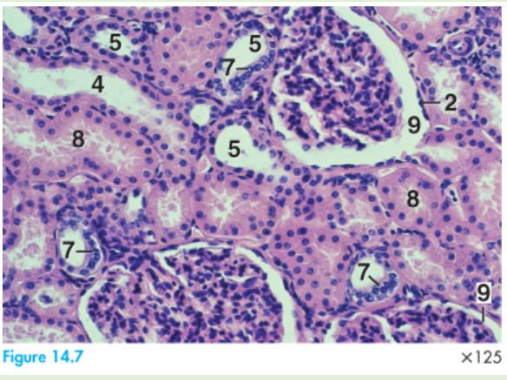
histology of cortex kidney of horse
9 - Bowman's space
2- Bowmans capsule parietal layer (oh so its parietal layer, bowmans space, then visceral layer, then glomerulus)
7- Macula densa
4- Collecting tubule
5- Distal convoluted tubule (because macula densa on here)
8- Proximal convoluted tubule — has brush border
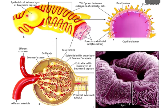
basal lamina made from 2 layers - podocytes + endothelium - protect against loss of leukocytes, erythrocytes, large molecules
mesangial cells-rovide structural support, regulate blood filtration, and perform immune-like cleanup
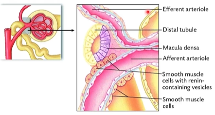
juxtaglomerular cells produce renin
podocytes are in the visceral layer of the bowmans capsule
bowman capsule is ultra filtrate
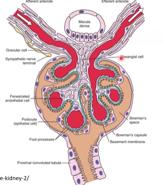
to summarise
juxtaglomerular apparatus: is just on the side, it is macular densa, distal convoluted tubule
renal corpuscle: is the main filtering blob thing we were talking about in this slidedeck, in the urinary system
bowman’s capsule: This is the outer, tan/blue "cup" or shell that forms the perimeter of the circle. It catches the fluid filtered from the red blood capillaries (the glomerulus) inside it.
Renal Corpuscle: The complete, entire filtering unit.
Bowman’s Capsule: Just the double-walled outer shell.
structures that make up the major filtration barrier
a. slit pore diaphragm
b. glomerular basal membrane
c. podocyte foot process
d. endothelial cell from the renal glomerulus
e. fenestrate or endothelial cell pore