Cardiovascular Assessment 46, Venous 58,
 Call Kai
 Learn
Learn Practice Test
 Spaced Repetition
 Match
Match Flashcards
 Knowt Play
1/109

There's no tags or description
Looks like no tags are added yet.
Name | Mastery | Learn | Test | Matching | Spaced | Call with Kai | Chat |
|---|
No analytics yet
Send a link to your students to track their progress
110 Terms
what history does the nurse need for cardiac?
Chest pain
Dyspnea, effects of dyspnea on lifestyle
Orthopnea
Shortness of breath, so severe that the person MUST sit up
Paroxysmal Nocturnal Dyspnea (PND) r/t heart failure
Shortness of breath after being recumbent awhile OR
Compensating by sleeping on several pillows or in a recliner
Fatigue with activity
Edema (fluid retention)
Blood lipid/cholesterol levels & results
what is Paroxysmal Nocturnal Dyspnea (PND)?
a sudden, severe feeling of shortness of breath that wakes a person from sleep, usually one to two hours after lying down
what does history of cardiac palpitations mention?
The heart feels like it skips beats or there are extra beats or a fluttering sensation in the chest
what does EKG history mention?
Diagnosed with an abnormal heart rate or rhythm
Pacemaker
Cardioverter Defibrillator
what does high blood pressure history mention?
Medications used for treatment, side effects of medications, & compliance
what does history of coronary artery disease?
Angina (chest pain with activity)
Heart Attack (Myocardial Infarction, MI)
Cardiac procedure (angioplasty, stenting) or surgery (bypass)
Heart failure (CHF)
Family history of heart disease prior to age 50, sudden death, diabetes, high blood pressure (hypertension, HTN), & high cholesterol
What other cardiac history does nurses need to know?
Heart murmur or rheumatic fever or heart valve problems or replacement
Antibiotic prophylaxis for invasive procedures with certain valvular problems
Diabetes, Heart Disease, & Stroke
What are some cardiac health hazards?
Smoking, cocaine, diet high in saturated fats, sedentary lifestyle, diabetes
how to auscalate for carotid arteries?
Auscultate for a Carotid Bruit (pronounced brew wee) with the bell of the stethoscope
Ask the patient to hold their breath
what are jugular veins facts?
Remove the pillow(s) if the neck is flexed
The external jugular veins are flat with the head of the bed elevated 30-45 degrees
Pulsations are visible above the clavicle & have 2 waves
what are jugular vein distention JVD facts?
Veins may bulge to the angle of the jaw
State the number of cm above the clavicle
Caused by fluid overload or heart (pump) failure
how to locate point of maximum impulse (PMI)?
May NOT be visible or palpable with the finger pads depending on body build
Located in the 5th left intercostal space (LICS) at the midclavicular line (MCL)
Normal size is < a “quarter”
Located in the 4th LICS left of the MCL up to age 7
Downward or lateral displacement indicates cardiac enlargement (cardiomegaly)
what is an abnormal palpitation of precordium?
Lifts or Heaves – Abnormal
what are thrills?
Associated with a heart murmur
Palpate with the palm or ulnar (pinkie) side of the fist
Vibrations that feel like a purring cat’s neck
what position is cardiac auscultation done?
45o supine
Sitting, leaning forward or left lateral decubitus
May facilitate hearing murmurs or distant (soft) heart sounds
are heart valves directly under auscultation sites?
Heart valves are NOT directly under the auscultation sites
what is AoRtic auscultation?
2nd right intercostal space (RICS) at the sternal border
what is a PuLmonic auscultation?
2nd left intercostal space (LICS) at the sternal border
what is Erb’s Point auscultation?
3rd LICS at the sternal border
what is an epigastric auscultation?
Just below the Xiphoid process at the midsternal line (MSL) to auscultate for an Aortic Bruit
what is the tricuspid auscultation?
4th or 5th LICS at the sternal border
what is mitral (apical) auscultation?
5th LICS at the midclavicular line (MCL)
what is the normal heart rate?
60-100 beats/minute, Regular Rhythm
what is irregular rhythm?
Premature beats cause occasional irregularity
what is atrial fibrillation?
upper chambers beat chaotically and out of sync in lower chambers
what is the S1?
Closure of Mitral & Tricuspid (atrioventricular) valves
Occurs simultaneously with the pulse
Louder at the Mitral (apex) & Tricuspid areas
S1 is louder than (>) S2
Occurs with the onset of systole
NOT normally split
what is S2?
Closure of the Aortic & Pulmonic (semilunar) valves
Does NOT occur simultaneously with the pulse
Louder at the Aortic & Pulmonic areas
S2 is louder than (>) S1
Occurs with the onset of diastole
Physiologic splitting (2 sounds close together) may occur during inspiration
what is distant heart sounds?
Soft & hard to hear
r/t obesity, emphysema or decreased cardiac output
what is S3 heart sound?
NORMAL in children
Indicates left ventricular failure or fluid overload in adults
Low pitched
Best heard with the bell in the mitral or tricuspid areas
Early diastolic sound
Mnemonic: Kentucky
LUB -duppa
what is S4 heart sound?
Low pitched
Best heard with bell in mitral or tricuspid areas
Late diastolic sound just before S1
Mnemonic: Tennessee
daLUB-dup
what is summation gallop?
S3 & S4 plus a rapid heart rate
Summation Gallop Sound
what is the cause of murmurs?
Turbulent blood flow r/t:
what is stenotic (narrowed) valve?
Think of a garden hose with a narrow spray
what is incompetent or regurgitant valve?
Does NOT close completely & leaks
what is abnormal passageway?
Patent Ductus Arteriosus
Ventricular Septal Defect (hole between the ventricles)
in which there is a mix between aorta and left pulmonary
what is dilated chamber?
Aortic Aneurysm (a bulge in the blood vessel)
Bruit (pronounced brew wee)
what increase the flow?
pregnancy
anemia
what are the best heart auscultation sites for a murmur?

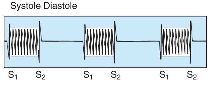
what is a systolic murmur?
Between S1 & S2
Equal / same
what is a Pan or Holosystolic Murmur?
Heard during the entire systolic or diastolic period
May obscure S1 or S2
longer
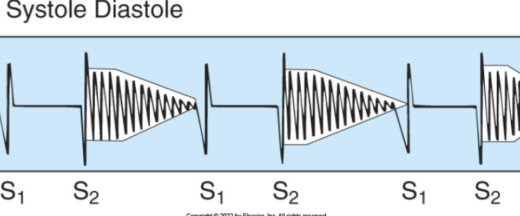
what is a diastolic murmur?
Between S2 & S1
what to mention for quality?
Blowing
Harsh
Machinery-Like (Patent Ductus Arteriosus)
Rumbling
Musical
Vibratory
what is the Grade (Scale of 1/6 or I/VI ?
1/6 - Very soft. May be missed
2/6 - Soft but heard with concentration
3/6 - Easily heard but NO thrill
4/6 - Easily heard, thrill present
5/6 - Heard with the stethoscope partially off the chest, thrill present
6/6 - Heard with the stethoscope completely off the chest, thrill present
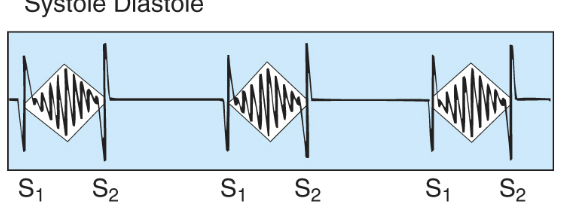
what is Crescendo/Decrescendo Murmur?
Know definitions & images
Crescendo Murmur-Increases in intensity
Decrescendo Murmur-Decreases in intensity
Crescendo/Decrescendo Murmur
Diamond shaped
what are Nursing Diagnoses r/t Cardiac Assessment?
Decreased cardiac output
Risk for decreased cardiac output
Excess fluid volume
what are symptoms in which require immediate response?
Chest Pain, Dyspnea, or Central Cyanosis
Call a Rapid Response or Code Chest for chest pain
Do NOT call the provider first
What is the history risk factors for venous problems?
Prolonged standing
Aching legs, especially at the end of day
Heredity (Family history)
Previous deep vein thrombosis (DVT, venous thromboembolism (VTE, blood clot) or pulmonary embolism (clot that travels to the lung & blocks pulmonary circulation)
Oral contraceptives & smoking (DVT risk)
Previous venous procedure or surgery for a venous problem
what are self-care activities for venous problems?
Elevating the legs
Walking
Wearing support hose
what is edema?
Swelling r/t fluid in the interstitial spaces r/t increased venous pressure
Pitting vs. non-pitting
Indentation remains with pitting edema
Generalized vs. dependent edema in body parts that are lower
what is unilateral edema?
Unilateral leg edema, redness, warmth, & discomfort
Possible deep vein thrombosis (deep clot - DVT) also referred to as a venous thromboembolism (VTE)
Measure the thigh & calf circumferences
what lymphedema?
Swelling, including the foot r/t impaired drainage of lymphatic fluid
how would you describe edema?
0-4+ scales are NOT standardized
Use your agency’s scale
how would you describe edema?
0-4+ scales are NOT standardized
Use your agency’s scale

what is Brownish Discoloration of the Shins?
r/t ↑ venous pressure causing RBCs to leak into the tissues
what are varicose veins?
enlarged, swollen, and twisted blood vessels that usually bulge just under the skin of the legs and feet
what does ankle ulcers look like?
Moist & weepy
what is Venous Stasis Dermatitis?
Edema, pruritus (itching), dryness, scaliness, brownish discoloration, & ankle ulcers
what is the Nursing Diagnoses r/t the Venous System?
risk for thrombosis
what is intermittent claudication?
Leg pain that occurs when walking r/t ischemia (insufficient blood flow (oxygen).
what to look out for in lower extremities of intermittent claudication?
Thin shiny skin
Absence of hair
Muscle atrophy
Paresthesias
Numbness, tingling, or burning sensations
what is elevated pallor?
paleness of the skin or soles of the feet when lifted above the heart
what is dependent rubor?
a light red or dusky-red discoloration of the foot or lower leg that appears when the limb hangs down in a dependent position, but fades to a normal color or paleness when the leg is elevated
describe Pallor r/t Raynaud’s Vasoconstriction?

describe Rubor r/t Raynaud’s Vasodilation?

describe Punched Out Toe Ulcer r/t Ischemia?

what is mottling?
Splotchy discoloration r/t impaired arterial circulation
what happens prior arterial surgery?
Arterial Bypass
Surgery that bypasses the blocked area in an artery
Amputation
what are self-care activities for peripheral arterial?
Foot inspection
Well fitting shoes
Protect the feet from injury
Podiatrist should trim thick nails
what to inspect and palpate of extremities?
Skin, Hair, & Temperature
capillary refill - Press on the nail bed for 5 seconds
Color should return in < 2 seconds
how to do temporal pulse?
Located in the hollow spot in front of the ear
how to do carotid pulse?
Located in the groove next to the trachea
Palpate 1 side at a time to prevent decreased blood flow to the brain
Why would you palpate both radial pulses?
to check for symmetry and compare the strength of blood flow in both arm
what is the femoral pulse?
Located in the crease in the groin
Decreased or absent in coarctation (narrowed area) of the aorta
Check babies & children, especially if the BP is elevated
what is the popliteal pulse?
Deep in the popliteal space & slightly lateral to the medial tendon
what is the posterior tibial pulse?
Medial (inner) ankle behind the medial malleolus
what is dorsalis pedis (pedal) pulse?
On top of the foot on a line between the big toe & second toe
Absent in some normal individuals
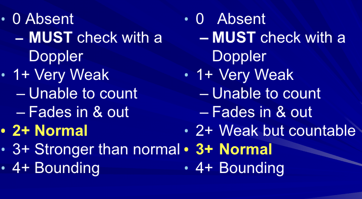
what is the pulse grading scales?
what is the allen test?
Tests radial/ulnar artery competence
Compress both arteries
Have the patient open & close the hand until it blanches (gets pale)
Release 1 artery
Normal
Immediate return of color upon release of arterial compression
Repeat & release the other artery
what is the ankle brachial index?
Ankle systolic/Brachial systolic
Use the Doppler for systolic blood pressure readings
For each leg, use the HIGHEST reading of the posterior tibial or dorsalis pedis BP
Use the HIGHEST brachial systolic reading
It does NOT need to be on the same side as the leg.
Decreased in Peripheral Artery Disease (PAD)
what are Nursing Diagnoses r/t the Peripheral Arterial System?
Ineffective tissue perfusion
Risk for ineffective tissue perfusion
Risk for peripheral neurovascular dysfunction
what is immediates response required for peripheral arterial?
Cold, pulseless, cyanotic extremity
how to do a brief scan of cardiovascular system?
involves assessment of level of consciousness,
colour changes,
sometimes pulse and blood pressure.
what position for carotid arteries assesment?
seated position or in the high-fowlers position with the client looking straight ahead.
For carotid auscultations, what are the steps?
ask patient to breathe in and out, hold their breathe
this is b/c a bruit can be similar to breathing
use bell, to listen
repeat on other side
what is important to remember for palpation of carotid arteries?
First, if you hear a bruit when auscultating the carotid arteries, do not palpate the carotid artery. Only proceed with palpation if you do not hear a bruit.
what does elevated central venous pressure (CVP) indicate?
fluid retention,
right ventricle contractile dysfunction that can be associated with heart failure, pericarditis, pulmonary hypertension,
and tricuspid stenosis.
what does low CVP indicate?
volume depletion (e.g., hemorrhage, dehydration).
what are ways to assess CVP?
an invasive procedure called pulmonary artery catheterization (critically ill)
A non-invasive way to assess the CVP is by inspecting the jugular veins
how to do a jugular veins inspection?
Assist the client into a supine position with the head of bed in a position at least 30 degrees and no higher than 45 degrees.
Remove the client’s pillow so their head is laying flat against the bed in a neutral position.
Stand on the right side of the bed and ask the client to gently turn their head to the left.
Use tangential lighting to highlight pulsations.
Begin by inspecting the right external jugular pulsation, as it is most visible. The vein should be flat or not visible; a bulging vein can suggest high CVP.
repeat on other side
what is jugular venous pulsation influenced by?
influenced by blood volume, cardiac function of the right ventricle, and venous compliance.
Venous is:
not palpable
double wave
varies with respiration
disappears when repositioned into sitting
arterial is:
palpable
one brisk wave
doesn’t vary with respiration
not affected by position change
limited location
what is the position of patient when doing a heart assessment?
The assessment is typically performed in a supine position with the client’s head on a pillow.
What type of approach should the nurse follow that focuses on physical and emotional safety when performing a cardiovascular assessment?
Trauma-informed approach.
involves integrating an understanding of the need for physical and emotional safety; choice and control; and empowerment
how to palpate for precordium?
Use the metacarpophalangeal surface of your fingers starting at the base of the heart. Leave them in place as long as you need to, but usually about two to three seconds and then move onto the sternal border and then the apex.
identify any vibrations
how to palpate for apical pulse?
put finger pads at 4th intercostal
Ask the client to hold their breath after taking a large breath in and out. You should feel the pulsation quite quickly if it is palpable.
Document: “Gentle tap of apical impulse, short duration, one smooth pulsation, 1–2 cm2 at apex.”
where is the apical pulse located?
5th intercostal space, left midclavicular line
what sound is best heard in apex?
S1 sound
what sound is best heard in base?
S2 sound
Peripheral vascular system - arteries
rounded appearance
thick walls
carries blood way from heart
lower oxygen concentration in pulmonary arteries
high pressure
valves not present