Parasitology SOLO 3
1/34
Earn XP
Description and Tags
Cestodes, trematodes, arthropods
Name | Mastery | Learn | Test | Matching | Spaced |
|---|
No study sessions yet.
35 Terms
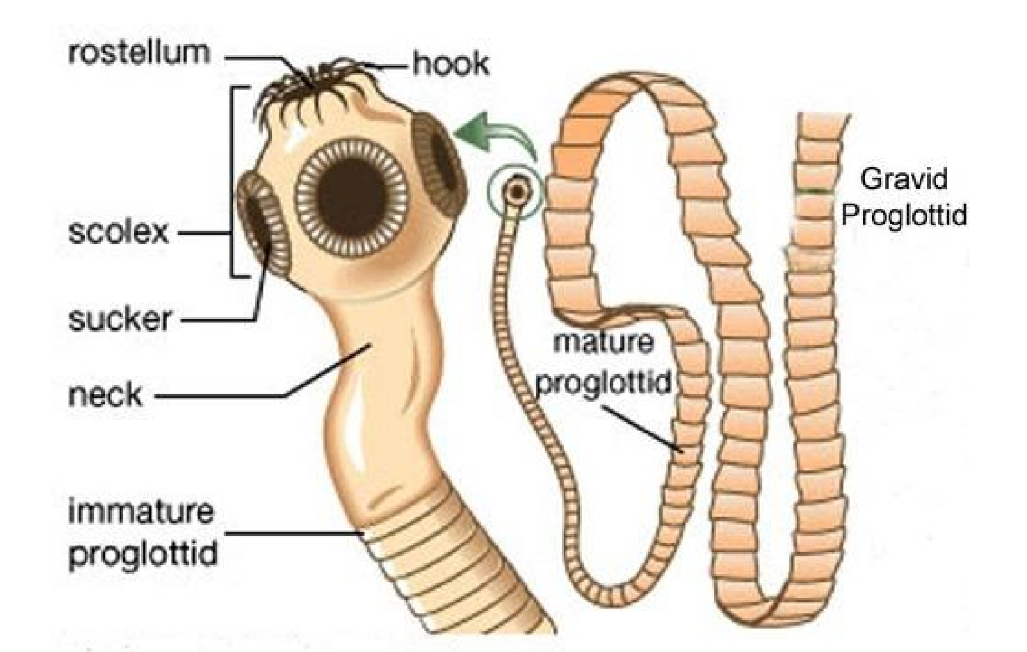
Tapeowrm bodies
flattened, segmented and ribbon-like with three sections
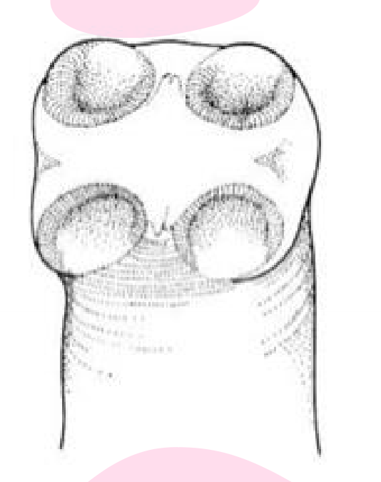
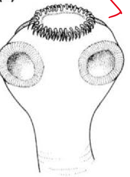
scolex
anterior end of worm (head)
modified for attachment to intestinal wall of host
very short
four cup-shaped suckers, sometimes hooks (used in attachment to host)
has a rostellum (fleshy anterior protuberances of the scolex, may bear circular row(s) of hooks and may be retractable)
neck
very short
just posterior to scolex
from which the rest of the worm grows
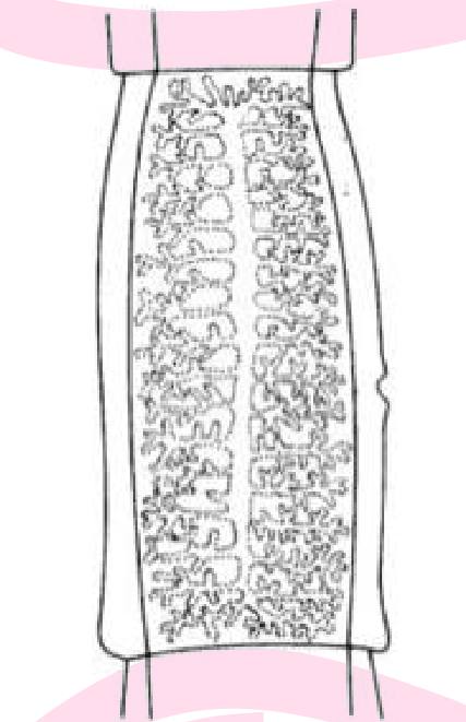
strobila
main body of worm
consists of a chain of segments known as proglottids
newest proglottids are near the neck and become older and more mature toward the end of the tail
Overview of tapeworms
hermaphroditic
each proglottid near the tail contains fully developed sex organs (gravid proglottids- uterus filled with fertilized eggs)
nutrients are absorbed through the outer surface of the body
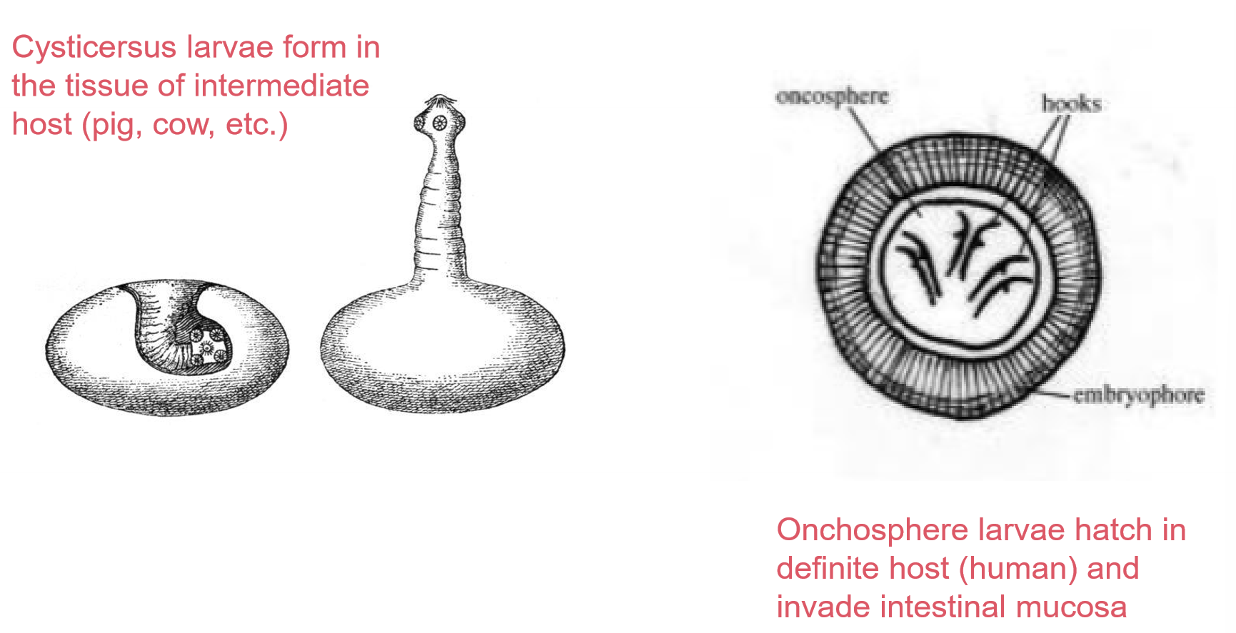
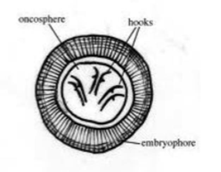
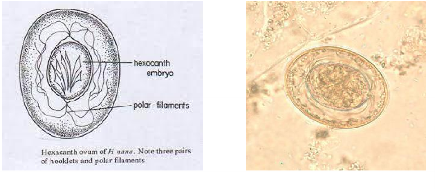
onchosphere (hexacanth larvae)
motile, first stage of certain cestodes
scolex has six hooklets
cysticercus larvae
thin-walled, fluid filled, bladder-like structure enclosing a scolex
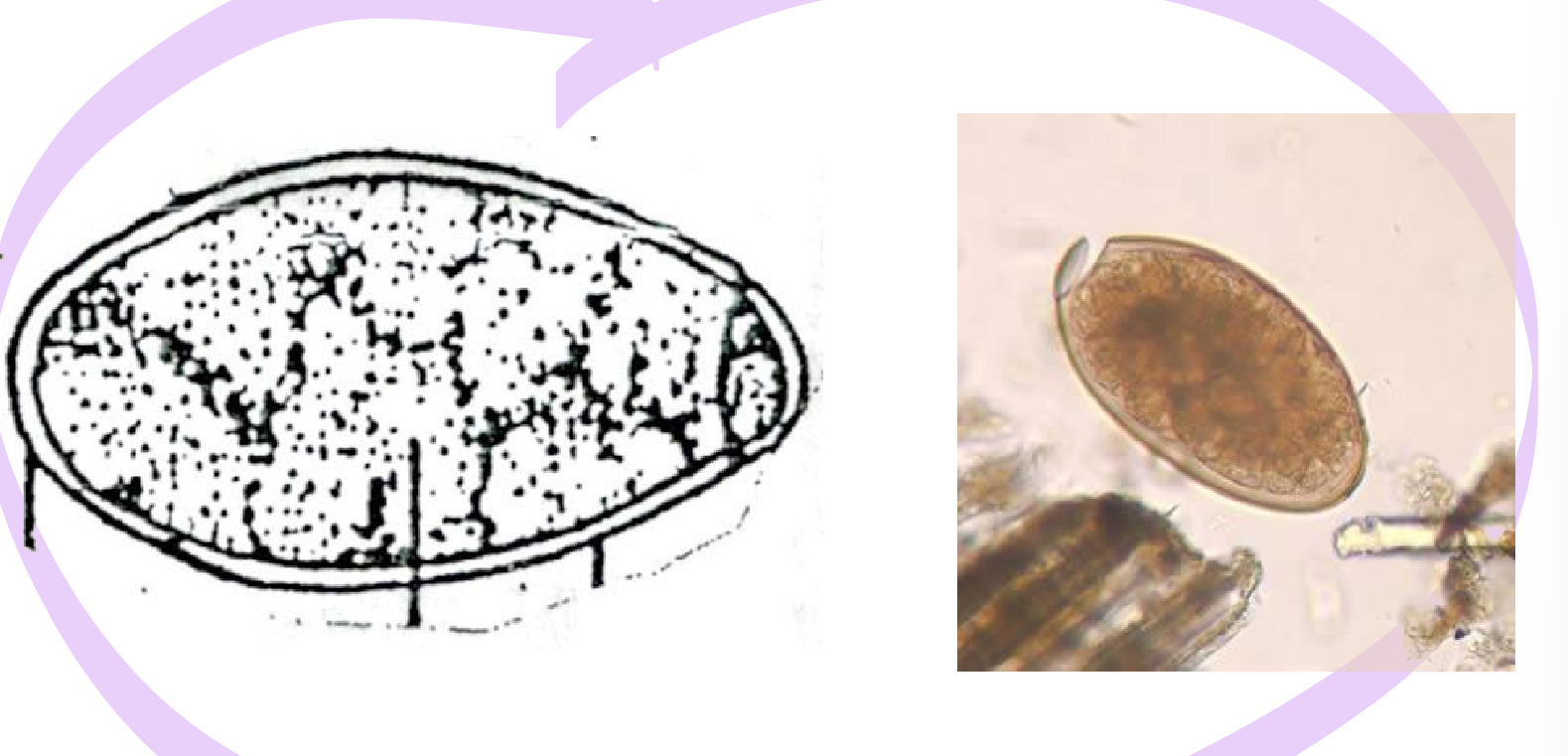
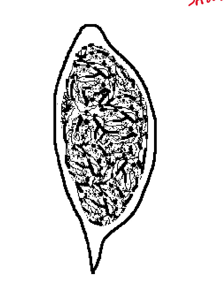
Taenia saginata
AKA beef tapeworm
disease
usually asymptomatic but sometimes see GI disease w/ abdominal pain/diarrhea
human location
small intestine
infective stage is cysticercus larvae in beef
diagnosis
identification of eggs, gravid proglottids, or sometimes scolices in stool
lifecycle
onchosphere larvae devlops into cysticercus larvae in tissue of itnermediate host
cysticercus larvae ingested by humans in raw or undercooked beef
larvae attaches to small intestine
matures to adult gravid proglottids
free eggs and scolices passed in stool
eggs eaten by intermediate host
morphology
gravid proglottids has 15-30 lateral branches
scolex has 4 cup-shaped suckers and no hooks
egg contains onchosphere embry with 3 pairs of hooklets
Taenia saginata onchosphere
Taenia saginata scolex
Taenia saginata gravid proglottid
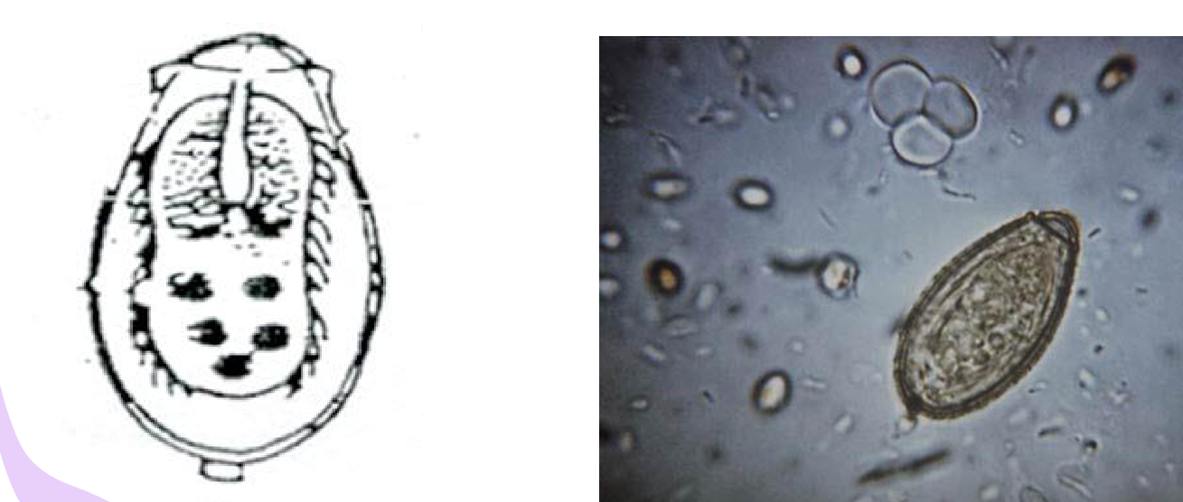
Taenia solium
AKA pork tapeworm
disease
mostly the same as saginata
cysticercosis (when egg accidentally ingested, cyst in muscle, brain, other tissues)
human location
adult in small intestine
occasionally cysticercus larvae in tissues
intermediate host is pig
diagnosis same as saginata
occasionally diagnosed by cysticercus larvae in stained biopsy tissue
morphology
eggs is same as T. saginata
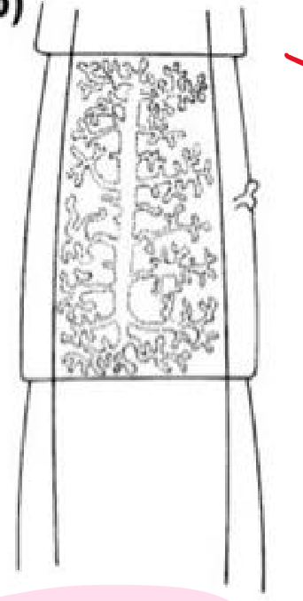
gravid proglottis has 7-12 uterine branches
scolex contains 4 suckers and a circular crown of hooks
Taenia solium scolex
Taenia solium gravid proglottid
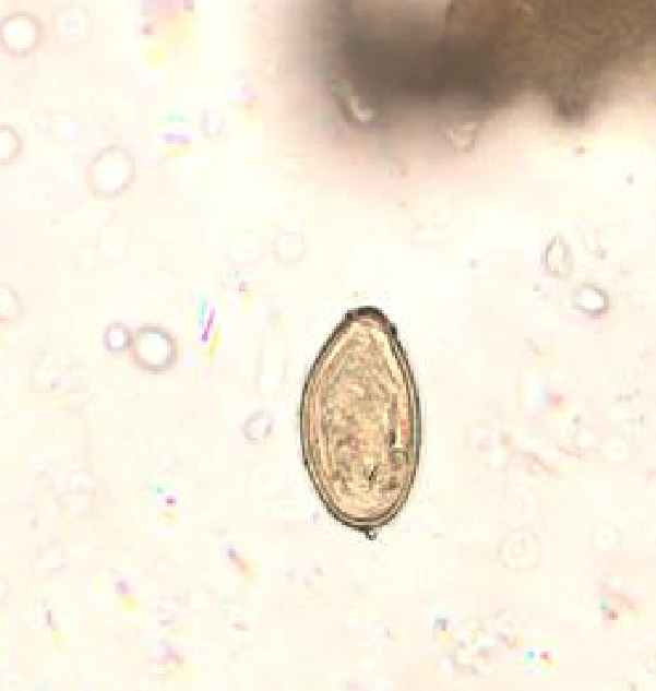
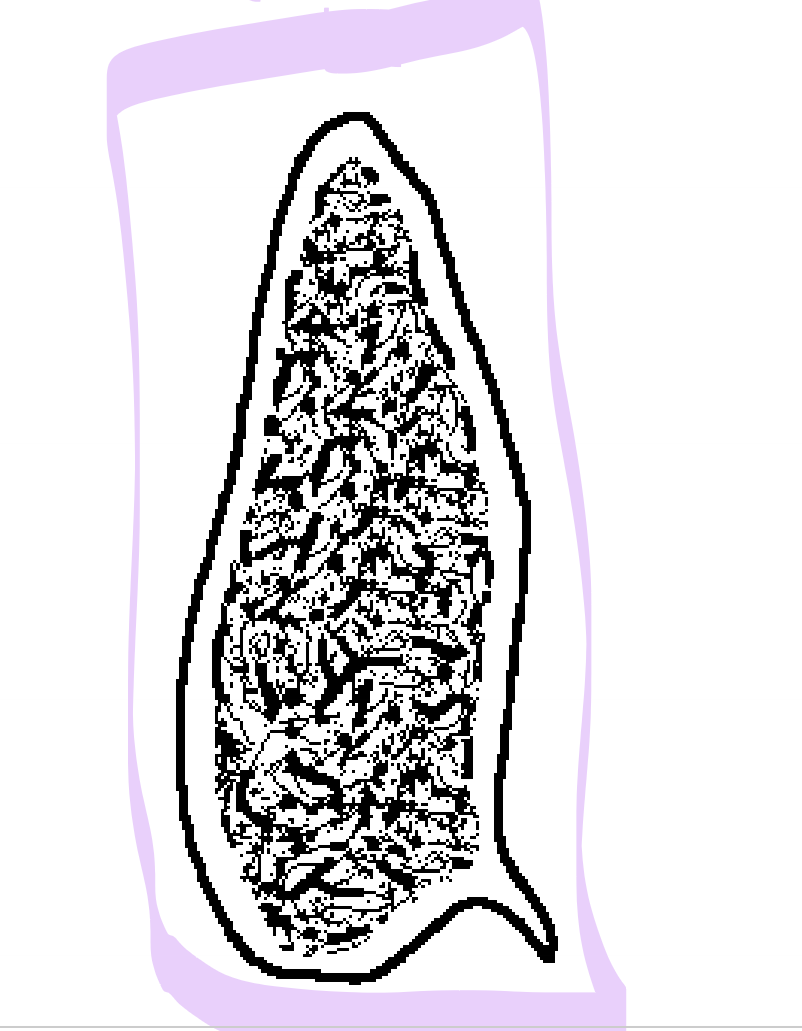
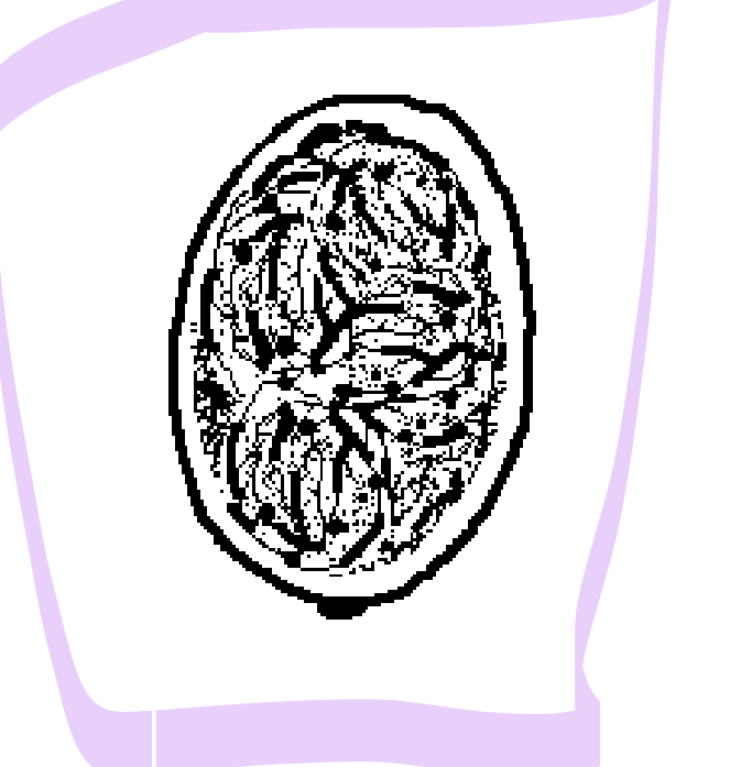
Diphyllobothrium latum
AKA broad fish tapeworm
disease
GI disease symptoms, macrocytic anemia and neurological disturbances due to B12 deficiency
sparganosis (procercoid larvae invades various tissues, symptoms ass, with inflammatory reaction)
human location
small intestine
diagnosis made by identification of eggs in stool
lifecycle
procercoid larvae develops into plaurocercoid larvae in fish muscle after crystacean is ingested by fish
plaurocercoid larvae ingested by human in raw or undercooked freshwater fish
plaurocercoid attaches to intestinal mucosa and matures to adult
morphology
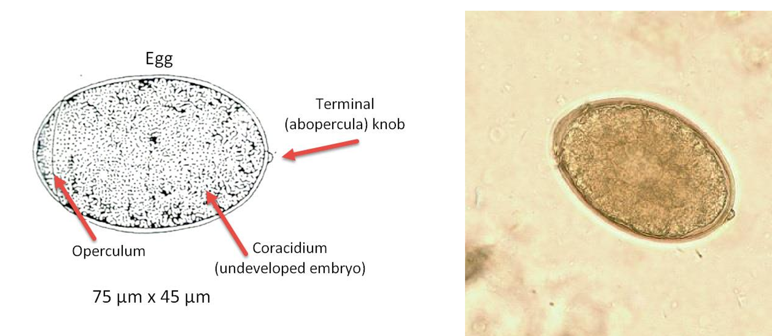
eggs is double walled and oval shaped
contains an operculum and a terminal knob
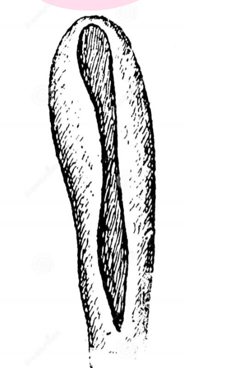
scolex has no hooks or suckers; has two long grooves called bothria which serve in attahcment to host
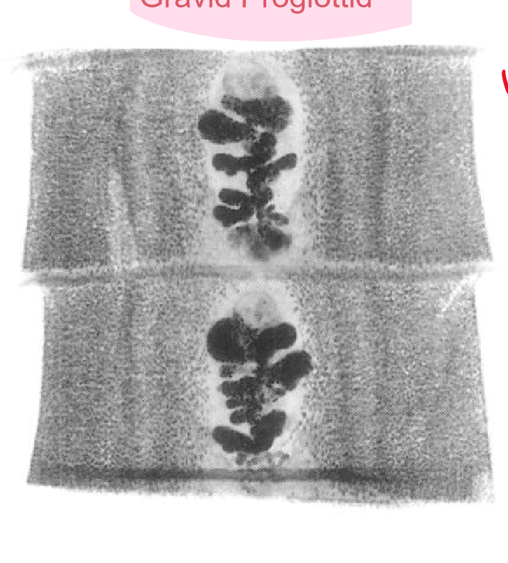
width of proglottid is greater than length; has rosette-shaped central uterus
Diphyllobothrium latum egg
Diphyllobothrium latum scolex
Diphyllobothrium latum gravid proglottid
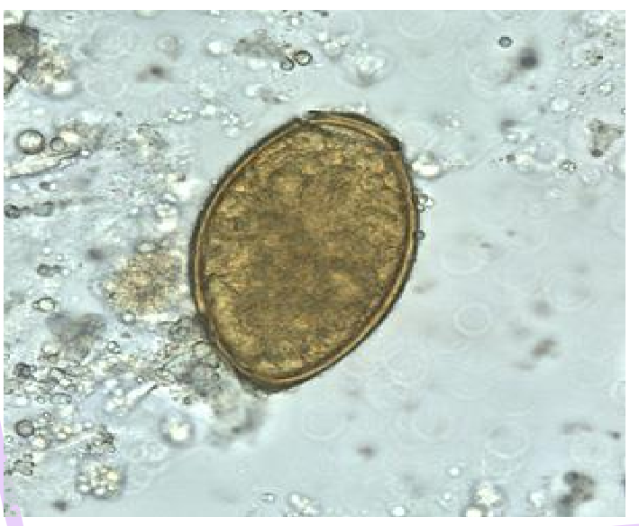
Hymenolepsis nana
AKA dwarf tapeworm
disease
light, asymptomatic infection
heavy infection including GI disease with abdominal pain and diarrhea
infective stage
egg
human location
small intestine
diagnosis
identification of eggs in feces
lifecycle
eggs are ingested in contaminated food or water
eggs hatch in small intestine
onchosphere larvae invade intestinal villi
cysticeroid larvae matures and emerges from larvae
scolex everts and attaches to mucosa
larvae matures to adult and eggs are release in disintegrating gravid proglottids
eggs passed in feces
can cause autoinfection
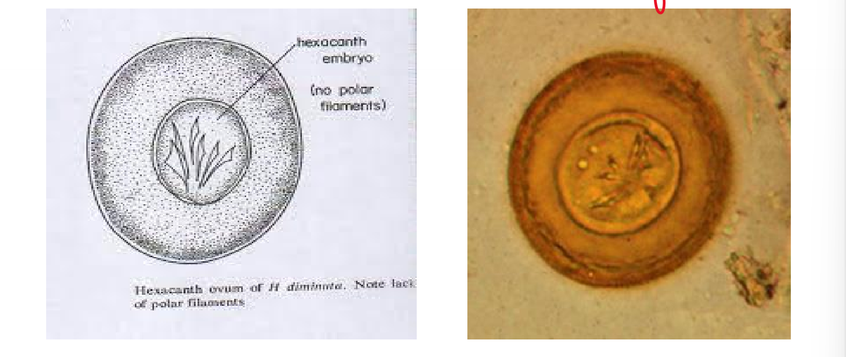
Hymenolepsis diminuta
disease
similar to H. nana
human location
small intestine
diagnosis
identification of eggs in stool
vector is rats
only occasionally infects humans
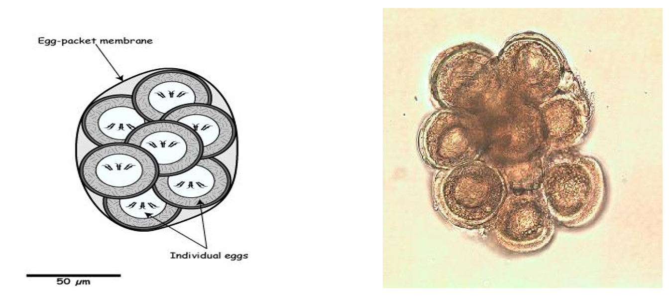
Dipylidium caninum
causes mild GI disease
vector is flea
rarely infects humans
main host is dogs or cats
flea infected with cysticercoid larvae is ingested
cysticercoid attaches to intestine and mature to adults
eggs released
uterus contains numerous egg packets
8 to 15 colorless eggs
contain onchosphere larvae with six hooklets
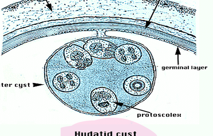
Echinococcus granulosis (dog tapeworm)
AKA hydatid cyst or hydatid disease
symptoms vary according to location and size of cyst
liver and lung are most common locations
usually no symptoms until cyst is very large
rupture of cysts lead to anaphylactic shock and death
sheep and other herbivores main intermediate host
diagnosis
serology, ultrasound, Xray
ID of scolices, hooks, etc in cyst fluid (not recommended due to anaphylactic shock)
lifecycle
eggs are ingested
larvae hatches and penetrates intestinal mucosa
larvae migrate to liver or lungs
hydatid cyst forms
viscera of infected herbivore eaater by dog
eggs passed in feces of canine
treatment includes removal of cyst without rupture
trematodes overview
AKA flukes or flatworms
flattened, nonsegmented, leaf-shpaed worms
adults have two suckers (acetabula); oral and ventral
tegument (body surface)
metabolically active, absorbs nutrients and releasess waste
digestive system
oral cavity is in center of oral sucker
intestinal tract ends blindly in one or two sacks
solid waste products regurgitated
two types of parasitic flukes in humans
organ flukes- found in intestine or other tissues; hermaphroditic
blood flukes- found in blood vessels; unisexual
larval stages of flukes
metacercaria
thickwalled, nonmotile, encysted
miricidium
ciliated, first-stage, free-swimming
sporocyst
encysted zygote in invertebrate host (snail)
rediae
small, sac-like larval form
cerecaria
adult form, free-swimming, has a tail
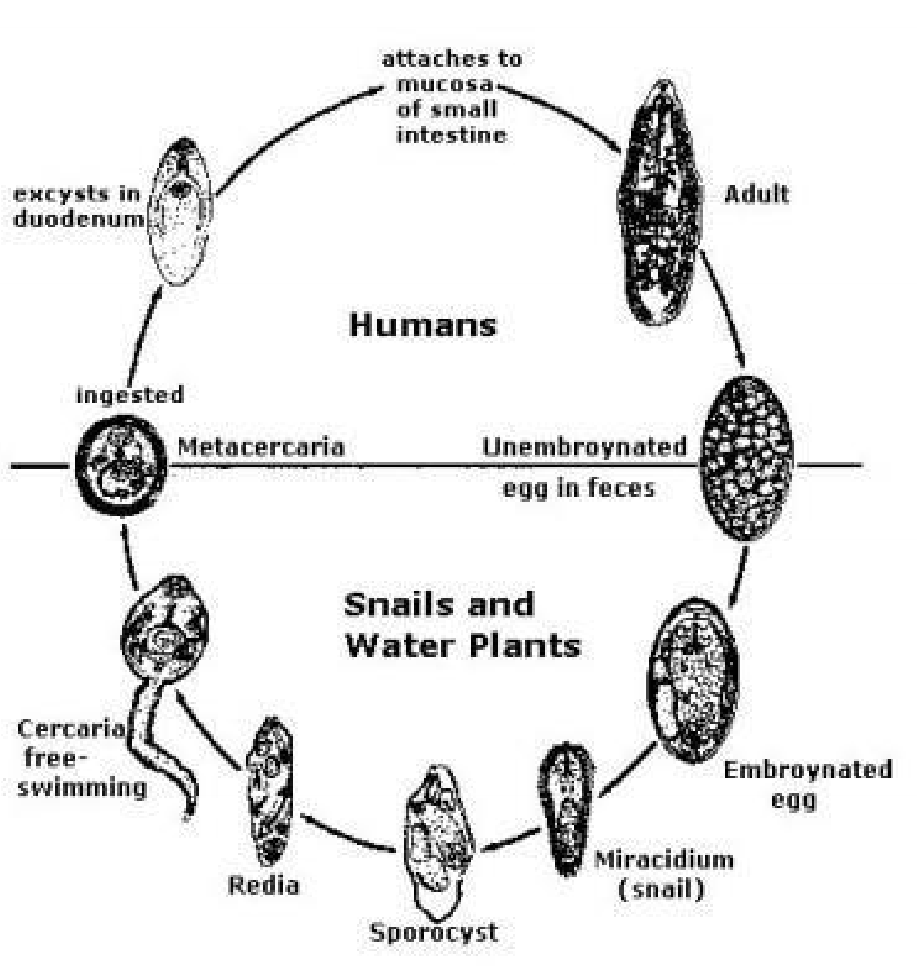
organ flukes infective stage
metacercaria is the infective stage
metacercaria ingested from water plant or aquatic creature
rediae develop into cercariae
blood fluke infective stage
cercaria is the infective stage
cercaria penetrate skin
sporocyst develops into cercariae
Fasciola hepatica
AKA sheep liver fluke
disease
fever, headache, chills, enlarged spleen, eosinophilia
severe infections cause biliary instruction, jaundice, and anemia
human location
bile duct
sheep is main host, but can be ingested by humans eating water plants
diagnosis
identification of eggs in stool

Fasciolopsis buski
AKA large intestine fluke
causes GI disease with nausea, diarrhea, and mucosal ulcers
heavy infections can be fatal due to intestinal obstruction
human location
small intestine
humans infected by ingesting encysted metacercariae on water plants
diagnosis
identification of eggs in stool
Clonorchis sinensis
AKA Chinese/oriental river fluke
disease
abdominal pain, diarrhea, enlarged liver, and jaundice
human location
bile duct
infective stage
metacercariae in tissue of undercooked fish
diagnosis
identification of eggs in stool
egg morphology
anterior end includes a thickened rime aorund the operculum
small knob at posterior end
Opisthorchis felineus
biliary tract fluke very similar to Clonorchis sinensis
Paragonimus westermani
AKA oriental lung fluke
disease
chest pain, cough, bloody sputum, and fever
human location
fibrous cysts in lung
infective stage
metacercariae in tissue of undercooked freshwater crabs or crayfish
diagnosis
identification of eggs in sputum or feces
xray (resemble TB) serology
egg morphology
includes an opercular rim on the anterior side and a terminal shell thickening at posterior end
Schistosoma species overview
AKA blood flukes
Schistocomiasis ranks second behind malaria as a cause of serious worldwide morbidity and mortality due to parasitic infection
all species cause dermatitis, then fever, chills and body aches
more specific disease caused by different species
can also cause “swimmer’s itch”
Schistosoma mansoni
causes cirrhossi of liver, bowel obstruction, bloody diarrhea, enlarged liver/spleen, and toxic reactions due to granulomas around eggs in tissues
eggs moves through blood vessels and become trapped in liver where they mature
adults found in liver sinuses and veins around intestinal tract
diagnosis is identification of eggs in stool
found in Africa, South and Central America and West Indes
eggs i more elongated with a lateral spine
Schistosoma japonicum
life cycle similar to S. mansoni
found in far east
eggs is more spherical with lateral knob
Schistosoma haematobium
causes urinary tract disease with leasions in bladder and bloody urine
also causes toxic reactions due to granulomas around eggs
adults are found in veins of urinary bladder
eggs can move through blood vessels and become trapped in other tissues
diagnosis is identification of eggs in concentrated urine
found in Africa, and Middle East
eggs are more elongated and has a terminal spine
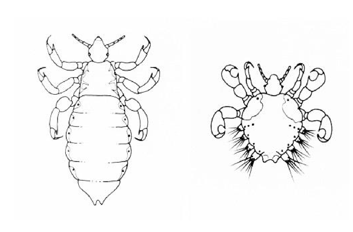
body louse/crab

scabies
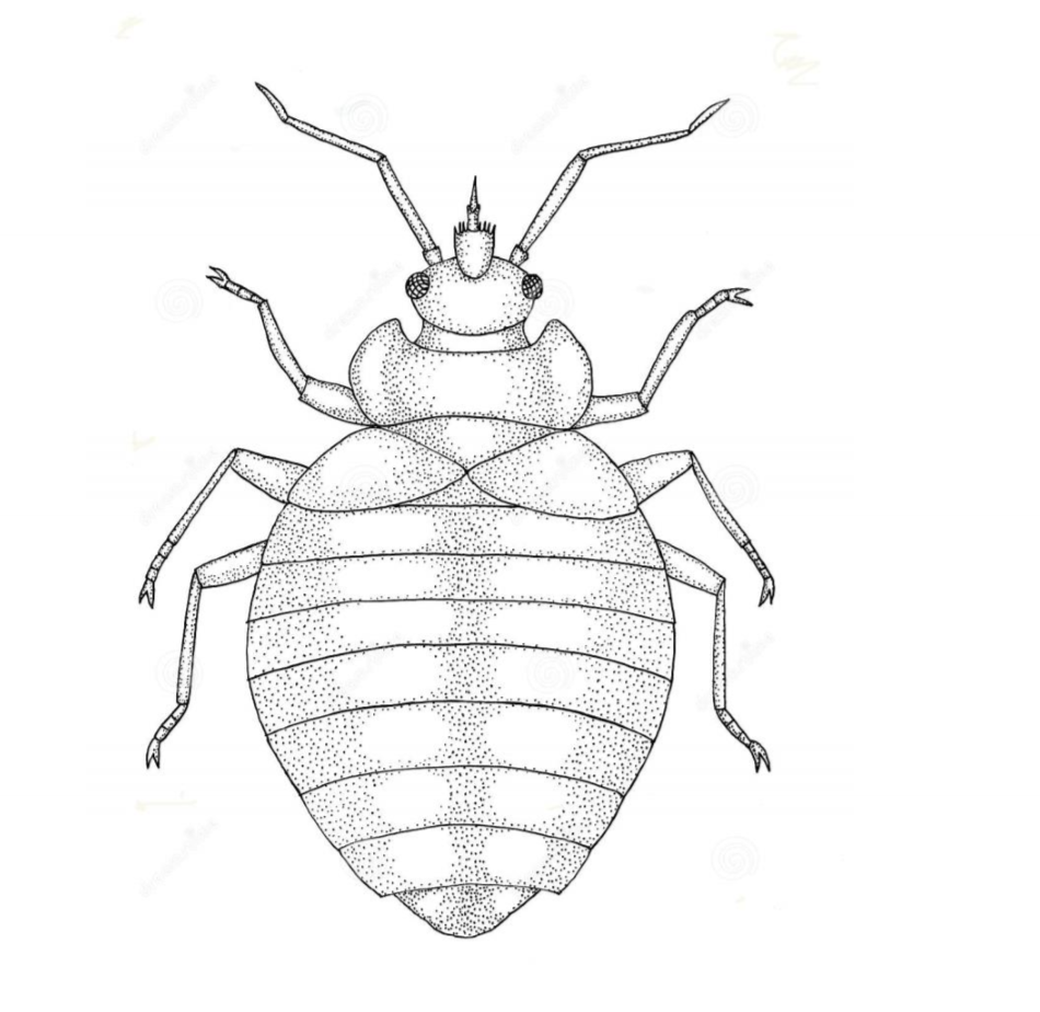
bed bug
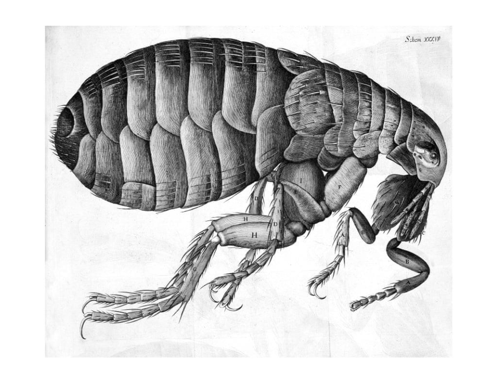
flea
tick