Lecture 11: Congenital Cardiac Disease
 Call Kai
 Learn
Learn Practice Test
 Spaced Repetition
 Match
Match Flashcards
 Knowt Play
1/33

There's no tags or description
Looks like no tags are added yet.
Name | Mastery | Learn | Test | Matching | Spaced | Call with Kai | Chat |
|---|
No analytics yet
Send a link to your students to track their progress
34 Terms
What is a patent ductus arteriosus?
connection between aorta and pulmonary artery (persistent patency of feta vessel connecting the two) resulting in left atrial & ventricular enlargement, Left heart volume overload, Pulmonary overcirculation
How is PDA diagnosed?
Physical exam
Most asymptomatic and cardiac murmur leads to workup
stunted size compared to littermates
Continuous murmur left heart base (washing machine)
Bounding arterial pulses
If CHF has developed, usually left heart failure with dyspnea, crackles
ECG: variable
Thoracic rads:
Cardiomegaly: can see generalized or mostly left heart enlargement
Pulmonary overcirculation
3 bumps: aorta at 1:00, pulmonary artery at 2:00, left auricular appendage at 2:00-3:00
Echo:
Left heart volume overload
Continuous flow main pulmonary artery
PDA visualized

What congenital defect is seen on this rad?
PDA
What is the treatment for PDA?
interventional closure:
minimally invasive coil or amplatz (ACDO) occlusion
thoracotomy
You are presented with a puppy who is the runt of the litter. Upon physical exam, you find left basilar continuous murmur and bounding femoral pulses. What congenital defect are you worried about? What potential complications of this defect?
PDA
Complications:
Left-sided congestive heart failure
Can cause secondary pulmonary hypertension → shunt reversal → differential cyanosis, erythrocytosis (can’t be repaired if reverses)
What is a ventricular septal defect?
communication between L & R ventricle due to a defect in the interventricular septum → results in left atrial and ventricular enlargement, left heart volume overload (L to R shunting), and pulmonary overcirculation
most common defects in horses, ruminants, cats (english bulldog, keeshond, maltese predisposed)
How are VSDs diagnosed?
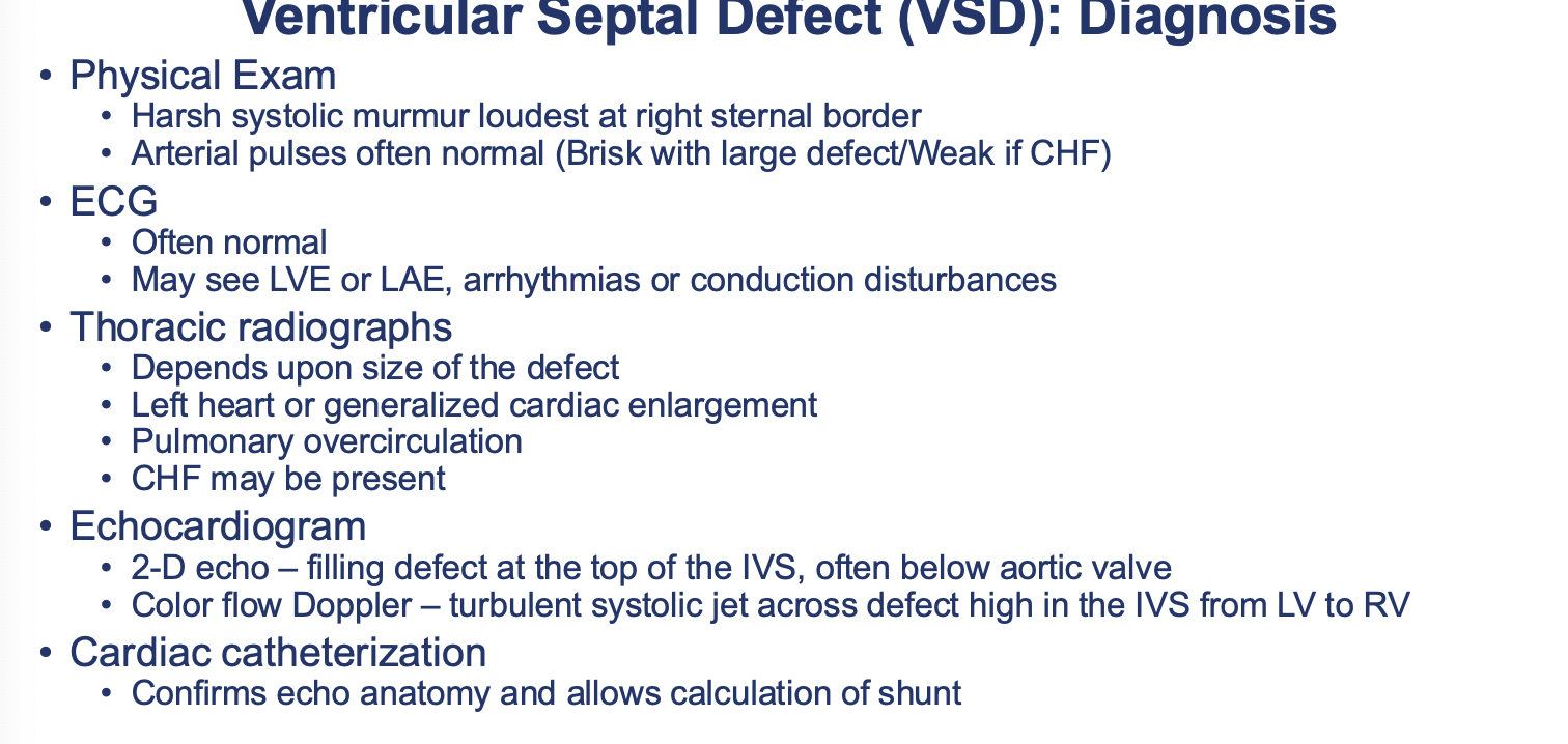


What defect is most likely affecting this cat? note the pulmonary overcirculation and cardiac enlargement.
VSD in a cat with CHF
What is the treatment for VSDs?
Medical therapy for CHF
Pulmonary Artery Banding
If muscular VSD then Amplatz occlusion possible (very uncommon)
Surgical Repair
Requires Cardiopulmonary Bypass
Patch applied over the VSD
Uncertain operative mortality
True or false: louder murmurs are usually associated with smaller VSDs?
true
What is an atrial septal defect?
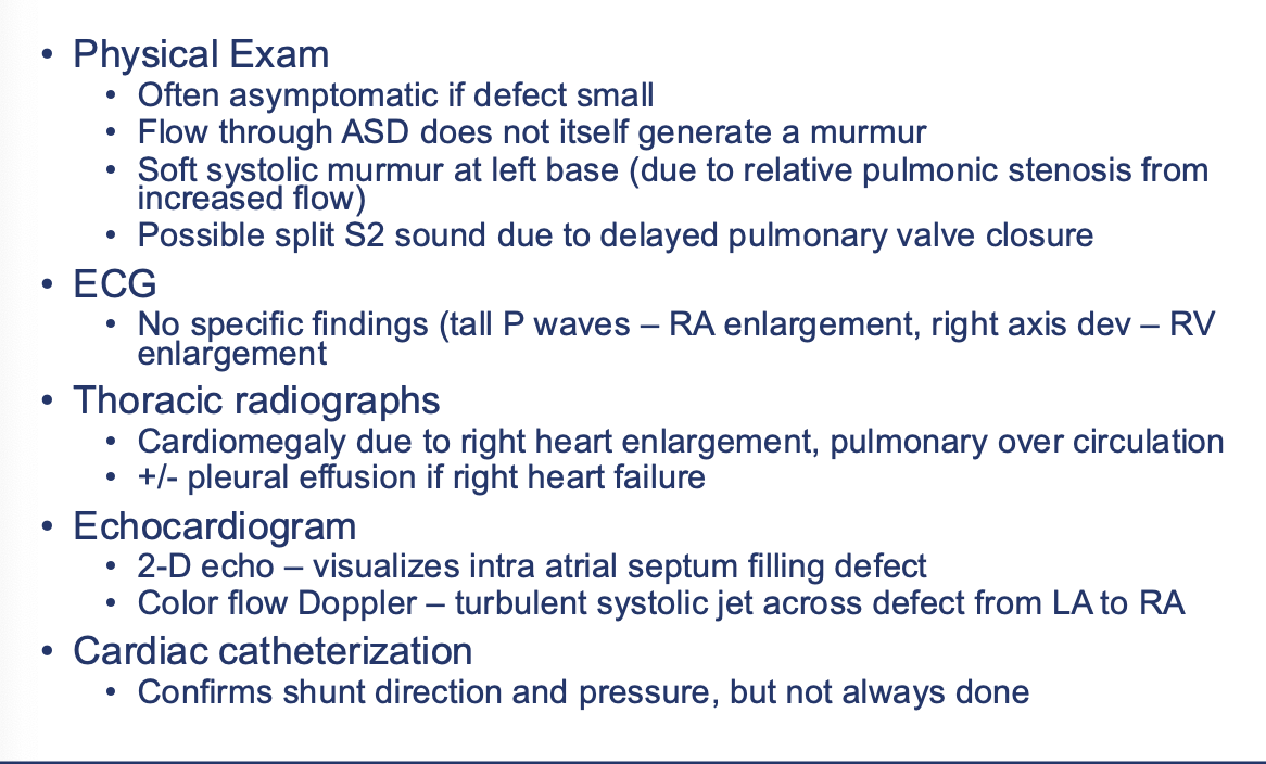
communication between L & R atrium due to a defect in the intra atria septum → results in right atrial and ventricular enlargement, right heart volume overload, and pulmonary overcirculation → may progress to pulmonary hypertension and right sided heart failure
True or false: ASDs are usually seen on their own.
false: they are most frequently seen with other defects
Who is predisposed to ASDs?
• Samoyed, Doberman, Standard poodle, Old English Sheepdog
• Cats, ruminants rarely
How are ASDs diagnosed?
What is the treatment for ASDs?
Medical therapy for CHF
Occluder devices
Surgical Repair
Requires Cardiopulmonary Bypass
Patch applied over the ASD
Uncertain operative mortality
Rare

What is pulmonic stenosis?
narrowing of the pulmonary outflow tract due to valvular stenosis, subvalvular stenosis, supravalvular stenosis → RV creates increased pressure to maintain normal PA pressure → RV concentric hypertrophy, post-stenotic dilation of pulmonary a., secondary tricuspid regurgitation can develop
Who is predisposed to pulmonic stenosis?
• English Bulldog, Schnauzer, Beagle, Chihuahua, Terrier breeds, Cocker Spaniels, Samoyed, Bassett hound
• English Bulldog & Boxer can have anomalous coronary vessel which encircles PA
• Cat: Isolated pulmonic stenosis rare
How is pulmonic stenosis diagnosed?

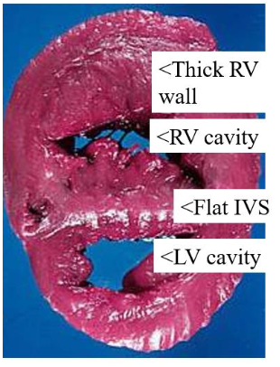
What defect has most likely caused these cardiac changes?
pulmonic stenosis
How is pulmonic stenosis treated?
Beta Blocker – reduce RV workload & risk for sudden death
Decr. HR = more RV filling time
Decr. Contractility = reduced pressure buildup near PS
Decreases risk for arrhythmias
Medical therapy for CHF
Balloon Valvuloplasty
What is the prognosis of pulmonic stenosis?
• Mild – excellent, monitoring only
• Moderate – good with monitoring or intervention
• Severe – guarded without intervention
What is subaortic stenosis
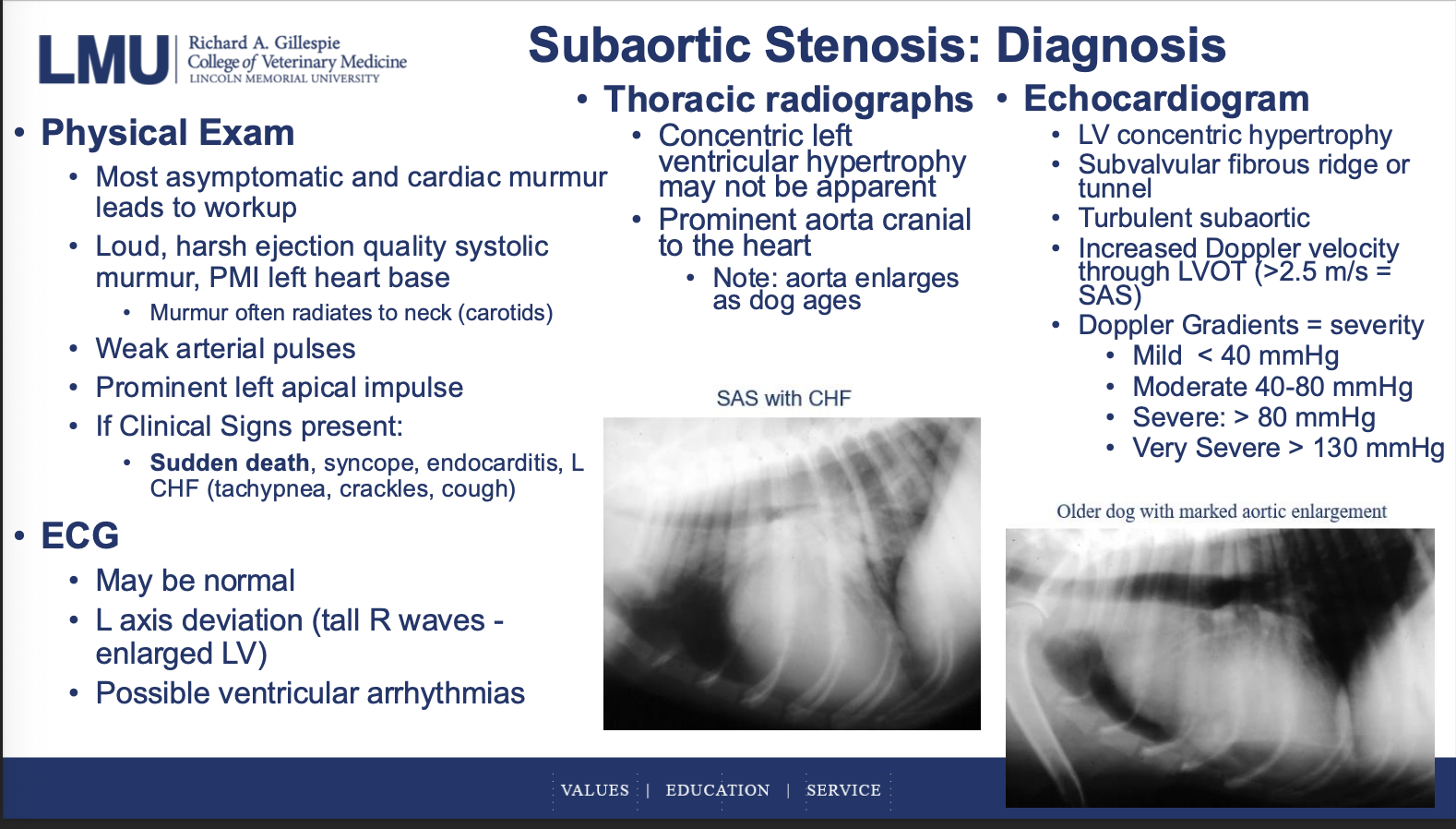
Narrowing of the aortic outflow tract: fibromuscular ridge below the aortic valve → high left ventricular pressures → left ventricular concentric hypertrophy
common in large breed dogs (Newfoundland, Goldens, Rottweilers)
How is subaortic stenosis diagnosed?
What is the treatment for subaortic stenosis?
Beta Blocker – reduce LV workload & risk for sudden death
Decr. HR = more LV filling time
Decr. Contractility = reduced pressure buildup near SAS
Decreases risk for arrhythmias
Medical therapy for CHF
Balloon valvuloplasty - high pressure balloon
Surgical removal of subvalvular fibrous ring - No clear-cut improved outcome
What is AV valve dysplasia?
Congenital malformation of the mitral or tricuspid valve
Results in regurgitation (+/- stenosis)
Volume (or pressure) overload of the affected atrium & ventricle
Valve leaflets thickened or fused
Abnormal chordae tendinae (short or long)
Abnormal papillary muscles: Malpositioned, Incomplete or absent, Abnormal attachment to ventricular wall
Common defect in the cat
Lab, Great Dane, GSD, Weimaraner
What are the physical exam findings of AV valve dysplasia?
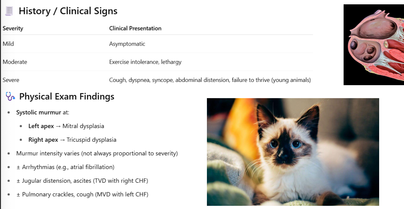
How is AV valve dysplasia diagnosed?
Thoracic rads:
cardiomegaly (LA or RA enlargement)
pulmonary venous congestion/edema (MVD)
pleural effusion, hepatomegaly, ascites (TVD)
ECG:
atrial enlargement (tall or wide P waves)
± ventricular enlargement and arrhythmias
Echo:
abnormal valve morphology (thickened, shortened, displaced leaflets)
color doppler: regurgitant jet or stenosis
How is AV valve dysplasia treated and what is the prognosis?
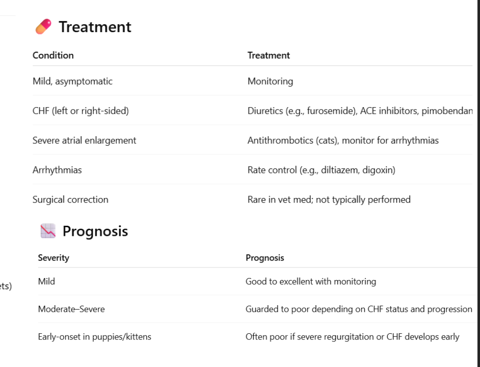
What are the 4 key abnormalities that together cause the right to left shunting and systemic hypoxemia of tetralogy of fallot?
• VSD
• Pulmonic stenosis
• RV hypertrophy
• Overriding aorta over VSD)
What are the forms of tetralogy of fallot?
Classic/Typical/Pulmonic stenosis dominant*
• Cyanosis, exercise intolerance, polycythemia
VSD Dominant
• Pulmonary overcirculation, no cyanosis
Balanced Tet
• Moderate PS and VSD
• Mild or intermittent cyanosis, subtle signs
How is tetralogy of fallot diagnosed?
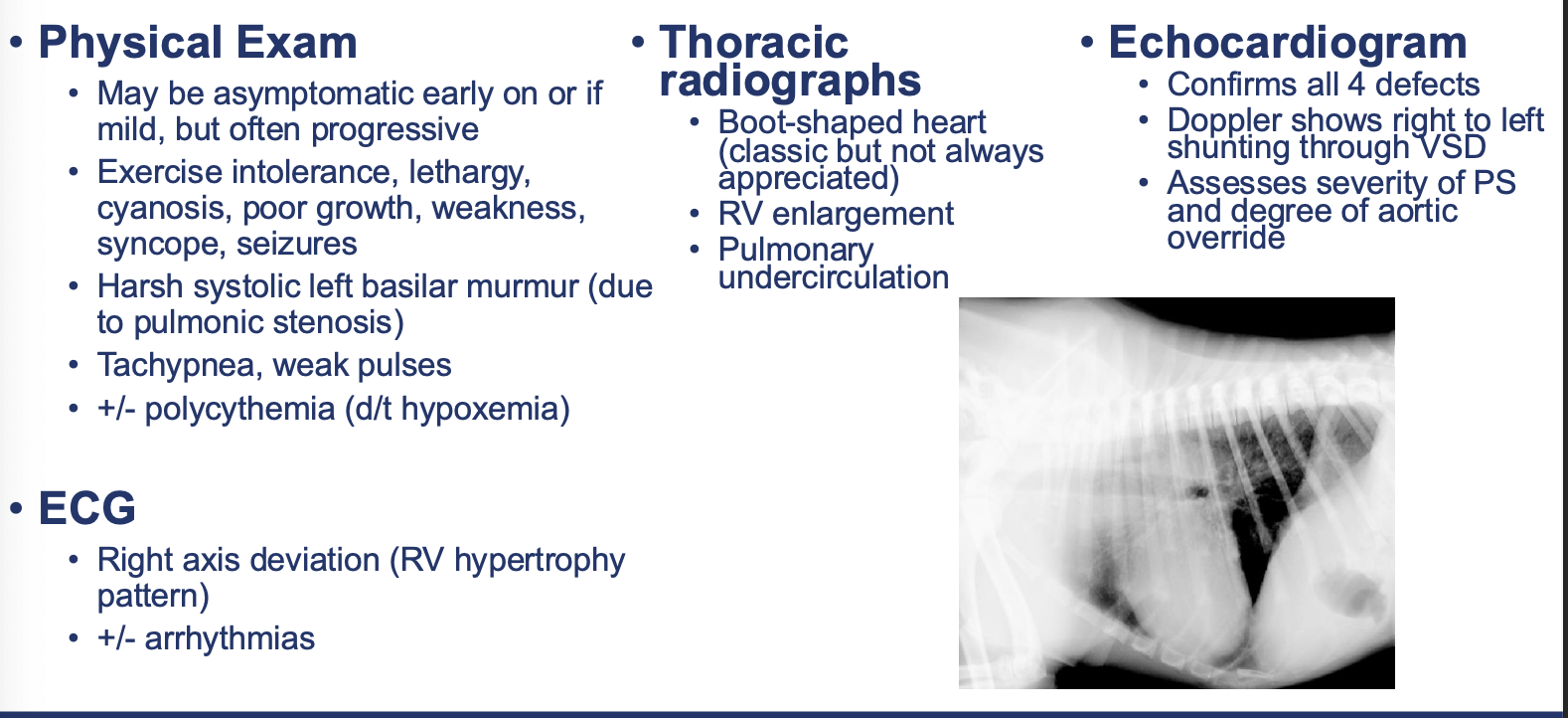
How is tetralogy of fallot treated?
• Beta Blocker – reduce dynamic RVOT obstruction and improve exercise tolerance
• Medical therapy for CHF
• Repeated phlebotomy or hydroxyurea for polycythemia
• Palliative surgery: Modified Blalock-Taussig shunt to increase pulmonary blood flow (uncommon)
What are the causes of right to left shunting?
Tetralogy of Fallot
Eisenmenger’s physiology (pulmonary hypertension)
• VSD with pulmonary hypertension (Eisenmenger’s syndrome)
• ASD with pulmonary hypertension
• PDA with pulmonary hypertension (differential cyanosis)
What are the clinical signs of right to left shunting?
Cyanosis
Reduced exercise tolerance (syncope)
Polycythemia
Neurologic signs, seizures
Capillary sludging due to polycythemia
Paradoxical embolism
Thrombus - infarct
Bacteria – CNS abscess