BIO1411L: Final Review (Chapter 1-4)
1/30
Earn XP
Description and Tags
Chapter 1: An Introduction to the Human Body, Chapter 2: The Chemical Level of Organization, Chapter 3: The Cellular Level or Organization, Chapter 4: The Tissue Level of Organization
Name | Mastery | Learn | Test | Matching | Spaced | Call with Kai |
|---|
No analytics yet
Send a link to your students to track their progress
31 Terms
(1.2) What are the six basic life processes that occur within the human body?
The six basic life processes that occur within the human body include:
1.) Metabolism is the sum of of all chemical processes that occur in the body. It can be further separated into two phases, being catabolism and anabolism.
-catabolism is the breakdown of complex chemical substances into simpler ones, resulting in the release of energy
-anabolism is the formation of complex chemical substances from smaller, simpler ones, resulting in the consumption of energy
2.) Responsiveness is the body’s ability to detect and respond to changes in its internal or external environment.
3.) Movement includes motion of the whole body or structures within the body.
4.) Growth is an increase in body size.
5.) Differentiation is the process in which unspecialized cells become specialized in their structure and function.
6.) Reproduction is the production of new cells either for (1) growth and repair in the body or (2) for production of a new individual.
(1.2) What is homeostasis? What are the four types of results of imbalances in homeostasis?
Homeostasis is the maintenance of relatively stable conditions in the body’s internal environment, despite changes that occur inside or outside the body.
-it is a dynamic process that involves constant monitoring and adjustments within normal limits
The four types of results from imbalances in homeostasis include:
1.) Disorder, which is any abnormality of structure or function
2.) Disease, which is a local or systemic illness w/ a specific set of signs and symptoms
3.) Signs, which are observable and measurable clinical changes
-can be visually observed by a physician
4.) Symptoms, which are subjective changes experienced by a patient
-can not be visually observed by a physician, only described by patient
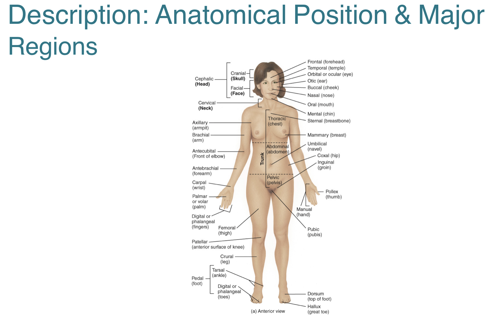
(1.4) What is the anatomical position and what are its features?
The anatomical position is the body’s standard position of reference with features that include:
-body erect, facing forward, head level
-feet flat and forward
-arms at sides with palms forward
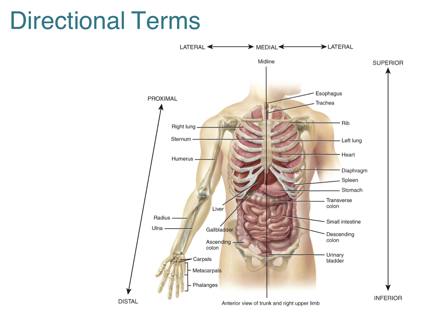
(1.4) What are directional terms and some examples of them?
Directional terms are words used to describe the position of one body part relative to another.
Examples of directional terms include:
-superior, which is the upper part of a structure
-inferior, which is the lower part of a structure
-anterior, which is closer to the front of the body
-posterior, which is closer to the back of the body
-medial, which is closer to the midline
-lateral, which is further from the midline
-intermediate, which is between two structures
-proximal, which is closer to point of attachment
-distal, which is further from point of attachment
-superficial, which is closer to the surface of the body
-deep, which is further from the surface of the body
-ipsilateral, which is on the same side of the body as another structure
-contralateral, which is on the opposite side of the body as another structure
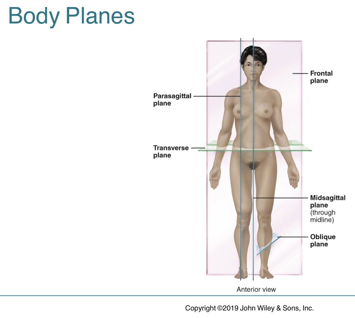
(1.4) What are body planes and some examples of them?
Body planes are imaginary flat surfaces that divide the body or body parts into sections.
Examples of body planes include:
-sagittal plane, which is a vertical plane that divides the body into right and left sides
-frontal plane (or coronal plane), which divides the body into anterior and posterior portions
-transverse plane, which divides the body into superior and inferior portions
-oblique plane, which passes through the body at an oblique angle or any angle other than a 90 angle
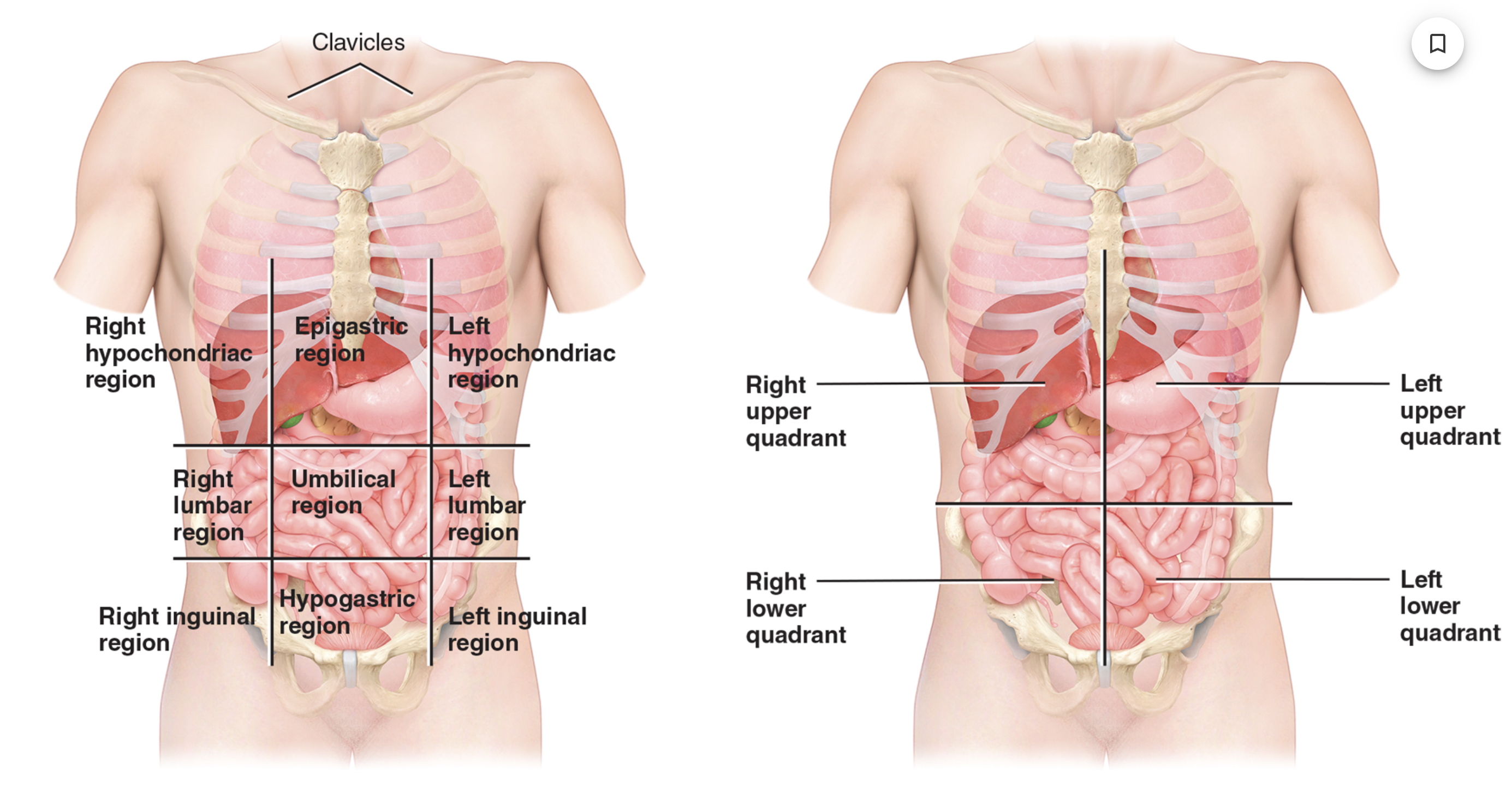
(1.7) What are the nine abdominopelvic regions? What are the four abdominopelvic quadrants?
The nine abdominopelvic regions are used for anatomical studies and includes:
-row #1: right hypochondriac region, epigastric region, left hypochondriac region
-row #2: right lumbar region, umbilical region, left lumbar region
-row #3: right inguinal region, hypogastric region, left inguinal region
The four abdominopelvic quadrants are used by clinicians to locate the site of pain, a tumor, or any other abnormalities. The quadrants include:
1.) right upper quadrant
2.) left upper quadrant
3.) right lower quadrant
4.) left lower quadrant
(2.1) What is matter? What are atoms? What are chemical elements?
Matter is anything that occupies space and exists as mass in one of three states including:
-solids (eg. bones, teeth)
-liquids (eg. blood plasma)
-gases (eg. oxygen, carbon dioxide)
Atoms are the smallest units of matter that retain the properties and characteristics of the element. The three main subatomic particles of an atom are:
1.) protons, which are positively charged
2.) neutrons, which have no charge
3.) electrons, which are negatively charged
Chemical elements refer to a type of atom characterized by a specific number of protons in its nucleus. Each element consists of one type of atom.
(2.1) What does the atomic number, mass number, and atomic mass of an atom indicate? What are isotopes?
The atomic number is the number of protons in the nucleus of an atom.
The mass number of an atom is the sum of its protons and neutrons.
The atomic mass of an atom is the average mass of all its naturally occurring isotopes.
-the atomic mass is close to the mass number of its most abundant isotope
Isotopes are atoms of an element that have different numbers of neutrons and therefore different mass numbers.
(2.4) What are inorganic compounds? Why is water such an important inorganic compound?
Inorganic compounds usually lack carbon and are structurally simple with ionic or covalent bonds.
-includes water, salts, acids, and bases
-exceptions are CO2 and HCO3
Water is the most important and abundant inorganic compound in the body. It serves as the medium for almost all of the body’s metabolic chemical reactions, and is considered the “universal solvent”. Its most important property is its polarity as it has a
-partial negative charge near oxygen atoms
-partial positive charge near hydrogen atoms
-bent shape, that allows each water molecule to interact with several adjacent ions or molecules
(2.4) What is a solvent? What is a solute? What is a solution? What are the two types of solutes?
A solvent is a substance in which some other substance can be dissolved.
A solute is a substance that is dissolved in the solvent.
A solution is the combination of a solvent and solute.
The two types of solutes include:
1.) hydrophilic, which are solutes containing polar covalent bonds and can be easily dissolved in water (“water-loving”)
2.) hydrophobic, which are solutes containing mainly nonpolar covalent bonds and cannot be easily dissolved in water (“water-fearing”)
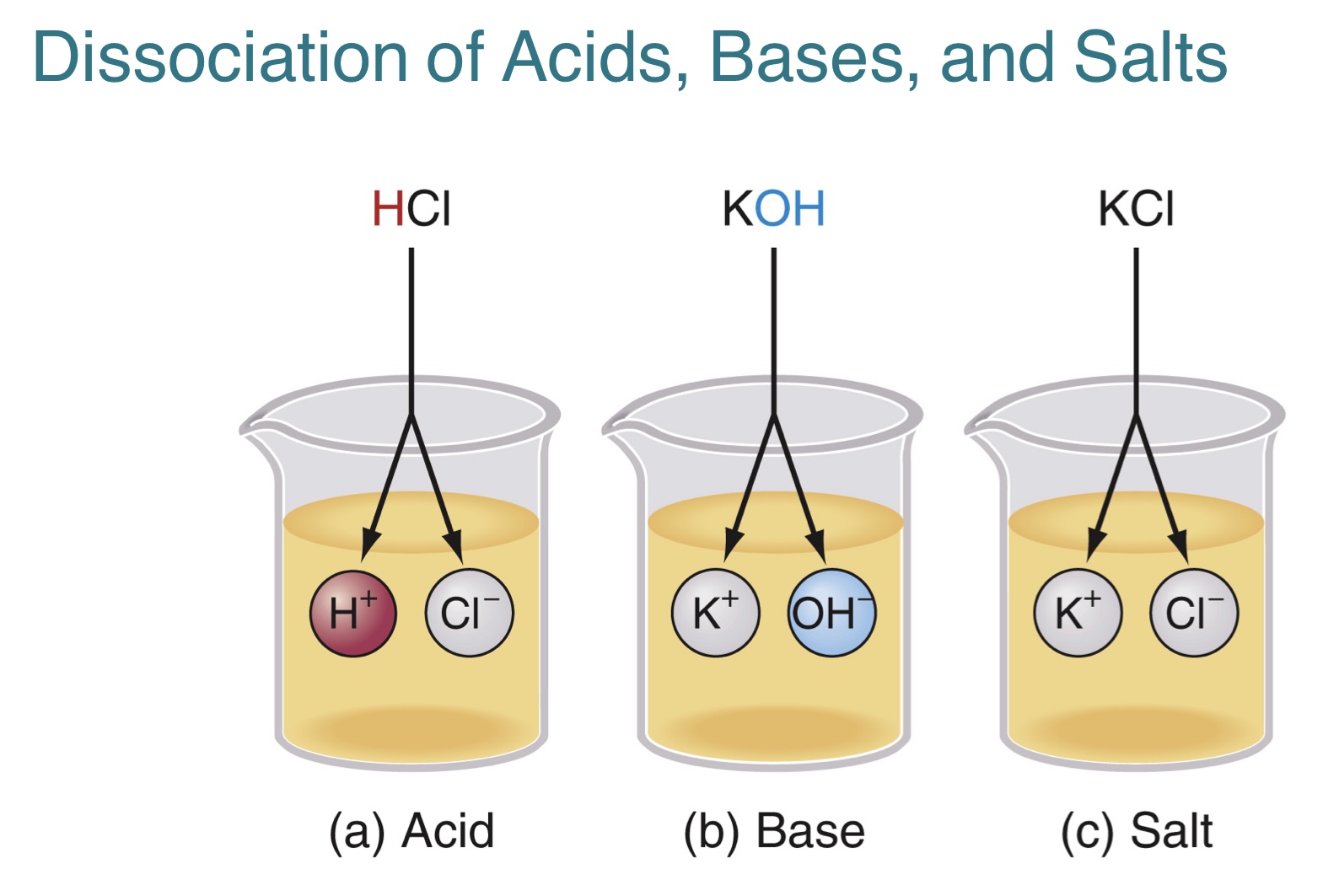
(2.4) What do acids, bases, and salts dissociate into when dissolved in water?
Acids dissociate into hydrogen ions (H+) and anions.
Bases dissociate into hydroxide ions (OH-) and cations.
Salts dissociate into cations and anions, other than H+ and OH-.
(2.4) What determines pH? What is the role of buffers in pH?
pH is a scale from 0-14 and is based on the concentration of hydrogen ions in a solution. The lower the number, the higher the hydrogen concentration. The three pH levels include:
1.) pH = 7, which is neutral and indicates that there is an equal number of H+ and OH-
2.) pH < 7, which is acidic and indicates that there is more H+ than OH-
3.) pH > 7, which is basic and indicates that there is more OH- than H+
The role of buffers is to help stabilize the pH of body fluids through:
-adding or removing H+ from solution
-converting strong acid to weak acid
-converting strong base to weak base
(2.5) What are organic compounds? What are the four major categories of organic compounds?
Organic compounds always contain carbon and are typically more complex than inorganic compounds.
-they are often covalently bonded into chains or bonded to other atoms like H,O,N
The four major categories of organic compounds include:
1.) carbohydrates
2.) lipids
3.) proteins
4.) nucleic acid (and ATP)
(2.5) What are monomers and polymers? What are macromolecules? How can macromolecules be formed or broken down?
Monomers are the basic building blocks of organic molecules.
Polymers are large molecule formed by the covalent bonding of many monomers.
Macromolecules are typically large polymers that are critical for biological functions.
Macromolecules are formed by combining two or more monomers into dimers or polymers, through dehydration synthesis.
-in dehydration synthesis, there is a loss of water molecules
Macromolecules are broken down by separating dimers or polymers into individual monomers, through hydrolysis.
-in hydrolysis, there is an addition of water molecules
(2.6) What are carbohydrates and its functions? What are the three major groups of carbohydrates?
Carbohydrates are organic molecules containing carbon, hydrogen, and oxygen.
They include sugars, glycogen, starches, and cellulose. It serves two functions that includes:
1.) major source of chemical energy for generating ATP, needed to drive metabolic reactions
2.) few structural units in the body, such as sugar in RNA and DNA
The three major groups of carbohydrates include:
1.) monosaccharides, which are the monomers of carbohydrates that contain 3-7 carbon atoms
2.) disaccharides, which are the combination of two monosaccharides by dehydration synthesis
3.) polysaccharides, which are the largest type of carbohydrates and contain tens to hundreds of monosaccharides
(2.6) What is the major polysaccharide in the human body and where is it found?
Glycogen is the major polysaccharide in the human body and it is made from glucose.
-it is stored in the liver and muscle cells, until it is needed by the body cells and is then converted back to glucose for use
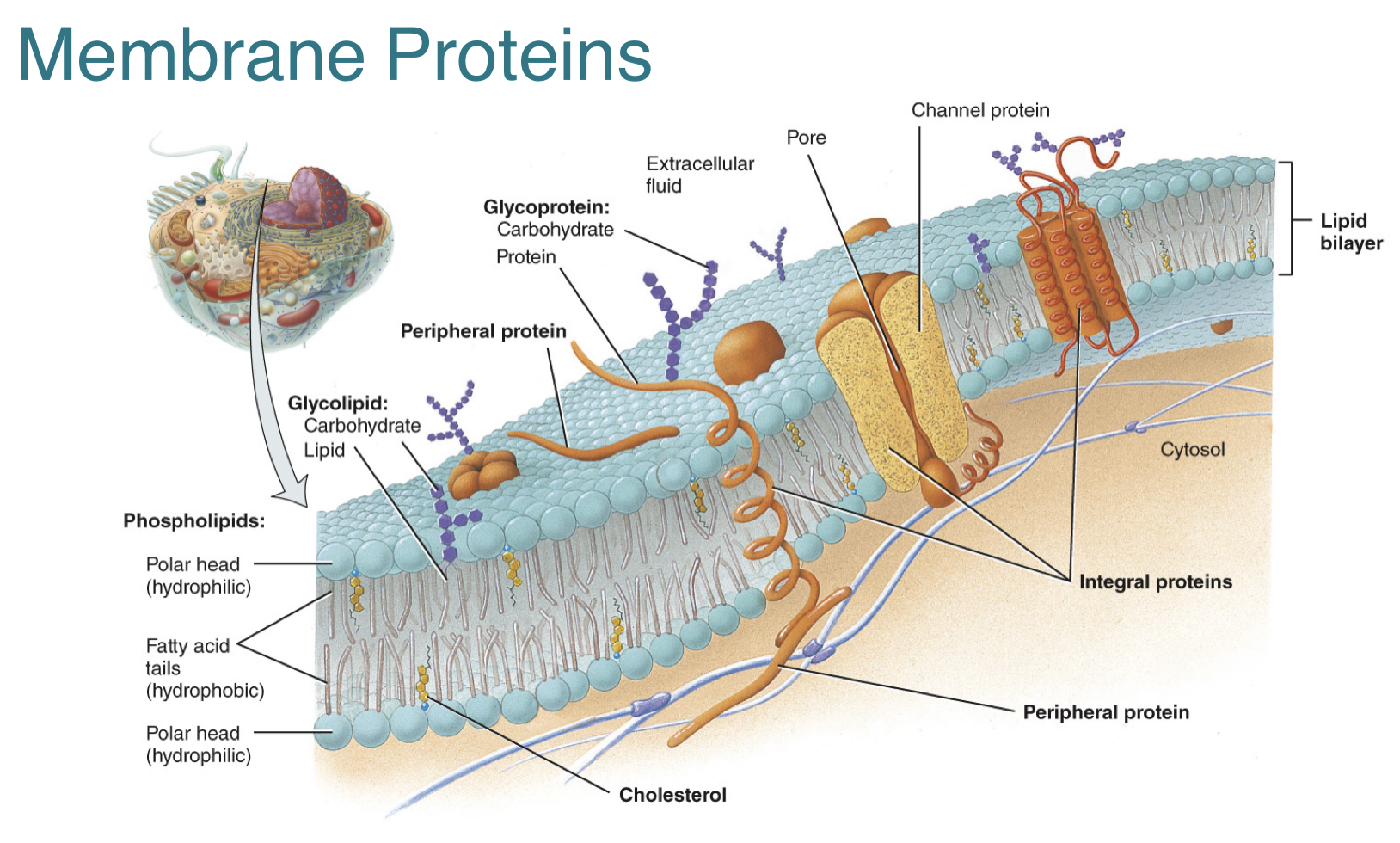
(3.2) What is the plasma membrane and its properties? What is the lipid bilayer and its properties? What makes the lipid bilayer selectively permeable?
The plasma membrane is a flexible, selective barrier between the cell and its environment.
The lipid bilayer is the basic structural framework of the plasma membrane, that is made up of phospholipids, cholesterol, and glycolipids. The bilayer arrangement occurs because the lipids have both polar and nonpolar parts that includes:
1.) hydrophilic head, which is polar and faces the extracellular fluid of cytosol
2.) two hydrophobic tails, which is nonpolar and forms the inner core
-allows passage of lipid-soluble molecules
-acts as a barrier to charged and polar molecules
The lipid bilayer portion of the membrane is:
1.) permeable to nonpolar, uncharged molecules (eg. oxygen, CO2, steroids)
2.) impermeable to ions and large, uncharged polar molecules (eg. glucose)
3.) slightly permeable to small, uncharged polar molecules (eg. water, urea)
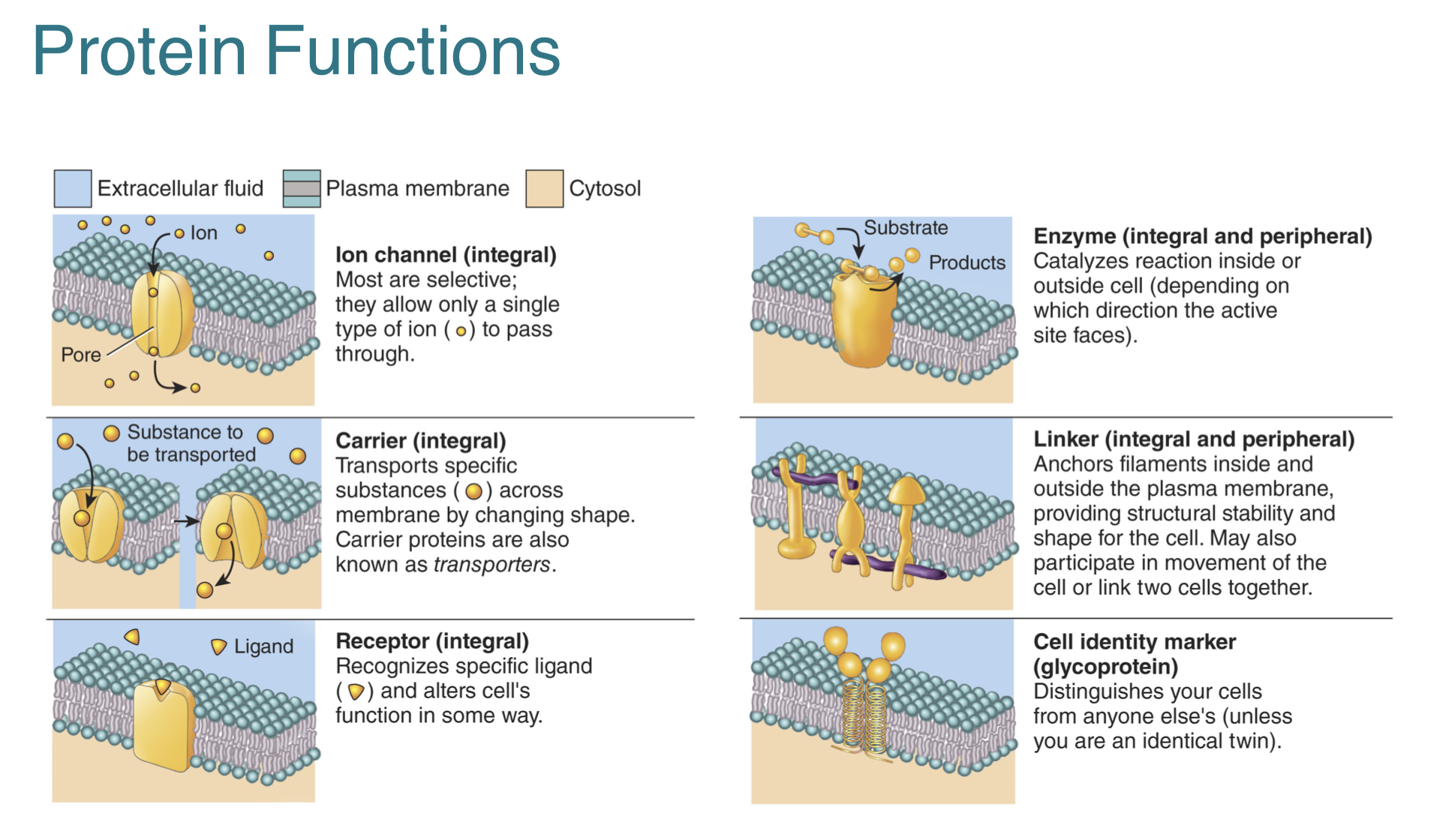
(3.2) What are membrane proteins and their six functions?
The plasma membrane contains an arrangement of membrane proteins, where some proteins are free to move and other proteins are anchored at specific locations.
The six types of functions for membrane proteins include:
1.) ion channels, which are pores allowing specific ions to move into or out of the cell
2.) carriers, which selectively move a polar substance or ion across the membrane by changing shape
3.) receptors, which are binding sites for specific ligands
4.) enzymes, which catalyze specific chemical reactions at the inside or outside surface of the cell
5.) linkers, which anchor neighboring cells or protein filaments inside and outside the cell
6.) cell-identity markers, which allow cell to cell recognition during tissue formation or respond to foreign cells
(3.2) What are the two types of classifications for membrane proteins?
Membrane proteins can be classified based on their location within the membrane. The two types of classifications for membrane proteins include:
1.) integral proteins, which are firmly embedded into the membrane
-many transmembrane proteins extend into both cytosol and extracellular fluid
-some glycoproteins w/ carbohydrate group extending into extracellular fluid
2.) peripheral proteins, which are not as firmly embedded into the membrane
-attached to polar heads of phosholipid
-attached to integral proteins on inner or outer surface of membrane
(3.2) What is a concentration gradient? What is a electrical gradient? What is an electrochemical gradient? What type of movements typically occur within these gradients?
A concentration gradient is the difference in the concentration of a chemical across the membrane.
A electrical gradient is the difference in electrical charges between two regions in the membrane.
-typically, the inner surface is more negatively charged while the outer surface is more positively charged
An electrochemical gradient is the combined influence of the concentration gradient and the electrical gradient on the movement of a particular ion.
In many cases, a substance will move across a plasma membrane “down its concentration gradient.” Some examples of this movement include:
-a substance will move to where it is less concentrated
-a positively charged substance will move toward a negatively charged area
-a negatively charged substance will move toward a positively charged area
(3.4) What is the cytoplasm? What are components of the cytoplasm (cytosol, cytoskeleton, organelles)?
The cytoplasm consists of the intracellular fluid and all the cellular contents between the plasma membrane and the nucleus.
The cytosol is the fluid portion of the cytoplasm that surrounds organelles. It consists of mostly water with various solutes (like ions, glucose, proteins, lipids, ATP, and waste products).
-it is the site of many chemical reactions inside the cell
The cytoskeleton consists of protein filaments within the cytosol that contribute to the cell’s structural framework.
Organelles are specialized structures within the cytosol that have specific shapes and perform specific functions.
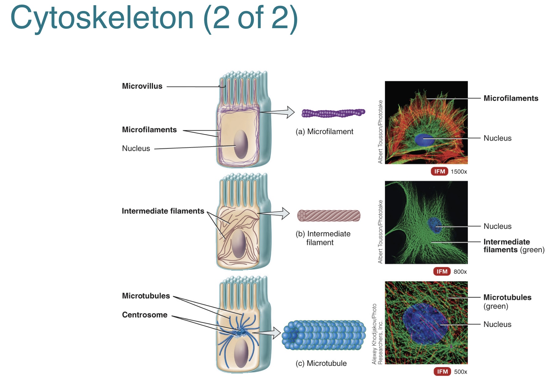
(3.4) What are the three types of cytoskeletal structures that can be found inside the cell, and their functions?
The three types of cytoskeletal structures that can be found inside the cell include:
1.) microfilaments, which are the thinnest
-provide mechanical strength to support cell shape and non-motile microvilli cell membrane projections
-generate cellular movements
2.) intermediate fibers
-stabilize position of organelles in cytosol
-resist stress and help attach cells to one another
3.) microtubules, which are the largest
-hollow protein tubes that determine cell shape
-move organelles in cytosol and specialized cell projections, such as cilia and flagella
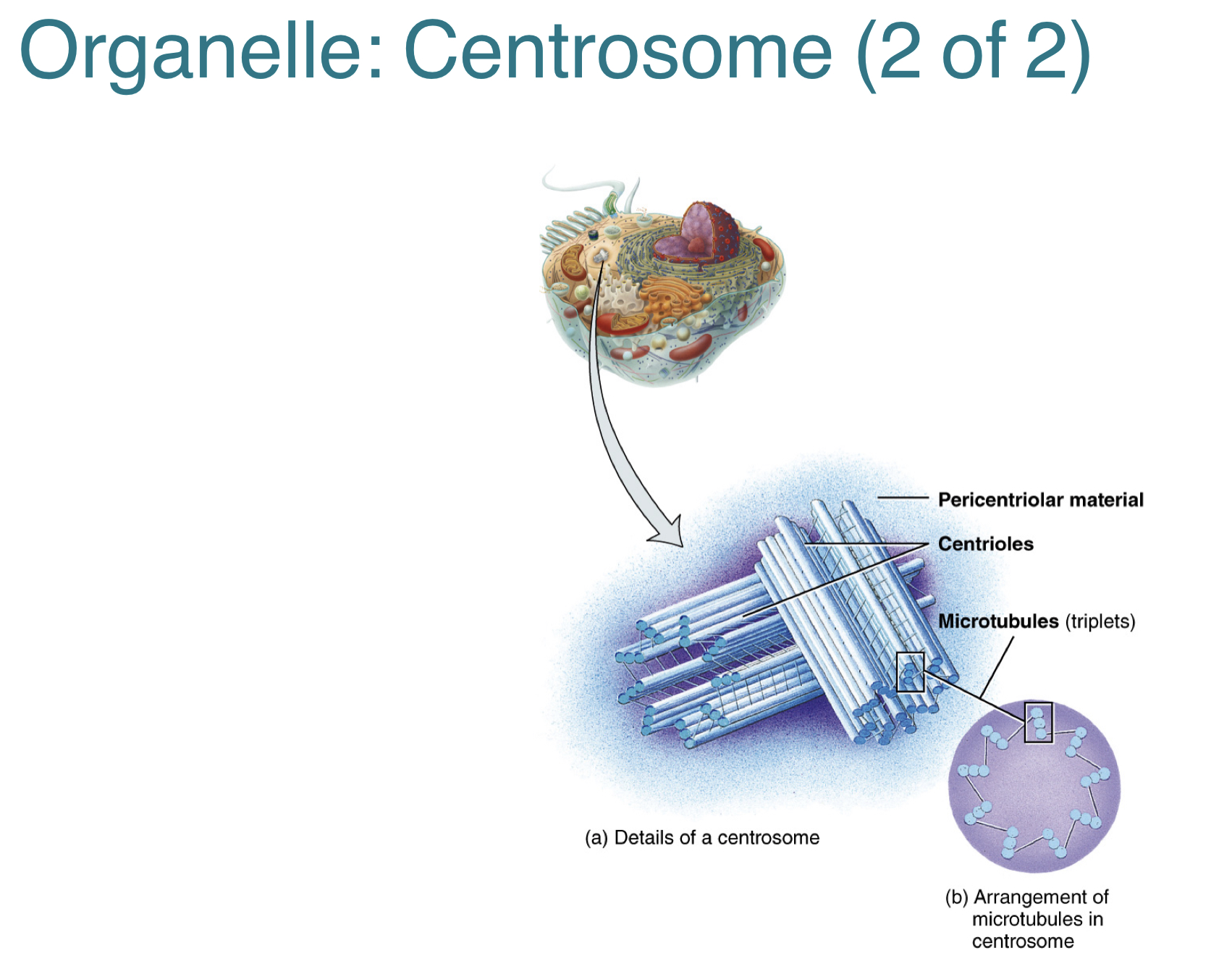
(3.4) What is a centrosome?
A centrosome is located near the nucleus and consists of two components that include:
1.) a pair of centrioles, which are cylindrical arrangements of nine clusters or three microtubules
2.) pericentriolar material, which are the tubulin protein around the two centrioles
-acts as the organizing center for microtubule formation and growth of mitotic spindle for cell division
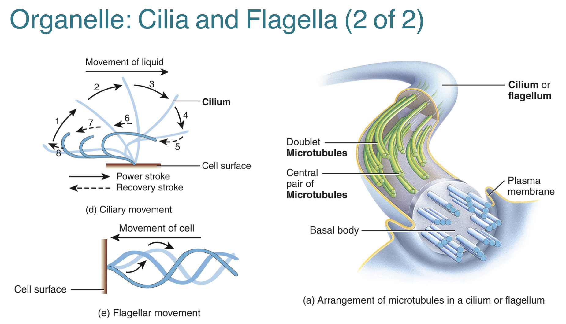
(3.4) What are cilia and flagella? What are the key differences between them?
The cell has two types of motile extensions of cell surface that contain microtubules and are still surrounded by the plasma membrane, which are the cilia and flagella.
-they are both cylindrical arrangement of nine clusters of two microtubules around central pair
-they are both anchored to basal body, just below plasma membrane
The key differences between the cilia and flagella are:
-Cilia are numerous, short, hair-like and move in a oar-like pattern within the extracellular liquid. Their function is to move fluids along the cell’s surface, helping to sweep away trapped foreign particles.
-Flagella are single, long, tail-like and move in a forward motion within the extracellular liquid. Their function is to move the entire cell. (eg. sperm propelling towards oocyte in uterine tube)
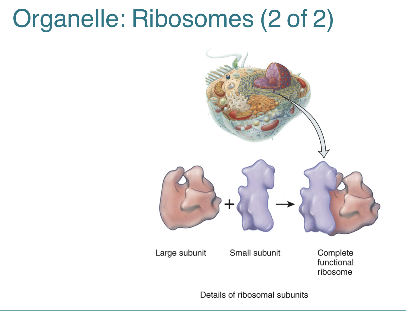
(3.4) What are ribosomes and their function? What are the two structural subunits of a ribosome?
Ribosomes are the sites of protein synthesis. They can either be attached to endoplasmic reticulum or unattached within the cytosol.
-when attached to endoplasmic reticulum, they synthesize proteins that are exported from the cell by exocytosis
-when unattached in cytosol, they synthesize proteins for use inside the cell
The two structural subunits of a ribosome include a small and a large subunit.
-they are synthesized in the nucleolus, then combine in the cytoplasm to function
-they contain ribosomal RNA
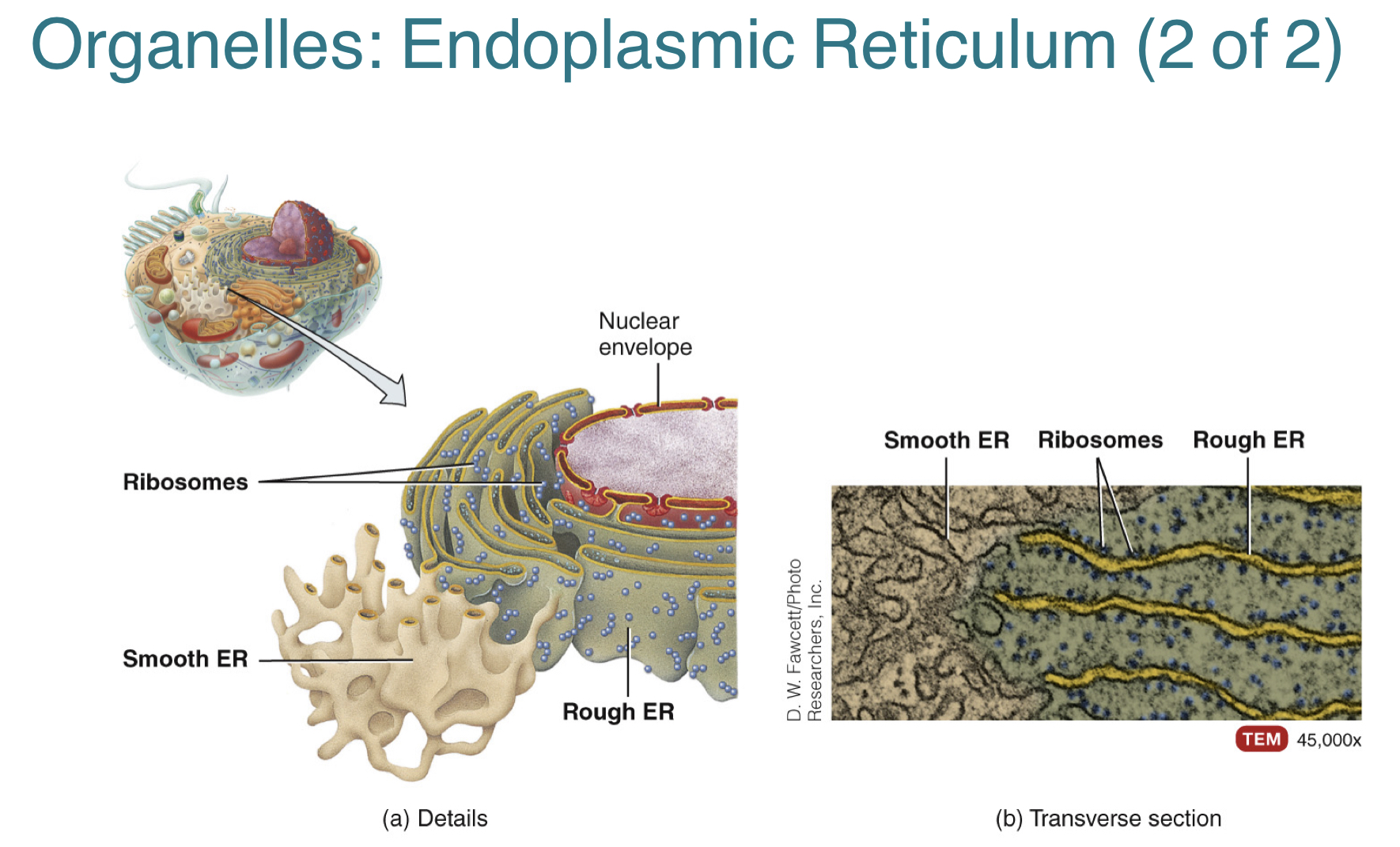
(3.4) What is the endoplasmic reticulum? What are the two types of endoplasmic reticulum?
Endoplasmic reticulum is a network of membrane-enclosed sacs or tubules that extend throughout the cytoplasm and connect to the nuclear envelope.
The two types of endoplasmic reticulum include:
1.) rough endoplasmic reticulum, which is continuous with the nuclear envelope and studded with ribosomes, thus “rough” appearance
-function: synthesizes proteins that enter spaces of the ER for processing and sorting to be inserted into other organelles or undergo exocytosis
2.) smooth endoplasmic reticulum, which extends from rough ER and lacks ribosomes
-function: synthesizes fatty acids and steroids, detoxifies drugs in liver cells, and stores calcium for contraction in muscle cells
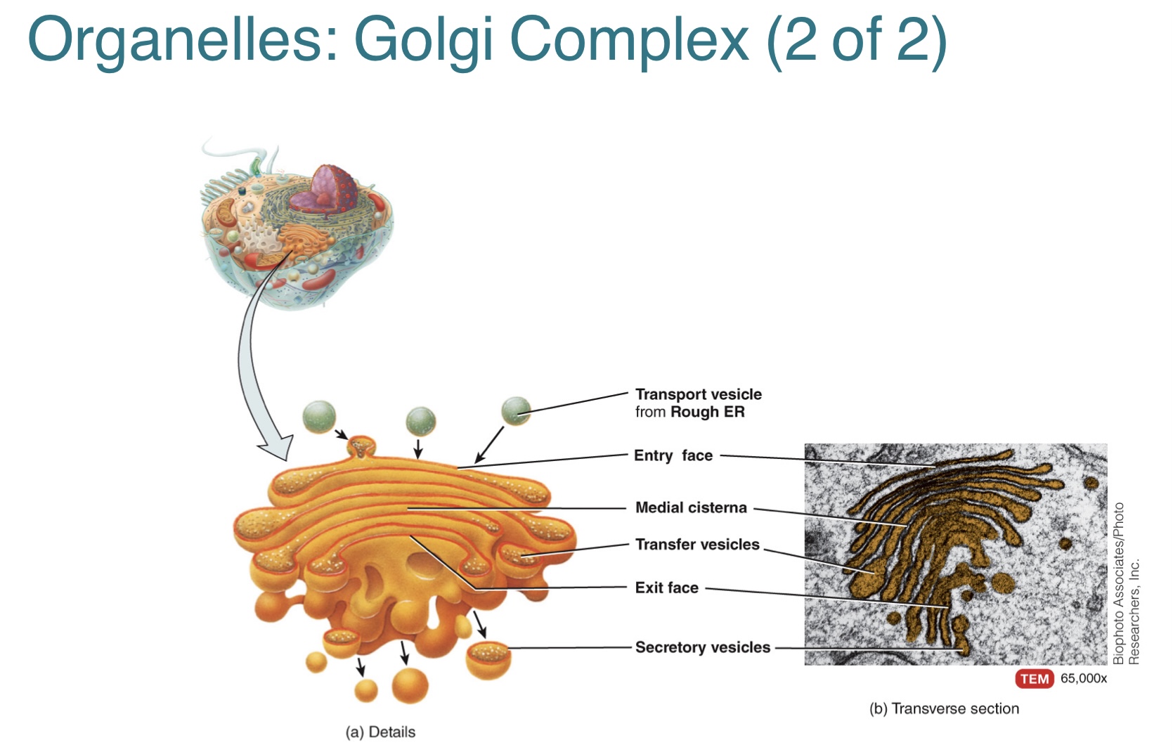
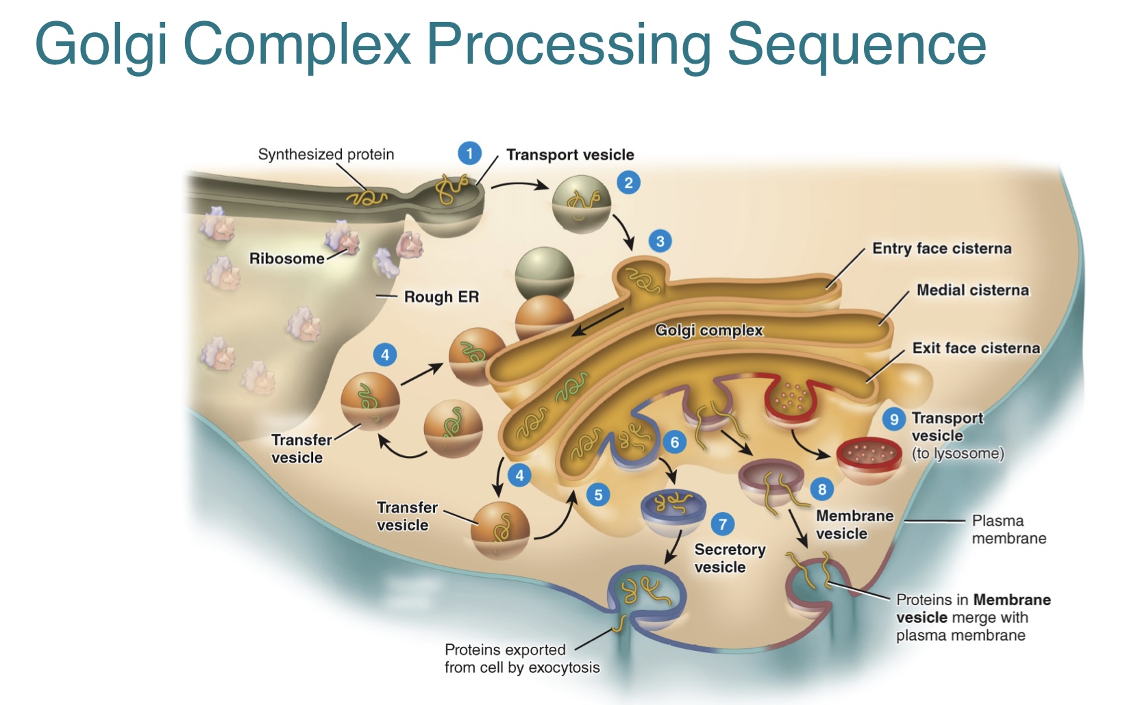
(3.4) What is the golgi complex? How do the processed proteins from rough ER leave the golgi complex?
The golgi complex is a stack of cisternae, which are flattened membranous sacs. It is located between the rough ER and plasma membrane.
-the enzymes in cisternae modify, sort, package, and transports proteins received from the rough ER
The processed proteins from the rough ER leave the golgi apparatus through three types of vesicles that includes:
1.) transport vesicles, which carry the proteins to other organelles in the cell
2.) membrane vesicles, which carry the proteins to merge with the plasma membrane
3.) secretory vesicles, which carry the proteins to the plasma membrane to undergo exocytosis and leave the cell
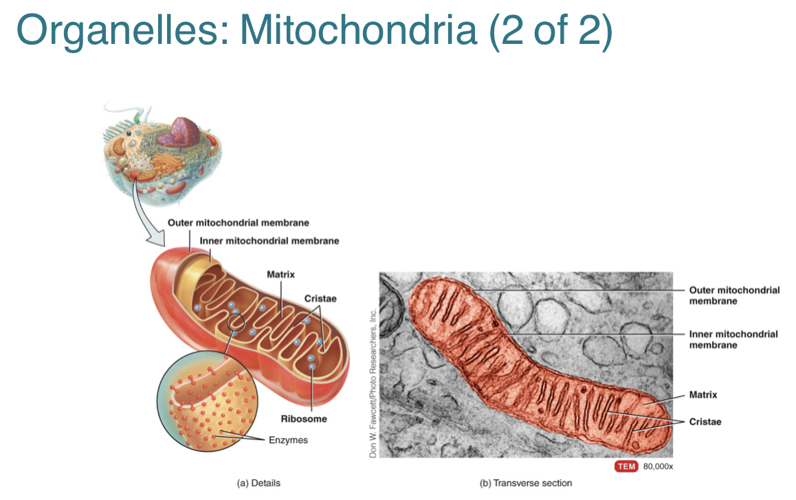
(3.4) What is the mitochondria, its properties, and its function?
The mitochondria is a double membrane organelle with fluid-filled spaces.
-outer membrane is smooth
-inner membrane is folded in cisternae, increasing surface area for aerobic cellular respiration
-fluid inside cisternae is matrix with enzymes
The function of the mitochondria is that it produces most of the cell’s ATP during the aerobic phase of cellular respiration.
-can also self replicate at times of increased energy demand or before cell division
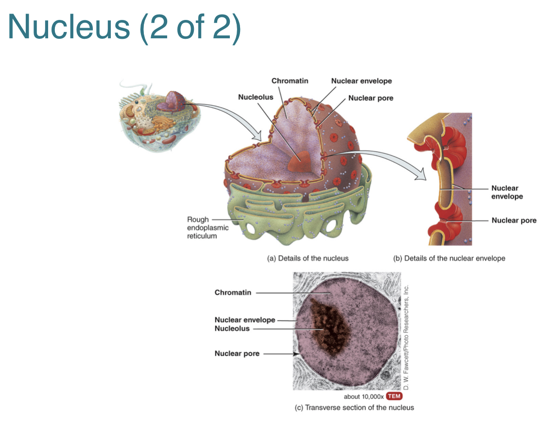
(3.5) What is the nucleus? What are some components of the nucleus.
The nucleus is a spherical or oval structure that is usually the most prominent feature of a cell. It acts as the control center for the cell and contains its genes.
Components of the nucleus include:
1.) nuclear envelope, which is a double membrane that separates the nucleus from the cytoplasm
2.) nuclear pores, which are openings in the envelope that control substance movement in and out of the nucleus
3.) nucleolus, which is a cluster of protein, DNA, and RNA, for synthesis of ribosomal subunits
4.) chromosomes, which contain genetic information
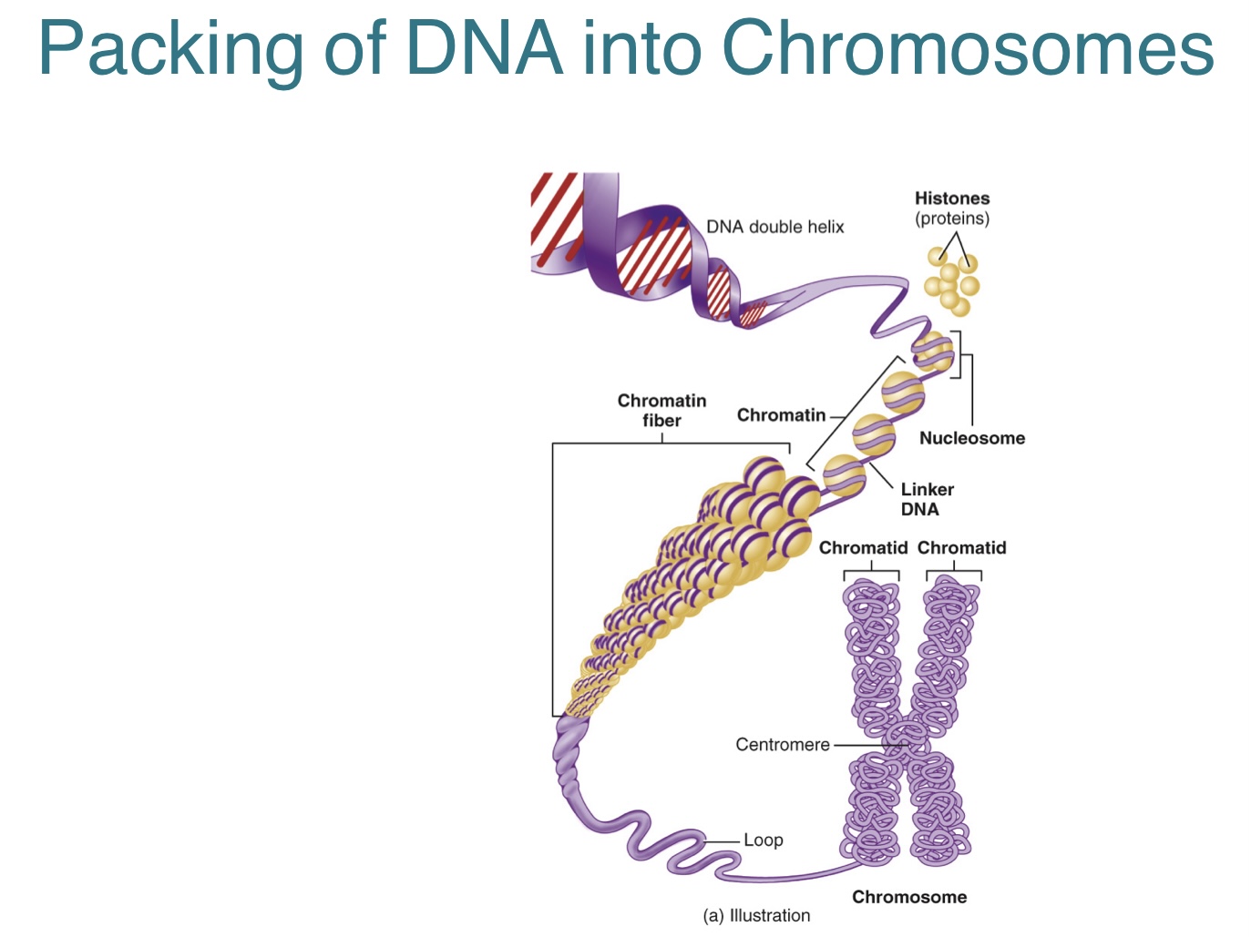
(3.5) What are genes? What are chromosomes and its components? How are chromosomes formed?
Genes are found within the nucleus, and contain most of the cell’s hereditary information. They control cellular structure and direct cellular activities.
-genes are arranged along chromosomes
Chromosomes are long DNA molecules coiled with proteins. The components of chromosomes consist of:
1.) histones, which are proteins that help package and organize DNA into nucleosomes
2.) linker DNA, which are the strands of DNA that holds adjacent nucleosomes together
3.) nucleosomes, which are the fundamental units found in chromosomes
Chromosomes are made up of chromatin, which are a complex of folded DNA and proteins before replication takes place. Once DNA replicates, a pair of chromatids are formed and are held together by a centromere. This leads to the formation of a chromosome.
(pair of chromatids = chromosome)
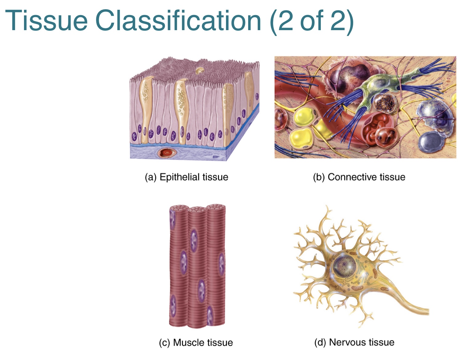
(4.1) What is a tissue? What are the four types of body tissues?
A tissue is a group of similar cells that have the same specialized function and embryonic origin.
The four types of body tissues include:
1.) epithelial tissue, which covers body surfaces, line cavities, and form glands
2.) connective tissue, which binds organs together, stores energy, and participates in body defenses
3.) muscle tissue, which produces the physical force for body movements
4.) nervous tissue, which detects and responds to changes in internal and external body conditions