mizzou anatomy exam 3
 Call Kai
 Learn
Learn Practice Test
 Spaced Repetition
 Match
Match Flashcards
 Knowt Play
1/235

There's no tags or description
Looks like no tags are added yet.
Name | Mastery | Learn | Test | Matching | Spaced | Call with Kai | Chat |
|---|
No analytics yet
Send a link to your students to track their progress
236 Terms
functions of the nervous system
1. collect information
2. proccess and evaluate info
3. respond to info
somatic sensory
receives sensory information from skin, fascia, joints, skeletal muscles, special senses
visceral sensory
sensation from organs, only stretch and tempature
somatic motor
"voluntary" nervous system: innervates skeletal muscle
autonomic motor
"involuntary" nervous system: innervates cardiac muscle, smooth muscle, glands
neurons
Cells that transmit electrical message from one area of the body to another area.
neuron =
nerve cell
dendrite
the bushy, branching extensions of a neuron that receive messages and conduct impulses toward the cell body
soma
cell body of a neuron
myelin
insulates axon, helps speed up signal through axon
pathway a nerve travels through neuron
dendrite, cell body, axon, axon terminal
synapase
where one neuron meets another
chemical synapse
use neurotransmitters
electrical synapse
rely on flow of ions at gap junctions
ependymal cells
produce cerebrospinal fluid
microglia cell
defense cells
oligodendrocytes cell
myelinated axons
astrocytes cell
blood brain barrier
most common
a myelinated axon
schwann cell
group of axons
nerve fasicles
group of nerve fasicles
nerve
nerve fascicles
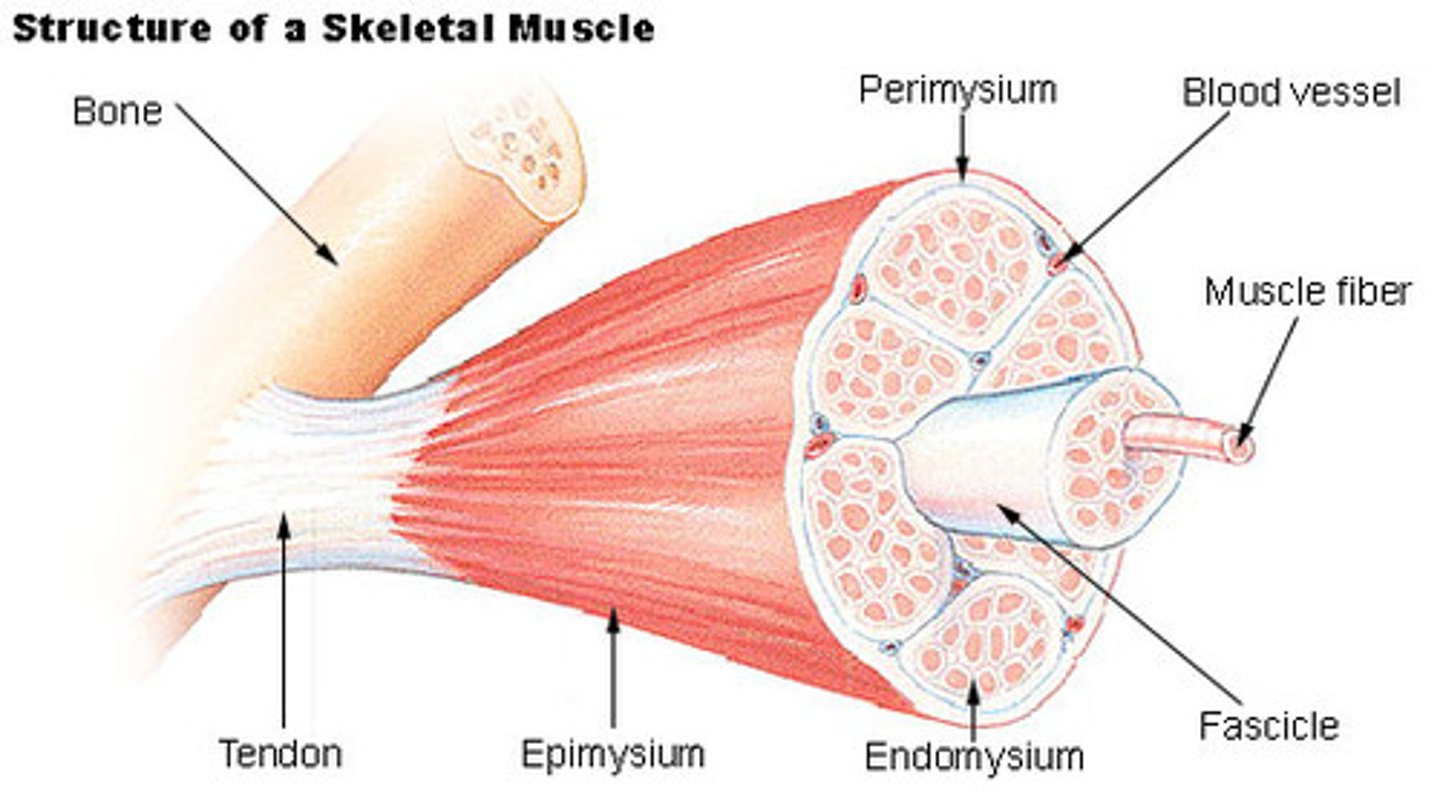
Endoeurium
inside the fasicle that covers each axon
deepest layer of nerve
perineurium
surrounds each fascicle
middle layer of nerve
epineurium
surrounds the entire nerve
most superfical layer
multiple sclerosis
a progressive autoimmune disorder characterized by inflammation that causes demyelination of the myelin sheath
functions of the brain
Controls heart rate, respiration and blood pressure and maintains homeostasis.
Innervation of head, neck and viscera.
High level tasks - intelligence, consciousness, memory, emotion, behavior
4 main parts of the brain
cerebellum
cerebrum
diencephalon
brainstem
gray mater
located in horn of spinal cord
unmyelinated
all over outside of brain
white mater
myelinated
outside of SC
inside of brain
diencephalon
contains hypothalmus and thalamus
know as inbetween brain
contains 3rd ventricle
make up the diencephalon
lateral walls - thalmus
floor- hypothalamus
roof- eplthalamus
thalmus
gateway to cerebral cortex
largest part of diencephalon
filters somatosenosory, visual and auditory info (not smell)
any part of brain that communicates with cerebral cortex must go through...
thalamus
functions of hypothalamus
Master control of the autonomic nervous system
Master control of the endocrine system
Regulation of body temperature
Control of emotional behavior
Control of food intake
Control of water intake
Regulation of sleep-wake (circadian) rhythms
epithalamus
contains the pineal gland
cerebrum
divied into left and right hemisphere
contains lobes of brain
gyri
ridges
sulci
shallow grooves
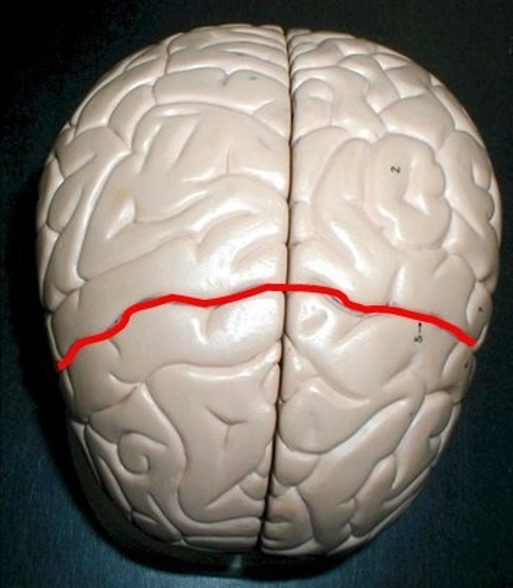
central sulcus
separates frontal and parietal lobes
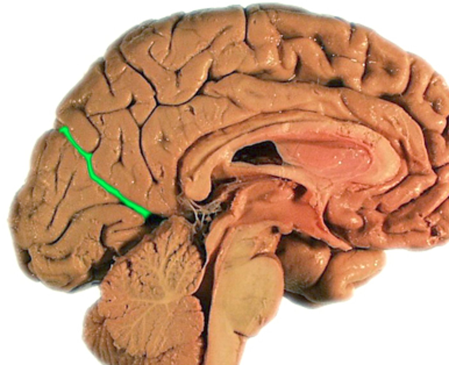
parietal-occipital sulcus
separates parietal and occipital lobes
lateral sulcus
Separates temporal lobe from parietal and frontal lobes
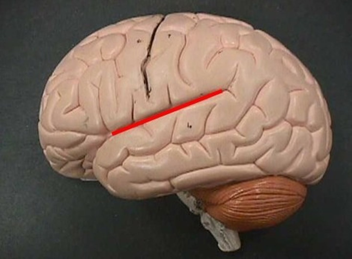
primary somatosensory area
postcentral gyrus
makes you aware that you are "sitting in a chair"
primary visual cortex
occipital lobe
primary auditory cortex
the region of the superior temporal lobe whose primary input is from the auditory system
primary motor cortex
precentral gyrus
controls skeletal muscle movement
brocas area
speech production
front lobe
wernickes area
speech comprehension
temporal lobe
corpus callosum
A thick band of axons that connects the two cerebral hemispheres and acts as a communication link between them. (white mater)
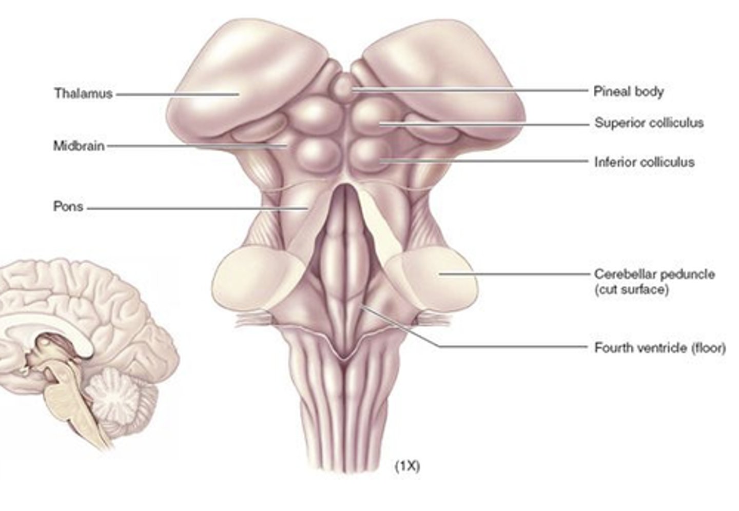
brainstem
consists of midbrain, pons, and medulla oblongata
functions of brainstem
1. connects cerebrum to spinal cord
2. start of cranial nerves for intervating the face
3. autonomic relfex center needed for survival
brainstem
cerebellum
second largest part of brain
coordinates body movements and balance
connected to brainstem
two hemispheres of cerebellum
connected by vermis
hippocampus
longterm memory
protect the brain
meninges, ventricles and CSF
meninges
membranes that cover the brain and spinal cord
CSF
cushions brain and circulate through ventricles
BBB
protects brain from exposure to toxins
pia mater
Innermost layer of the meninges
covers the brain
arachnoid mater
middle layer of the meninges
appears like a spider web
subarachnoid space
a space in the meninges beneath the arachnoid membrane and above the pia mater that contains the cerebrospinal fluid
dura mater
thick, outermost layer of the meninges surrounding and protecting the brain and spinal cord
from outermost to inward meninge layers
dura
arachnoid
subarachoid
pia
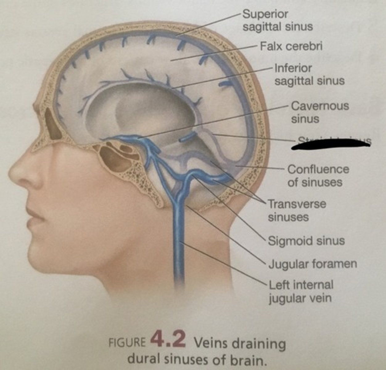
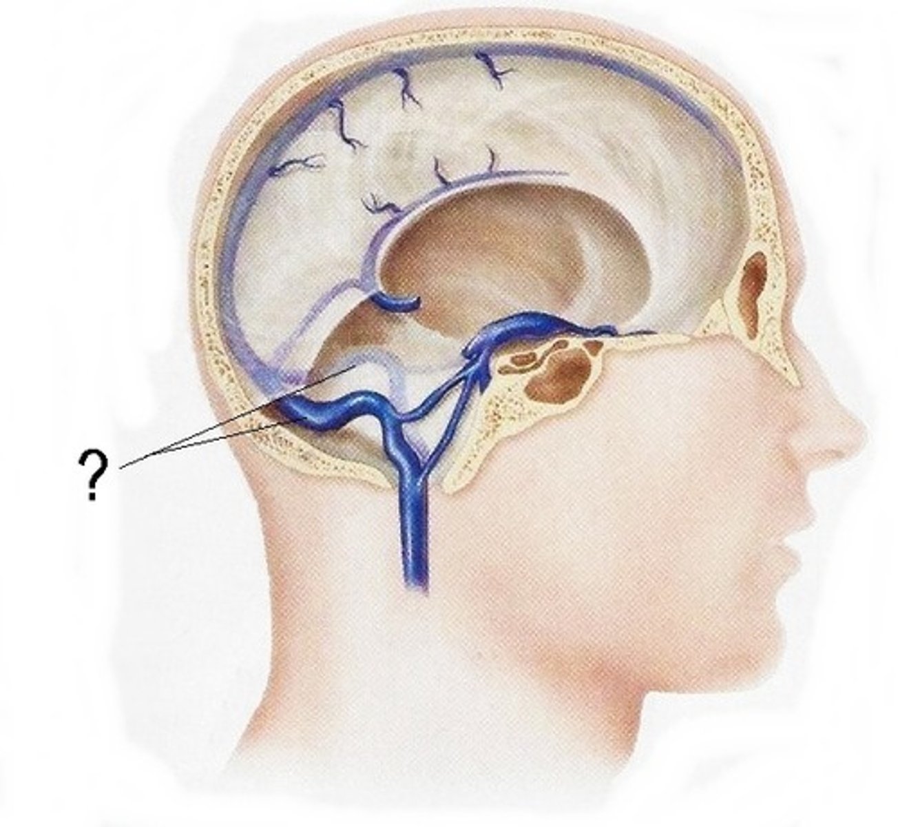
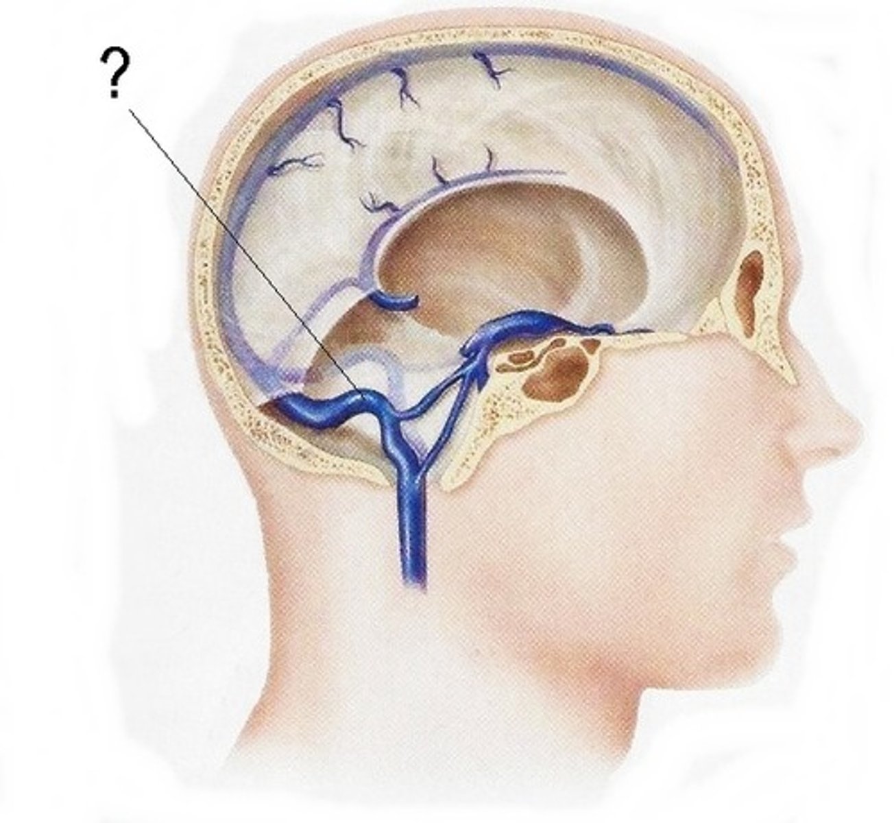
dural venous sinuses
drain blood from brain and drain into the internal jugular veins
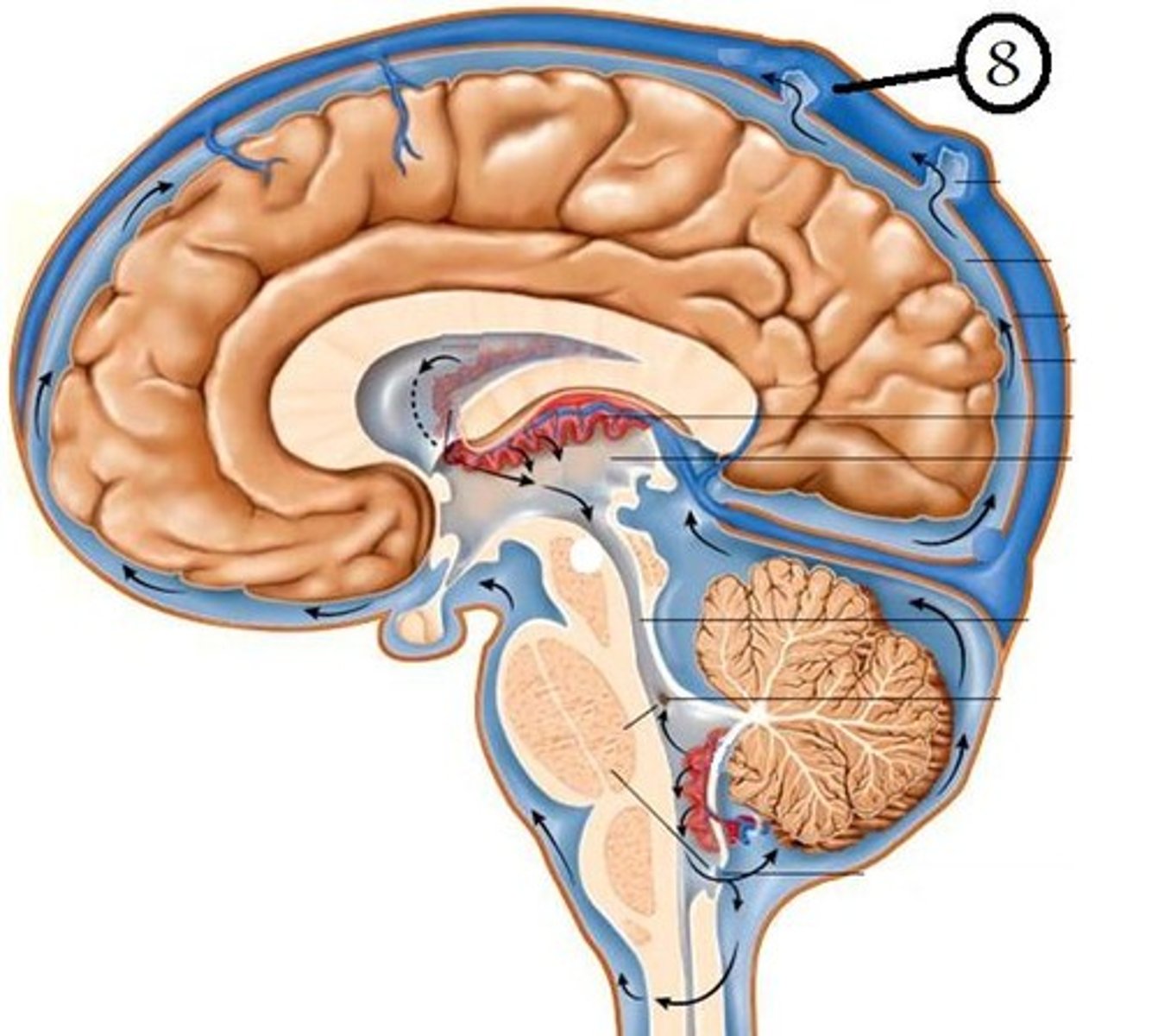
superior sagittal sinus
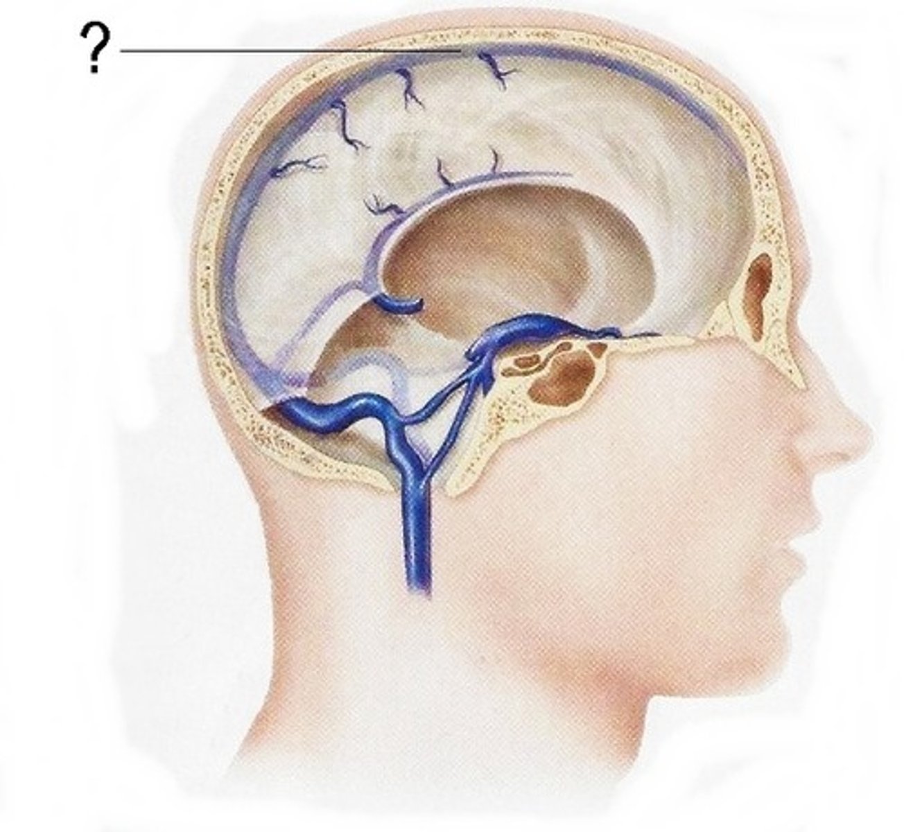
Striaght sinus
transverse sinus
sigmoid sinus
function of ventricles
making and circulating CSF
lateral ventricles
A set of paired ventricles lying within the cerebral hemispheres.
where is CSF made
choroid plexus
4 types of nerve plexus
cervical
brachial
lumbar
sacral
list cranial nerves in order
olfactory
optic
oculomotor
trochlear
trigeminal
abducens
facial
vestibulocochlear
glossopharyngeal
vagus
spinal accessory
hypoglassal
sensory or motor
s
s
m
m
b
m
b
s
b
b
m
m
optic chiasma
the crossing of the optic nerves from the two eyes at the base of the brain
eyeball problems
strabismus
ptosis
diplopia
strabismus
eyes are not parallel
cross eyed
ptosis
drooping eye
Diplopia
double vision
functions of cranial nerves
-smell
-vision
-eye movement
-superior oblique (EM)
- v1- eye, v2 cheek v3 jaw
2/3 of tongue not taste
- lateral rectus (EM)
- taste 2/3 front and facial expressions
- hearing and balance
- 1/3 taste and swallowing
- organ sensation and swallowing, speech ??
- moves head and neck
- tongue muscle movement
origin of cranial nerves
nasal cavity
retina
midbrain
midbrain
sensory receptors
pons
pons
inner ear
medulla
medulla
rootless of cervical plexus
medulla
`damage to CN 12
deviated tongue, tongue steers toward affected side
damage to CN 7
Bell's Palsy, half of face droops and has weakness
diagram of tongue
know where CN are
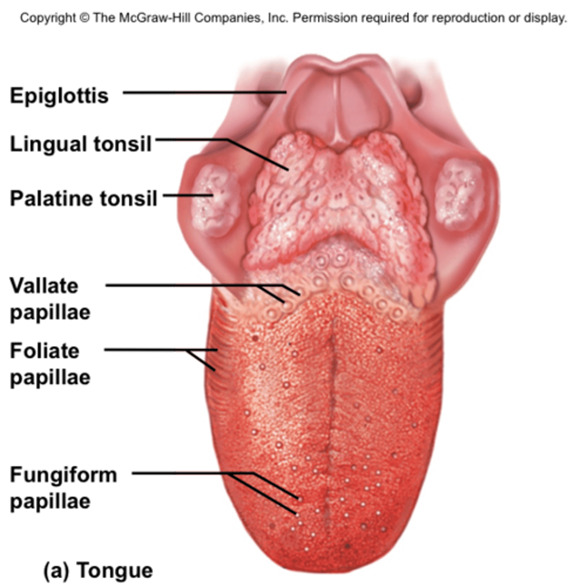
visceral motor cranial nerves
CN3- controls pupil size
CN 7 -lacrimal and salivary glands (produce tears and saliva)
CN 9- salivary glands (parotid)
CN 10 -movement of organs
function of external ear
receives sound waves
auricle or pinna and external auditory meatus
middle ear
contains malleus, incus and stapes or ( hammer, anvil and stirrup)
malleus
hammer; first of the three auditory ossicles of the middle ear
incus
anvil; middle of the three auditory ossicles of the middle ear
stapes
stirrup; last of the three auditory ossicles of the middle ear, connected to oval window
what prevents the ear from vibrating too much
tensor tymaoni (attaches to malleus) and stapedius (attaches to stapes)
Pharynogotympanic tube
equalizes pressure
ear popping on planes
causes otitis media
otits media
inflammation of the middle ear, middle ear infection
fluid buildup in ear
more common in kids because of horizontal membrane
inner ear (labyrinth)
contains bony labyrinth and membranous labyrinth
bony labyrinth
passageways in temporal bone, cavity with semicircular canals and vestibule and cochlea
membranous labyrinth
walls and sacs within the bony labyrinth
inner ear is divided into 3 parts
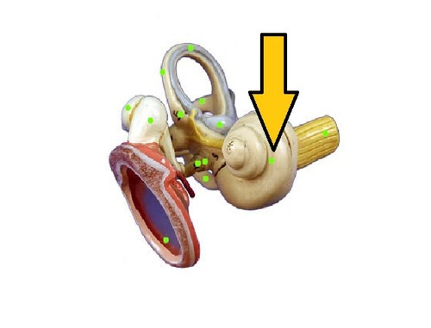
cochlea
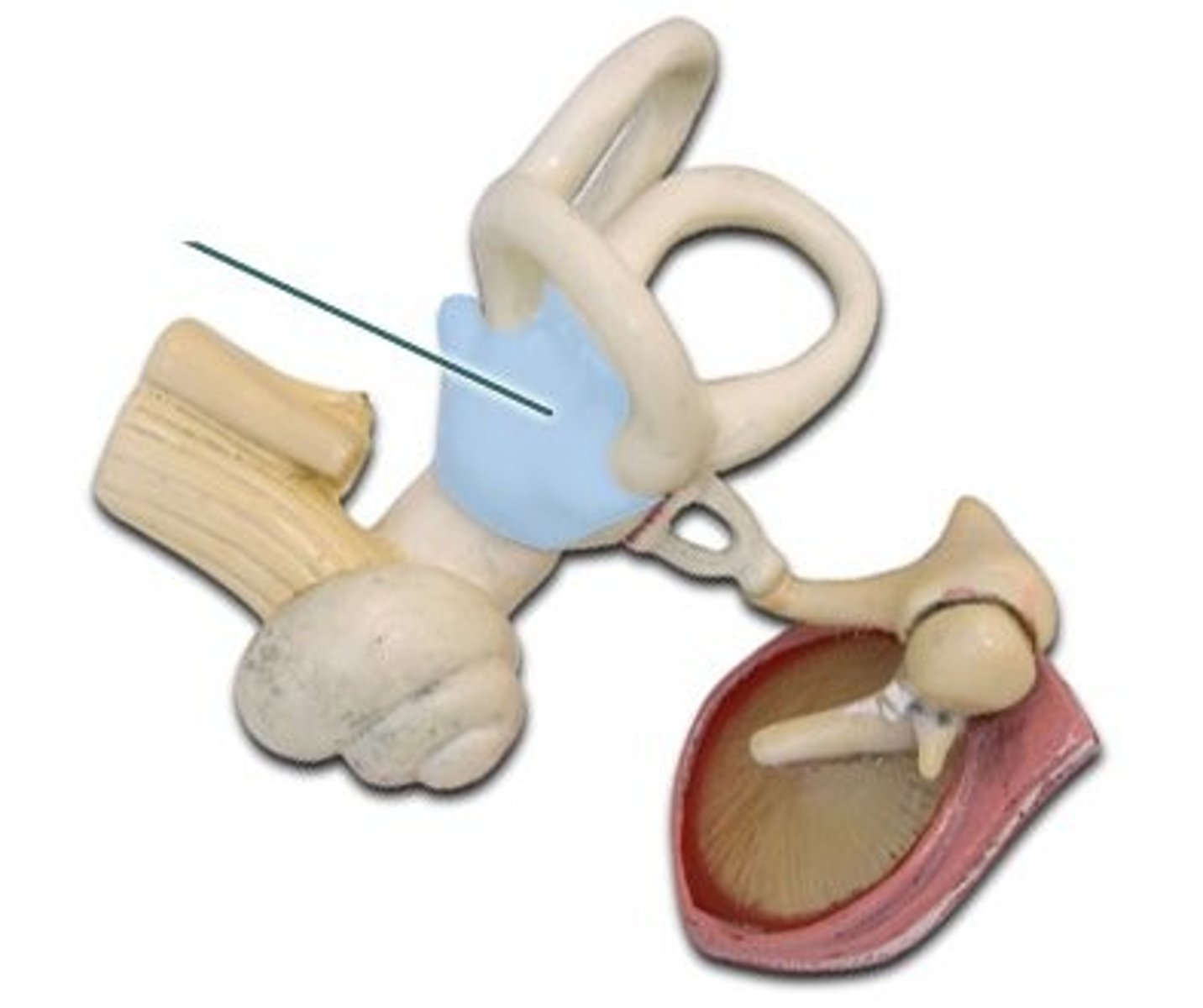
vestibule
semicircular canals
cochlea
hearing
vestibule
equilibrium starting and stopping
enterance to middle ear