HS140 — Lab Exam
1/43
Earn XP
Description and Tags
Memorize, understand the diagram!
Name | Mastery | Learn | Test | Matching | Spaced | Call with Kai |
|---|
No analytics yet
Send a link to your students to track their progress
44 Terms
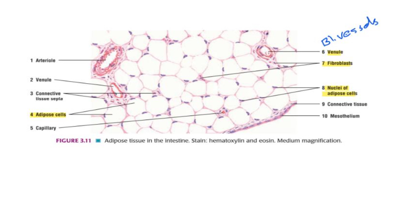
White Adipose Tissue // Adipose tissue in the intestine. Stain: hematoxylin and eosin. Medium magnification.
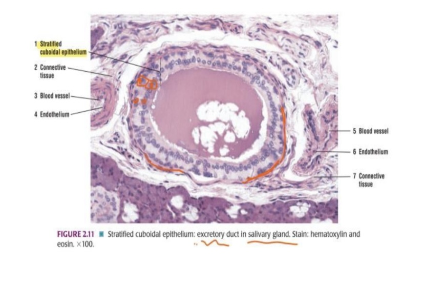
Stratified cuboidal epithelium: excretory duct in salivary gland.
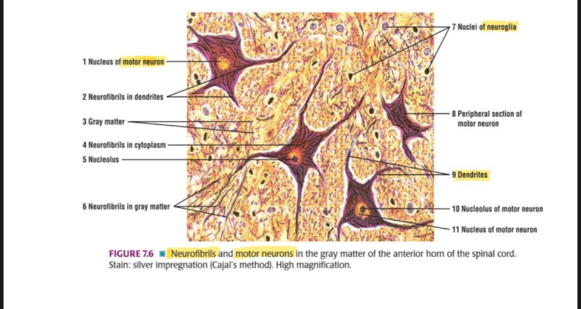
Neurofibrils and motor neurons in the gray matter of the anterior horn of the spinal cord.
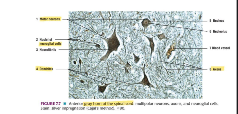
Anterior gray horn of the spinal cord: multipolar neurons, axons, and neuroglial cells.
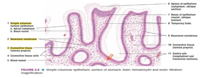
Simple columnar epithelium: surface of stomach.
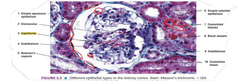
Different epithelial types in the kidney cortex.
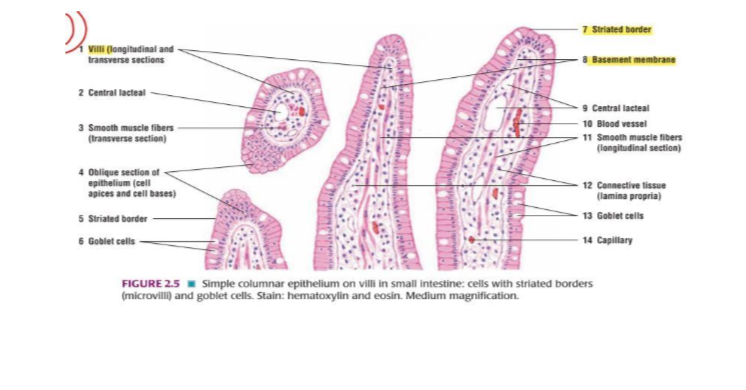
Simple columnar epithelium on villi in small intestine: cells with striated borders
(microvill) and godiet cels.
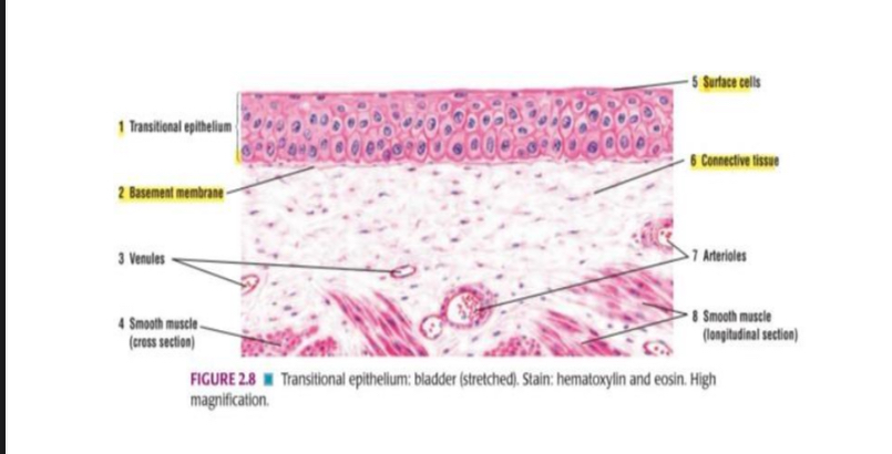
Transitional epithellum: bladder (stretched).
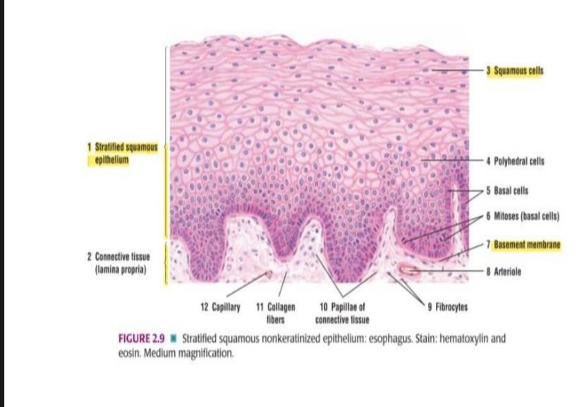
Stratified squamous nonkeratinized epithelium: esophagus.

Stratified cuboidal epithelium: excretory duct in salivary gland.
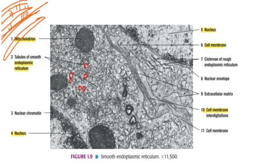
Smooth endoplasmic reticulum.
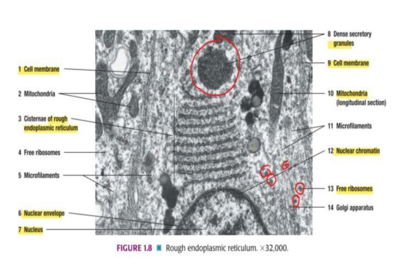
Rough endoplasmic reticulum.
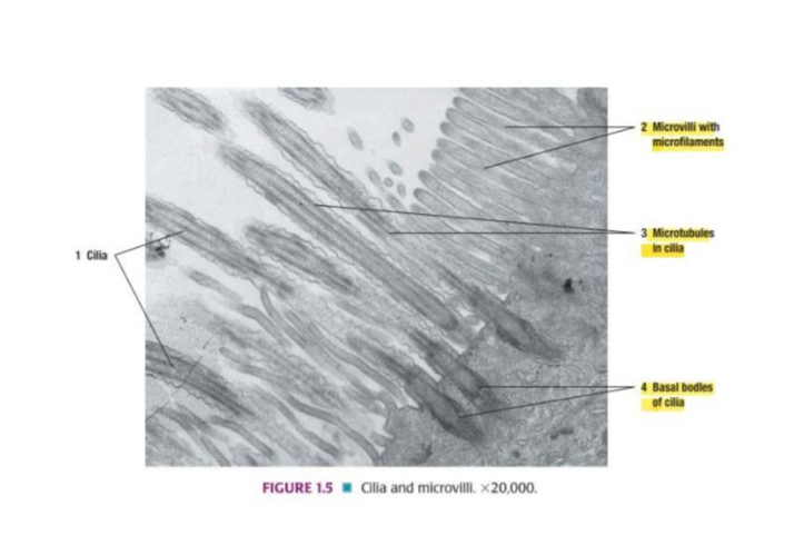
Cilia and microvilli.
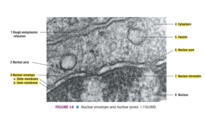
Nuclear envelope and nuclear pores.
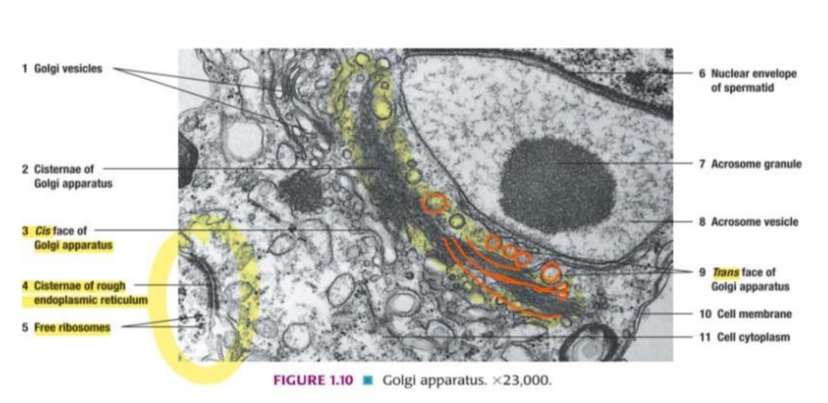
Golgi apparatus.
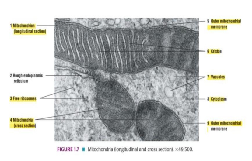
Mitochondria
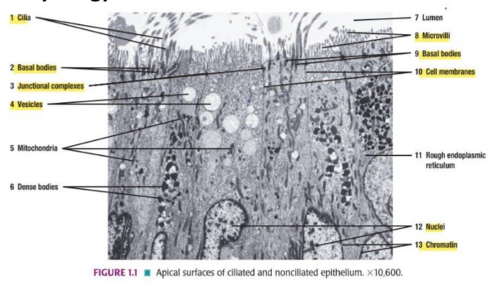
Apical surfaces of ciliated and nonciliated epithelium.
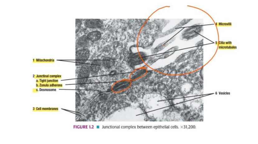
Junctional complex between epithelial cells.
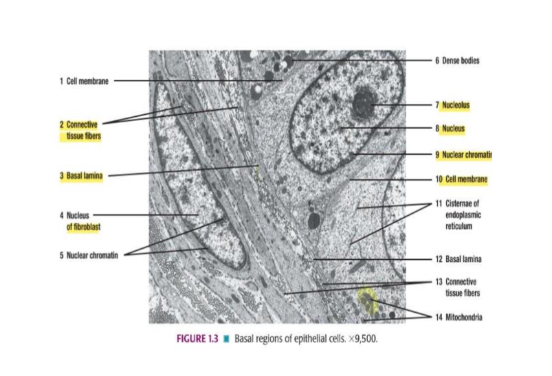
Basal regions of epithelial cells.
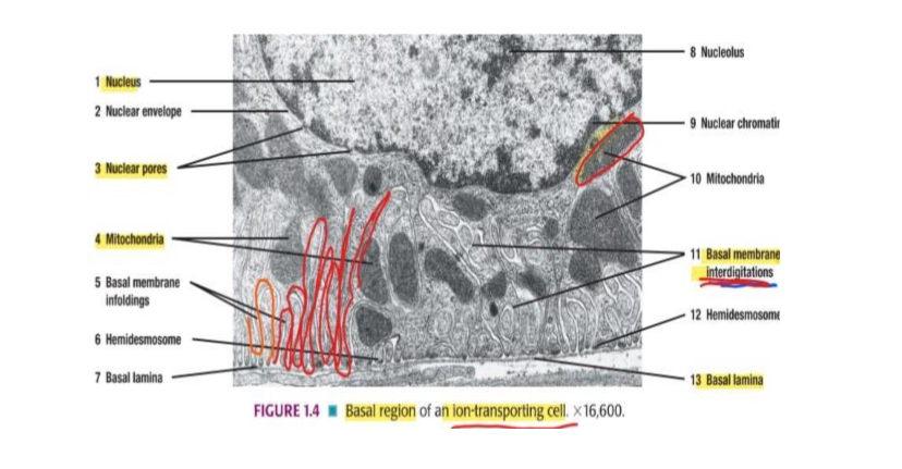
Basal region of an ion-transporting cell.
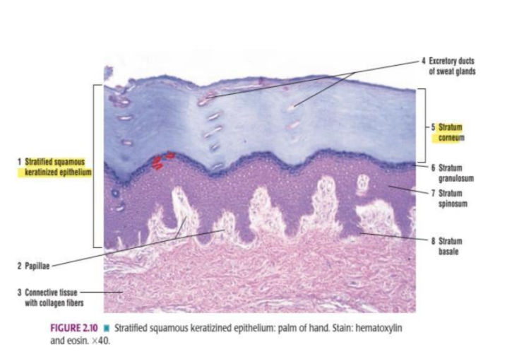
Stratified squamous keratizined epithelium: palm of hand.
What type of tissue is this?
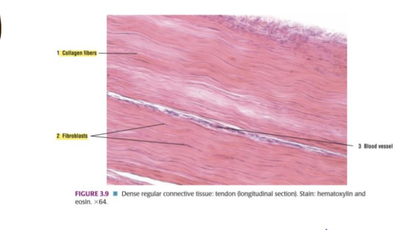
Dense Regular connective tissue: tendon (longitudinal section). Stain: hematoxylin and eosin. Medium Magnification.
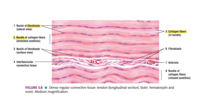
What type of tissue is this?
Dense regular connective tissue: tendon (longitudinal section). Stain: hematoxylin and eosin.
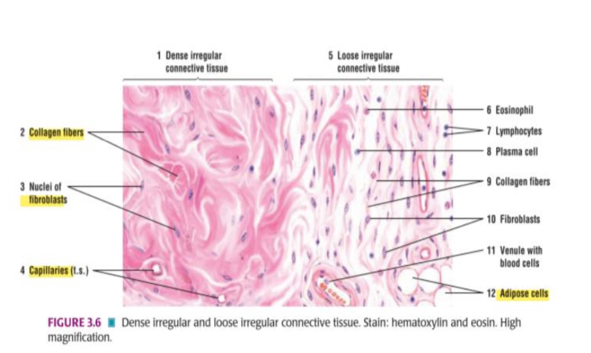
What type of tissue is this?
Dense irregular and loose irregular connective tissue. Stain: hematoxylin and eosin. High Magnification.
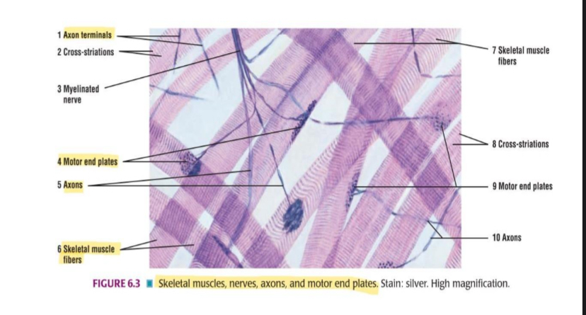
Skeletal muscles, nerves, axons, and motor end plates. Stain: silver. High magnification.
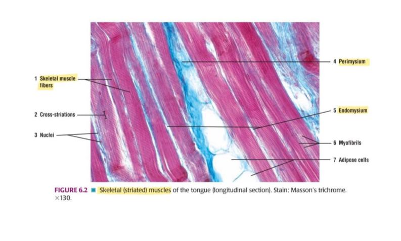
Skeletal (striated) muscles of the tongue (longitudinal section). Stain: Masson's trichrome.
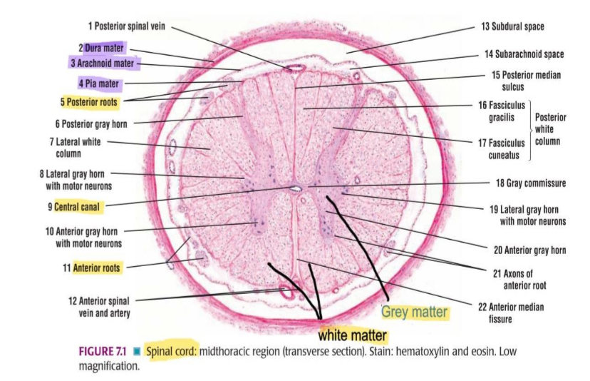
Spinal cord: midthoracic region (transverse section). Stain: hematoxylin and eosin. Low magnification.
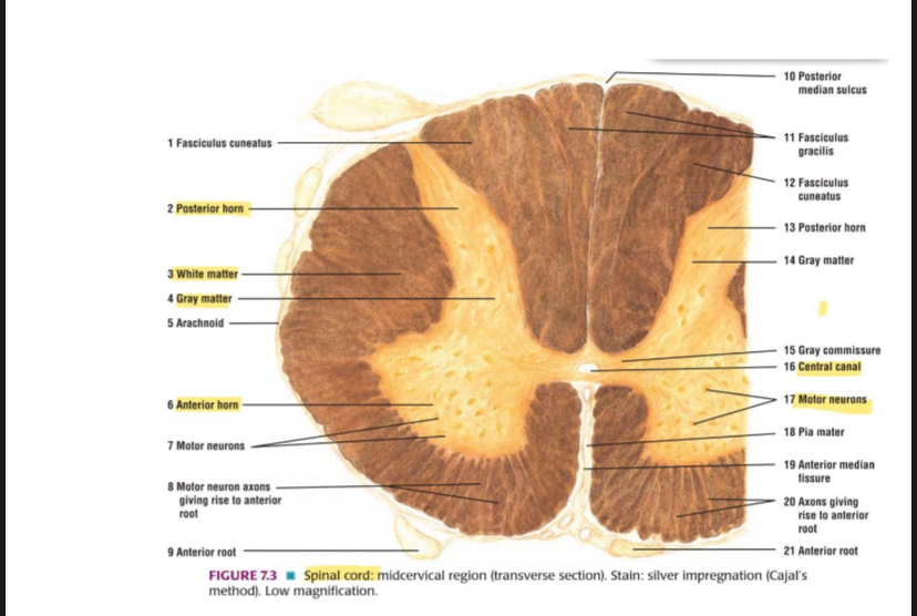
Spinal cord: midcervical region (transverse section). Stain: silver impregnation (Cajal's
method). Low magnification.
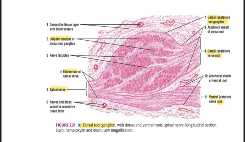
Dorsal root ganglion, with dorsal and ventral roots, spinal nerve (longitudinal section.
Stain: hematoxylin and eosin. Low magnification.
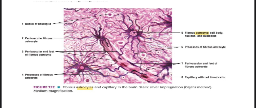
Fibrous astrocytes and capillary in the brain. Stain: Stiver impregnation

Microglia of the brain. Stain: Hortega's method. Medium magnification.
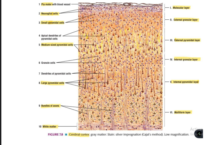
Cerebral cortex: gray matter: Stain: silver impregnation
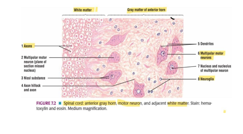
Spinal cord: anterior gray horn, motor neuron, and adjacent white matter.
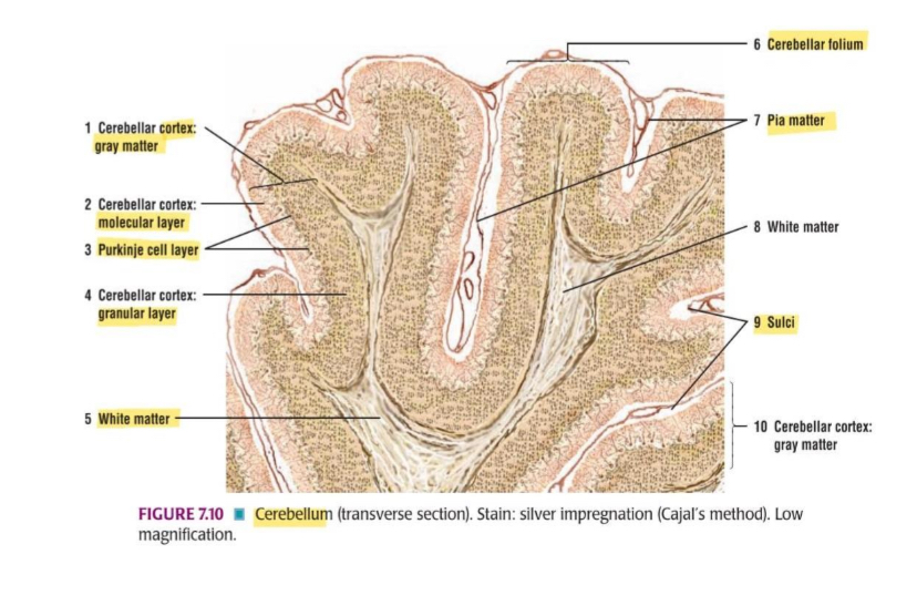
Cerebellum (transverse section). Stain: silver impregnation
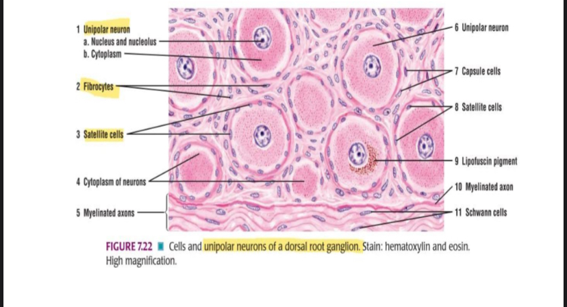
Cells and unipolar neurons of a dorsal root ganglion.
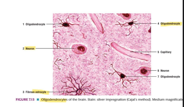
Oligodendrocytes of the brain. Stain: silver impregnation
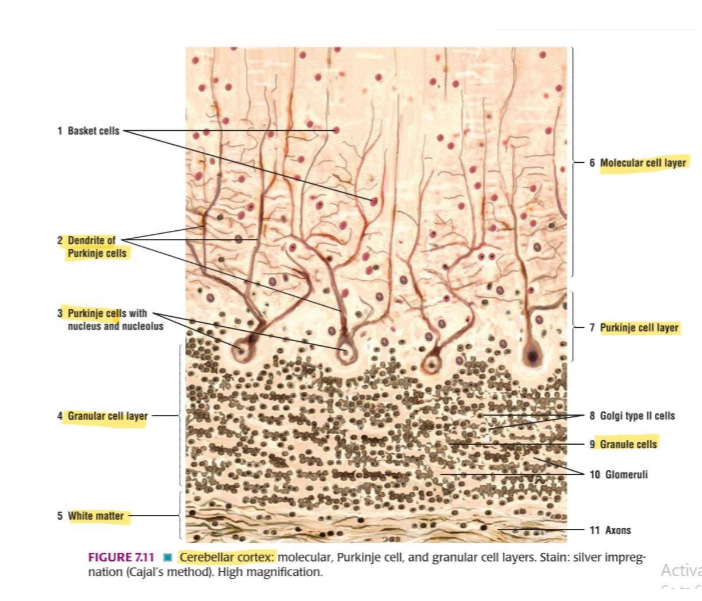
Cerebellar cortex: molecular, Purkinje cell, and granular cell layers.
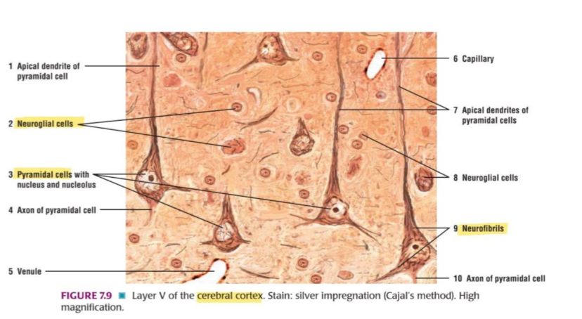
Layer V of the cerebral cortex.

Anterior gray horn of the spinal cord: multipolar neurons, axons, and neuroglial cells.
What type of tissue is this?
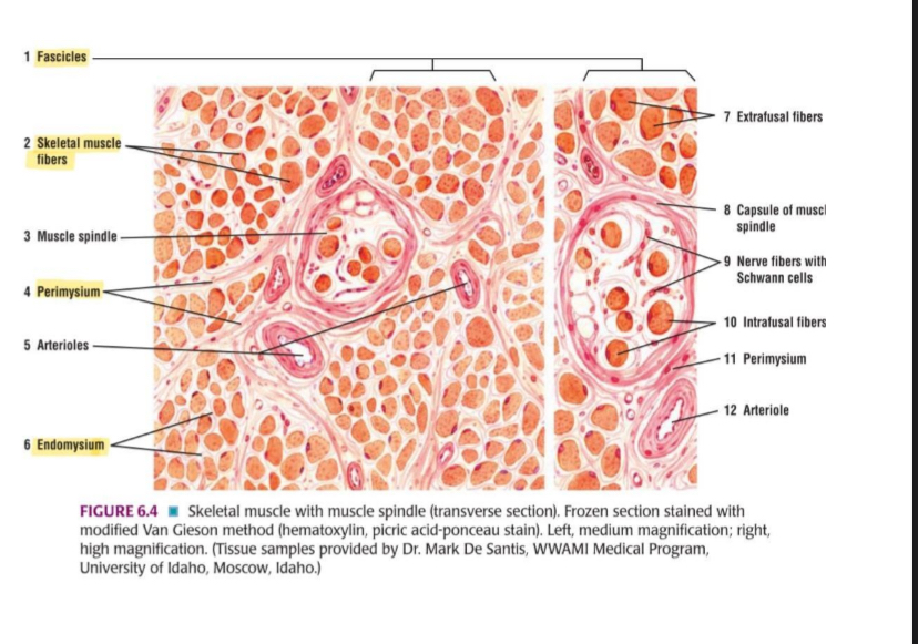
Skeletal muscle with muscle spindle (transverse section). Frozen section stained with
modified Van Gieson method (hematoxylin, picric acid-ponceau stain). Left, medium magnification; right, high magnification.