Pt Care 3: Module 1 - 4 - Introduction to CV Modules, Hypertension, Dyslipidemia, Ischemic Heart Disease
 Call Kai
 Learn
Learn Practice Test
 Spaced Repetition
 Match
Match Flashcards
 Knowt Play
1/167

There's no tags or description
Looks like no tags are added yet.
Name | Mastery | Learn | Test | Matching | Spaced | Call with Kai | Chat |
|---|
No analytics yet
Send a link to your students to track their progress
168 Terms
What are the CVD Risk Factors?
• HTN
• Hyperlipidemia
• Smoking
• Diabetes
• Overweight and obesity
• Poor diet
• Physical inactivity
• Excessive alcohol use
What is one drawback with guidelines?
Different organizations within the same field can produce guidelines resulting in discordance
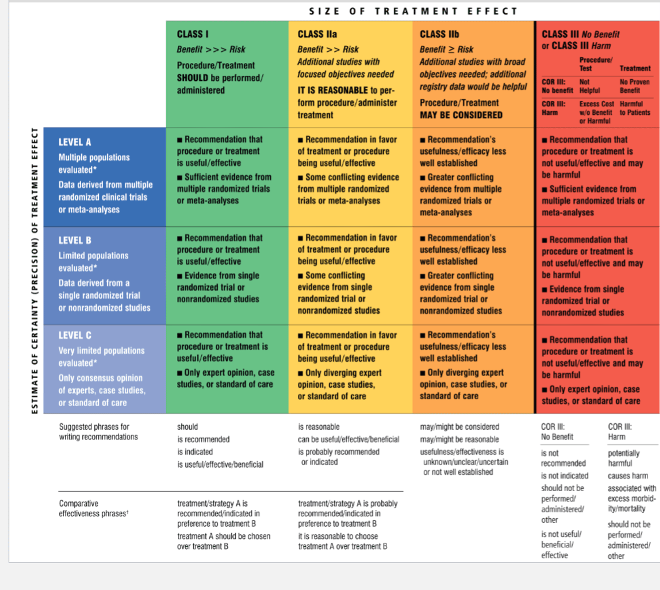
Recommendation Grading System
• Class: Benefit vs. Risk
• Level: Type of evidence and populations evaluated
Relative Risk Reduction (RRR)
= 1 - HR (or OR)
HR - Hazard Ratio
OR - Odds Ratio
Ex. HR = 0.75, then RRR is 25%
"Treatment X provides a 25% reduction in the risk for outcome A compared to treatment Y"
What is more important, relative change or absolute change?
Absolute change since relative change can be misleading
Number Needed to Treat/Harm (NNT/NNH)
= 1/ARD (Absolute Risk Difference in decimal form)
• Gives an estimate of how many patients are required to be treated for a given period of time for one to have a benefit
• In general, for major CV outcomes, numbers around 25 are amazing, 50 are good-to-great, and 75-100 or less can be acceptable depending on the outcome
• Want a lower NNT, but for more serious outcomes, you are willing to accept a higher #
Ex. CV death vs. runny nose
Example of NNT
• ARD = Rate in Grp 1 - Rate in Grp 2 = 11.7% – 9.8% = 1.9%
• NNT = 1 / (1.9% / 100 ) = 53
• NNT 53 over 12 months
• “ 1 additional CV event avoided per 53 patients treated ticagrelor instead of clopidogrel over 12 months treatment"
Heart rate is determined by the spontaneous depolarization of the _______ ____________.
SA Node
(The heart can beat without a signal from the nervous system; however, the autonomic nervous system can regulate the rate.)
How does the SNS work to increase heart rate?
• Release of NE works on B1 adrenergic receptors of the heart SA node and increases Na+ and Ca2+ channel permeabilities so depolarization is achieved faster
• Happens through HCN channels using Gs/cAMP modulation of funny current

How does the PNS work to decrease heart rate?
• Release of Acetylcholine works on M2 receptors of the heart SA node and increases K+ channel efflux so hyperpolarization deters subsequent action potentials
• Happens through HCN channels using Gi/cAMP modulation of funny current

Arrhythmias
Abnormal heart rhythms outside of the normal 60 - 100 bpm
Heart Failure
• Condition in which there is an inability of the heart to pump enough blood through the body to supply the tissues and organs with nutrients and oxygen
• Heart can no longer maintain Normal Sinus Rhythm (BPM 60 - 100) due to myocyte pathology (either signal transduction slow or weak myocytes)
Autonomic Dysfunction
• An abnormality of the involuntary aspect of the nervous system.
• POTS – Postural Orthostatic Tachycardia Syndrome
• Orthostatic hypotension
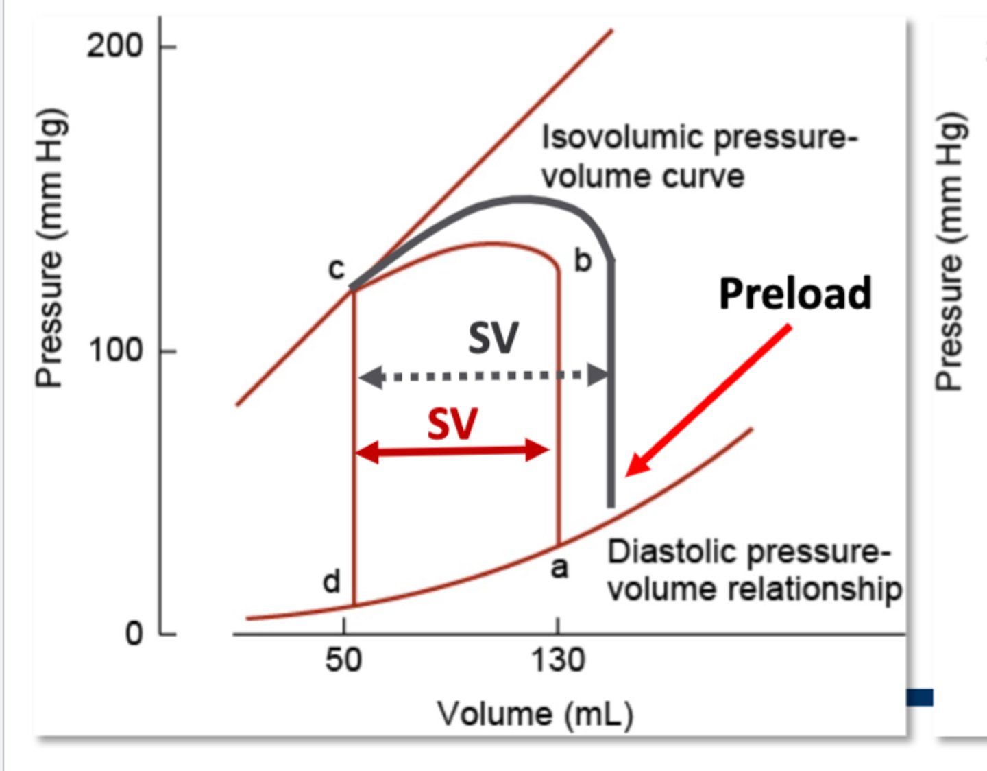
Preload
Degree of myocardium stretch before contraction
Afterload
The resistance against systolic contraction (Aortic/Arterial Pressure)
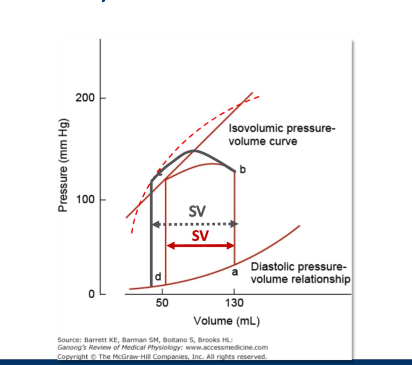
Increased Contractability Causes Increased Stroke Volume (Extrinsic Control)
Explanation: In the graph, you start with 130 mL of blood as the end-diastolic volume (denoted by letter a). Then, the pressure increases in the ventricle without the volume changing (you see the red line going straight up), until there is enough pressure for the aortic valve to open (denoted by letter b), and now oxygenated blood can start going into the rest of the body, so the blood volume starts decreasing (going from b to c). What the figure is trying to show is that when there is increased contractibility from SNS activation, the ventricles are able to contact with more strength, so more blood leaves through the aorta. So the red line resembles a normal contraction, and 80 mL of blood leaves (130 mL - 50 mL). But the gray line represents a heart with SNS activation and hence increased contractility, so ~90 mLs of blood leaves (130 mL - ~40 mL). So with the extra contractibility, more blood is ejected out and the SV increases.
Increased Preload Causes Increased Stroke Volume (Intrinsic Control)
Explanation: Preload refers to the amount of blood you start with (end-diastolic volume). In the previous example, we started with 130 mL of blood. But the gray line shows us what would happen if there was a higher preload, so 150 mL of blood instead. When you start with more blood, you can pump out more since you want to end up with the same volume after ventricular ejection is finished (this is called the end-systolic volume). In this case, the end-systolic volume is 50 mL. So if you start with 150 mLs of blood instead of 130 mL, and want to end up with 50 mL, you end up ejecting 100 mLs of blood instead of 80 mLs, so there is a higher stroke volume again.
Increased Afterload Causes Decreased Stroke Volume (Intrinsic Control)
Explanation: Afterload represents the pressure in the arteries that the ventricles must overcome to open the aortic valve and push blood into the systemic circulation. So afterload is fighting against ventricular contraction (systole) and keeps the aortic valve from opening. In this case, you start with the same 130 mL of blood, but now you need the ventricles to generate more force to get the aortic valve to open at point B. So for instance, in the normal red cycle, the afterload is ~120 mm Hg to get the aortic valve to open. But now in the gray, you have an increased afterload of ~150 mm Hg of pressure to get the aortic valve to open. So the ventricles need to generate more force to overcome the higher afterload, and additional time is needed for the ventricles to contract and generate adequate force. Since you only have a set amount of time for the heart to go through the cardiac cycle, there's less time for the heart to eject blood into the systemic circulation since it was so busy trying to just get the aortic valve to open, so for that reason, rather than ejecting 80 mLs of blood, the heart was only able to eject out ~60 mLs of blood, hence the stroke volume is decreased (which is different from the two examples above).

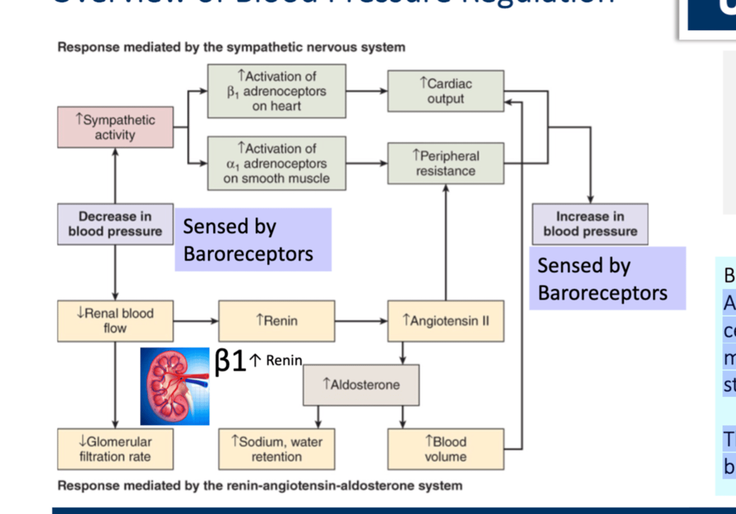
Baroreceptors
• Located in Carotid Bodies, Aortic Arch, and Juxtaglomerular cells in the kidney are mechanoreceptors activated by stretch.
• They are constantly regulating blood pressure and volume and responds to acute minute-to-minute changes
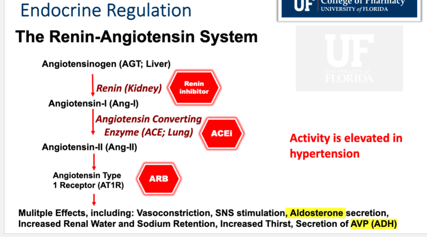
The Renin-Angiotensin System (RAS)
Angiotensinogen (AGT; Liver) -Renin→ Angiotensin-I (Ang-i) -Angiotensin Converting Enzyme (ACE; Lung)→ Angiotensin-II (Ang-II) → Angiotensin Type 1 Receptor (AT1R) → Multiple effects include:
• Vasoconstriction
• SNS simulation
• Aldosterone secretion
• Increased Renal Water and Sodium Retention
• Increased Thirst
• Secretion of AVP (ADH)
What enzymes works to counteract RAS?
• Natriuretic peptides like atrial and brain natriuretic peptides
• Natriuretic causes excretion of Na+
• There are receptors in the kidney, vasculature, heart, adrenals and brain
Does oxygen cause vasoconstriction or vasodilation?
Vasoconstriction, since it means there's no need for dilation which will bring even more oxygenated blood
Factors affecting vascular tone
• Damage to endothelium: reduced NO generation limits ability to dilate
– Especially deleterious in the heart, and other tissues with high metabolic activity (eg exercising skeletal muscle,
brain)
• Vessel wall fibrosis: limits ability to respond to flow and results in higher vessel wall stress with increasing pressure Increases systolic peak P, leads to greater damage to vessel over time. (Arteriosclerosis)
• Vessel inflammation/ Production of reactive oxygen species: with diabetes, smoking, inflammatory disease disrupt normal endothelial and vascular smooth muscle function
Vasodilators
• Metabolites - CO2, Adenosine, H+, cAMP
• Paracrine Factors - NO, Bradykinin (ANGIOEDEMA), Prostaglandins and Prostacyclins can do both depending on the specific type
• Histamine - associated with allergic and immune responses
End-Organ Damage
Damage to the vascular tissues of the heart, kidneys, brain, eyes, and other organs; caused by a continuing increase in systolic and diastolic blood pressure.

Is a pt with HF considered as having clinical ASCVD?
NO, Clinical ASCVD =
• Acute coronary syndrome (NSTEMI, STEMI, unstable angina)
• Myocardial infarction
• Stable or unstable angina
• Coronary or other arterial revascularization
• Stroke
• Transient ischemic attack (TIA)
• Peripheral arterial disease (including aortic aneurysm)
What does ASCVD stand for?
• AtheroSclerotic CardioVascular Disease
• Atherosclerosis: hardening/thickening of the walls of the arteries
– Coronary heart disease (CHD): Fatal or nonfatal myocardial infarction (MI), angina pectoris
– Cerebrovascular disease: Fatal or nonfatal stroke and transient ischemic attack (TIA)
– Peripheral artery disease: Intermittent claudication and critical limb ischemia
– Aortic atherosclerotic disease (abdominal aortic aneurysm or descending thoracic aneurysm)
ASCVD Risk Categories
• Low risk → less than 5%
• Borderline risk→ 5% to 7.4%
• Intermediate risk → 7.5% to 19.9%
• High risk → greater than 20%
Should we use carotid intima-media thickness to estaimate ASCVD risk?
NO! It is not an accurate measure.
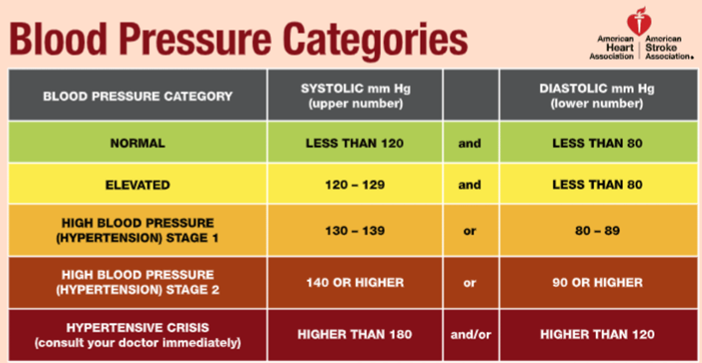
Blood Pressure Categories
• Normal: <120/<80
• Prehypertensive: 120-129/<80
• Stage 1 HTN: 130-139/ 80-89
• Stage 2 HTN: >/= 140/ >/= 90
• HTN Crisis: >180 and/or > 120
How many readings are required to diagnose a pt with HTN?
Requires 3 consecutive in office (although some other places 2 or greater...) readings of high blood pressure
What are non-dippers?
• Non-Dippers have normal day time pressures, but do not have the "night-time dip"
Blood pressure is ________ in the morning and _______ at night.
highest; lowest
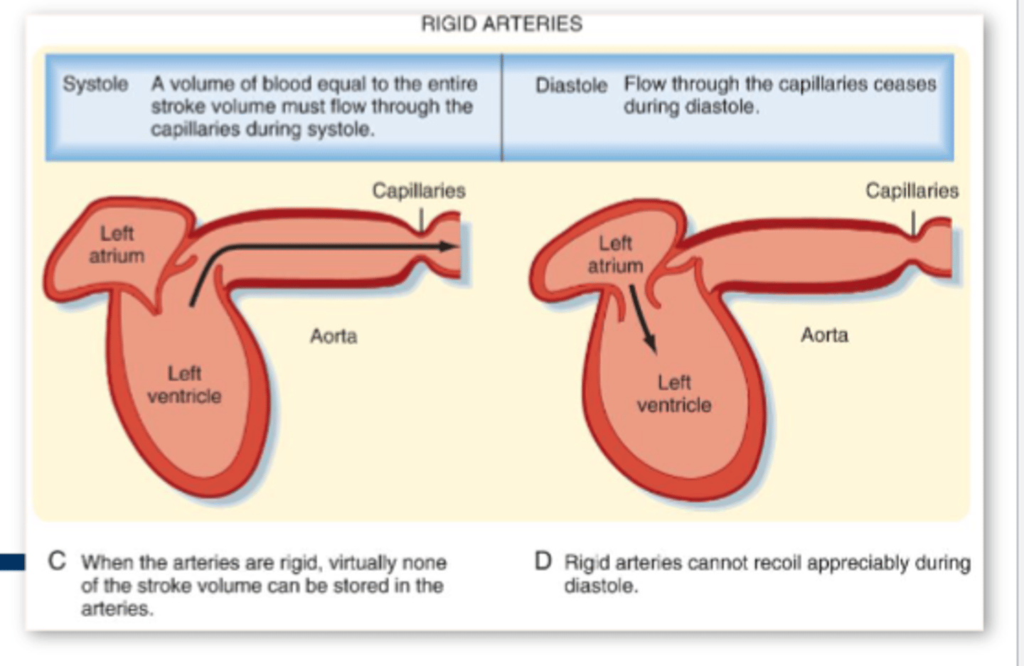
Isolated Systolic Hypertension
• Systolic pressure >130 with diastolic < 80mmHg
• Systolic blood pressure (SBP) is the strongest predictor of CV events in adults over 50
• SBP >>> DBP reflects arterial stiffness, generally caused by aging
• Stiffness increases the risk of CV morbidity and mortality
• Takeaway: Could be dangerous since if there is so much stiffness, we are no longer getting rebound during diastole which can lead to static blood & HF
What are some sources of secondary HTP?
- Obstructive sleep apnea
- Drugs (
- Parathyroid disease (↑ Plasma Ca2+)
- Pheochromocytoma (↑Epinephrine)
- Primary aldosteronism (↑Aldosterone)
- Thyroid disease (↑ SNS activity)
- Renovascular disease: decreased nephron number, decreased GFR (↑Na+ retention)
BP =
CO x TPR

MAP =
2/3 DP + 1/3 SP
(Heart spends about 2/3 time in diastole, and 1/3 time in systole)

What are potential mechanisms of essential hypertension?
1. Defect in functional vasoconstriction or damage to the vessel wall (increased vasoconstriction or loss of vasodilation)
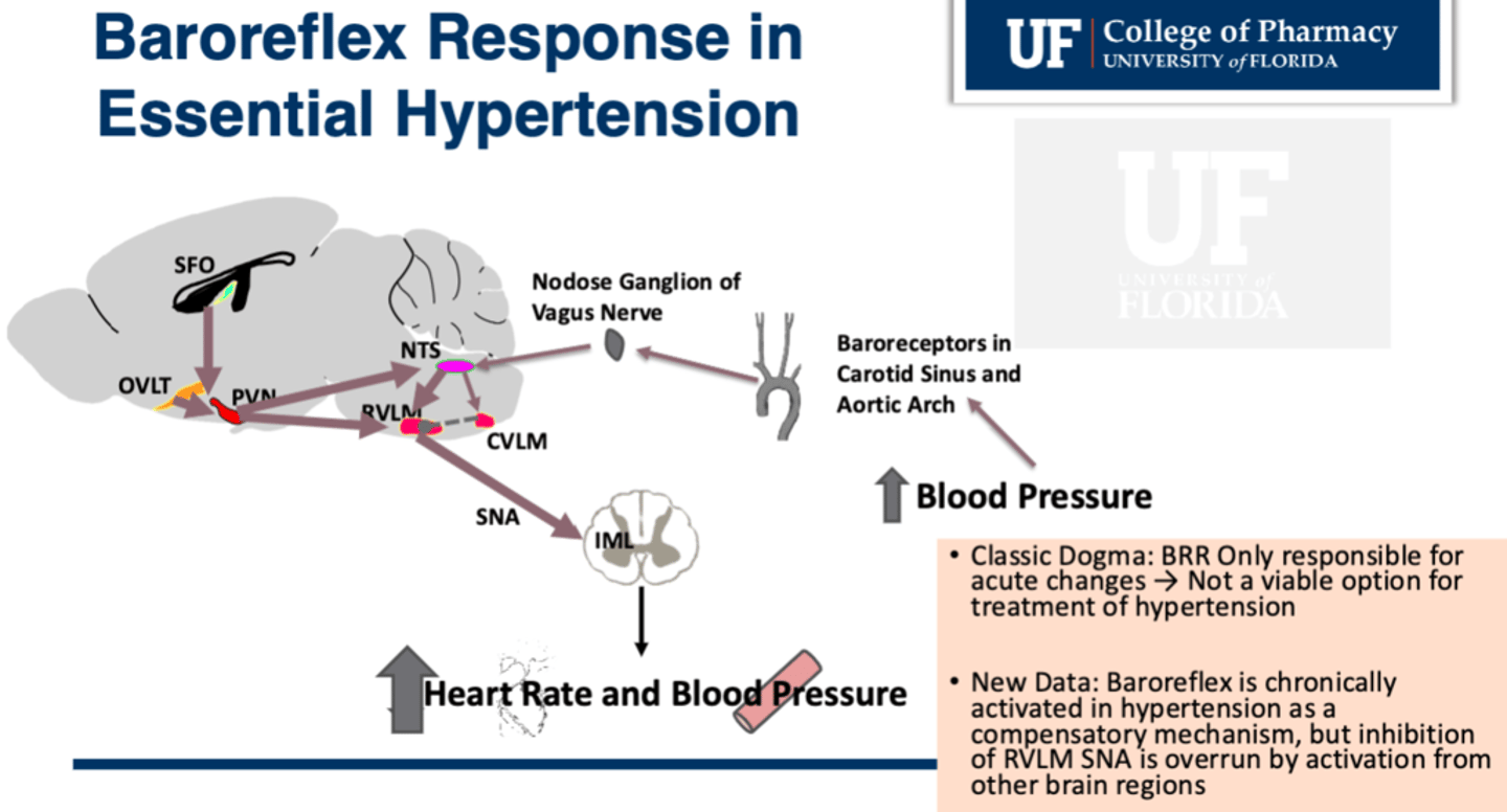
2. Neurogenic: Increased SNS activity or impaired baroreflex response
3. Defects in Renal Sodium Homeostasis (sodium retention)

Three Theories of Neuronal Etiology of Hypertension
• Baroreceptors reset at higher blood pressure
• Baroreceptors lose sensitivity
• Stress components from the Cortex overwhelm the Baroreflex signal in the Medulla at the RVLM (rostral ventrolateral medulla) so the SNA is stimulated (sympathetic nerve activity is controlled by RVLM)
- Usually baroceptors inhibit RVLM
- But the SFO (another part of the brain called the subfornical organ) activates RVLM when the body is stressed out and will overwhelm the baroreceptor inhibition of the RVLM
Why do hypertension rates increase with age?
• May be due to reduced renal function with age
- Loss of almost 50% of nephrons by age 75
• May be associated with impaired baroreflex sensitivity
- Neurons are slower to fire and effectors are slower to activate
• May be isolated systolic HTP
- Stiffening of vessels with age reduces compliance
Arteriosclerosis is worsened by excess ______ and atherosclerosis is worsened by excess _______ in the blood.
sugar; lipids
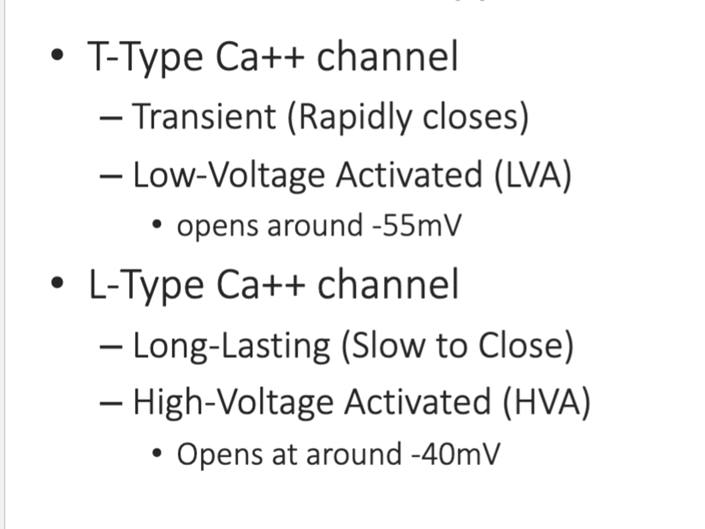
Which VGCCs channel opens first?
T-type since they are Low Voltage Activated compared to L-type
Action Potentials in the Nodes
Phase 4 (”Resting” TMP) TMP:
1. Ih/If – the “funny” current
• Opens in response to hyperpolarization
• Slow depolarization
• Slow Na+ ion channel (channel permeable to both Na+ and K+)
• Start of Prepotential
• Beta blockers work here
2.• ICaT – Transient Ca+ Current
• Opens in response to slow depolarization from “funny current”
• Completes Prepotential
Phase 0 (Depolarization)
1. IcaL Long-lasting Ca+ Channel opens
• Transmembrane Potential (TMP) threshold is reached
• Depolarization
• Calcium channel blockers work here mostly
Phase 3 (Repolarization)
1. Ik – delayed rectifier K+ Channel
• Opens to counteract increase in Ca+
• Causes brief plateau before return to resting TMP as Ca+ channels close

Funny current
Nodal AP: its called funny because its activated by HYPERPOLARIZATION (not depolarization)

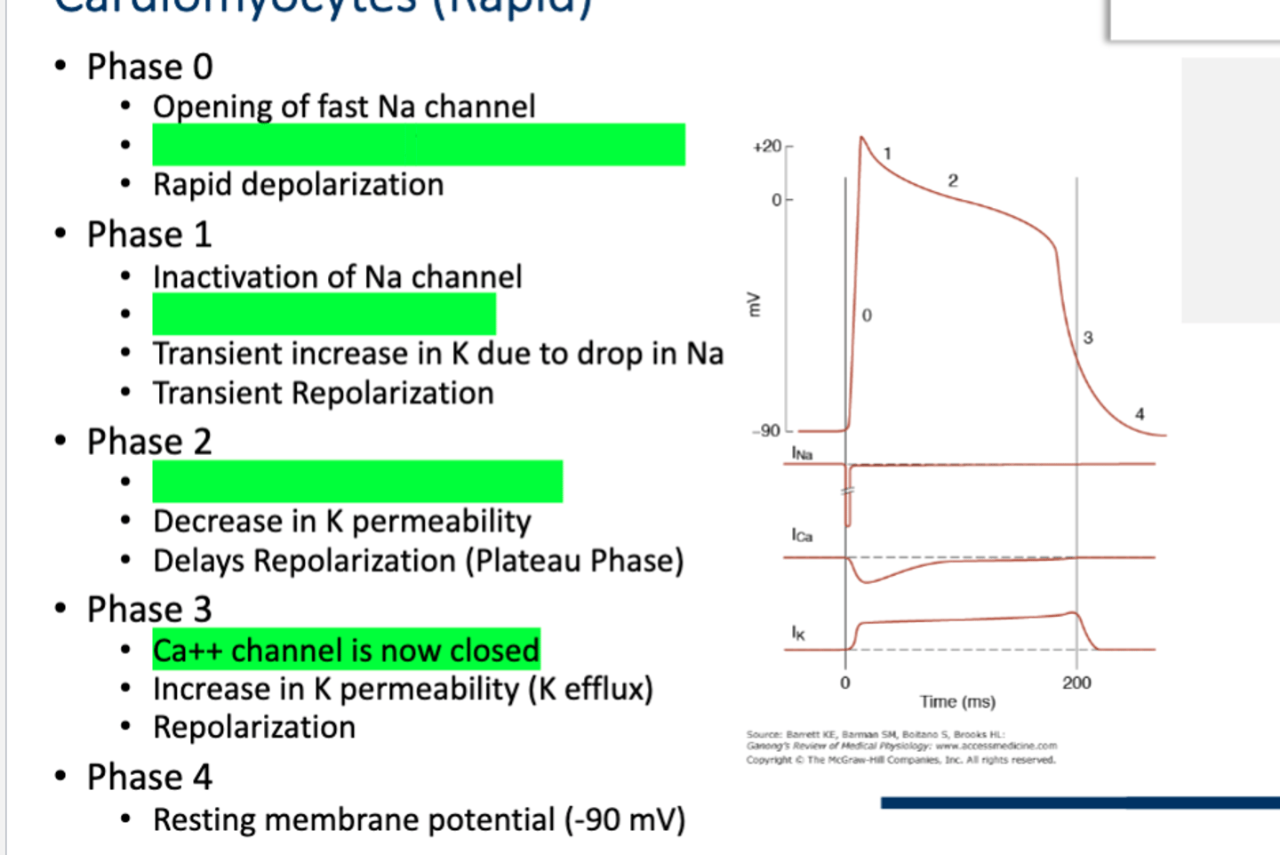
Action Potentials in the Cardiomyocytes
Phase 0
• Opening of fast Na channel
• Opening of slow L-Type Ca++ Channel
• Rapid depolarization
Phase 1
• Inactivation of Na channel
• Increase in Ca++ current
• Transient increase in K due to drop in Na
• Transient Repolarization
Phase 2
• Ca++ channel is slow to close
• Decrease in K permeability
• Delays Repolarization (Plateau Phase)
Phase 3
• Ca++ channel is now closed
• Increase in K permeability (K efflux)
• Repolarization
Phase 4
• Resting membrane potential (-90 mV)
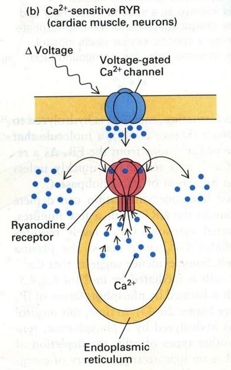
Is L-Type Ca2+ channel is mechanically linked to RyR on SR?
No, Cardiomyocyte contraction relies on Extracellular Ca2+.
• Lack of Mechanical Linkage: In some muscle types (especially cardiac muscle), the L-type Ca²⁺ channels and RyRs do not physically connect or directly interact. Instead, they operate through a process called calcium-induced calcium release (CICR). The entry of Ca²⁺ through L-type channels increases the local concentration of calcium near RyRs, triggering them to open and release more calcium from the SR. In skeletal muscle cells, they are physcally linked.
Calcium Induced Calcium Release
• When in cardiac and smooth muscle cells the excitation-induced entry of a small amount of Ca2+ from the ECF through voltage-gated surface membrane receptors triggers the opening of Ca2+-release channels in the sarcoplasmic reticulum, causing a much larger release of Ca2+ into the cytosol from this intracellular store
• Lack of Mechanical Linkage: In some muscle types (especially cardiac muscle), the L-type Ca²⁺ channels and RyRs do not physically connect or directly interact. Instead, they operate through a process called calcium-induced calcium release (CICR). The entry of Ca²⁺ through L-type channels increases the local concentration of calcium near RyRs, triggering them to open and release more calcium from the SR.
Ryanodine receptors
Receptors on the sarcoplasmic reticulum that bind with dihydropyridine receptors on the adjoining T tubule and serve as Ca2+-release channels during excitation-contraction coupling using RyR on SR
Smooth Muscle Excitation Contraction Coupling
• Ca2+ enters through L-Type Ca2+ channel
• Ca2+ activates Calmodulin
• Calmodulin activates Myosin Light Chain Kinase (MLCK).
• MLCK phosphorylates and activates Myosin allowing it to bind actin
• Release of ADP+Pi causes power stroke
• ATP binding leads to release from actin
• Myosin Light Chain Phosphatase dephosphorylates and inactivates myosin causing the cell to relax
Pharmacomechanical Coupling
• Agents like hormones (ex. NE, Ang II, NO, ANP, Beta-agonist) can cause a muscle contract without having to influence the membrane potential via Ca2+, but can act on different enzymes used that mediate/moderate smooth muscle contract like MLCK, MLCP, or Caldesmon/Calponin (tonic inhibitors of ATPase activity in absence ofCa2+/Calmodulin)

What are the different effects of CCBs (specifically since they target L-type?
• Negative chronotropic (HR decreases)
• Negative ionotropic (contraction)
- Less constriction in smooth muscle due to less MLCK activation since calmodulin activation requires Ca2+ binding
• Less aldosterone synthesis since Ca++ is a key step for it since both T-type and L-type VGCCs are found in aldosterone producing cells in the adrenal cortex
- Causes a decrease in blood pressure because
1. Less water retention due to a decrease in Na+ reabsorption
2. Since aldosterone is a vasoconstrictor, less aldosterone means decreased resistance
General ADME of CCBs
• Absorption
– Nearly complete (low secretion) but most have significant ‘first-pass effect’
• Distribution
– Generally have high protein binding (70-98%)
• Metabolism
– Many CCBs metabolized by cytochrome P450 (CYP) 3A4
• Elimination
– Half-life vary widely from 1.3-64 hrs
– Have various formulations available
Are short or long half-lives preferred for CCBs?
Long, since CCBs with short half-lives cause rapid change in BP
– Can be associated with dizziness and flushing, and can also lead to reflex tachycardia
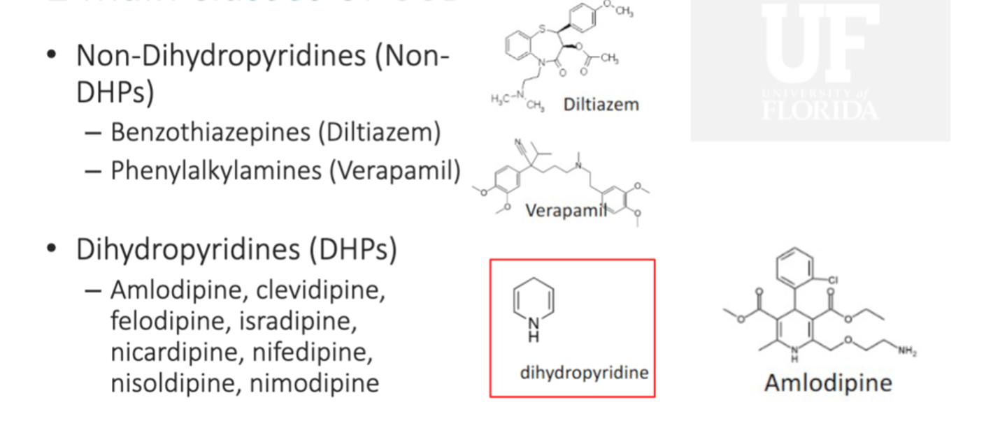
Non-dihydropyridines (Non-DHPs) Drugs
CCBs including verpamil (phenylakylamines) and diltiazem (benzothiazepines), do affect the hearts conduction system and can lead to AV blocks/bradycardia

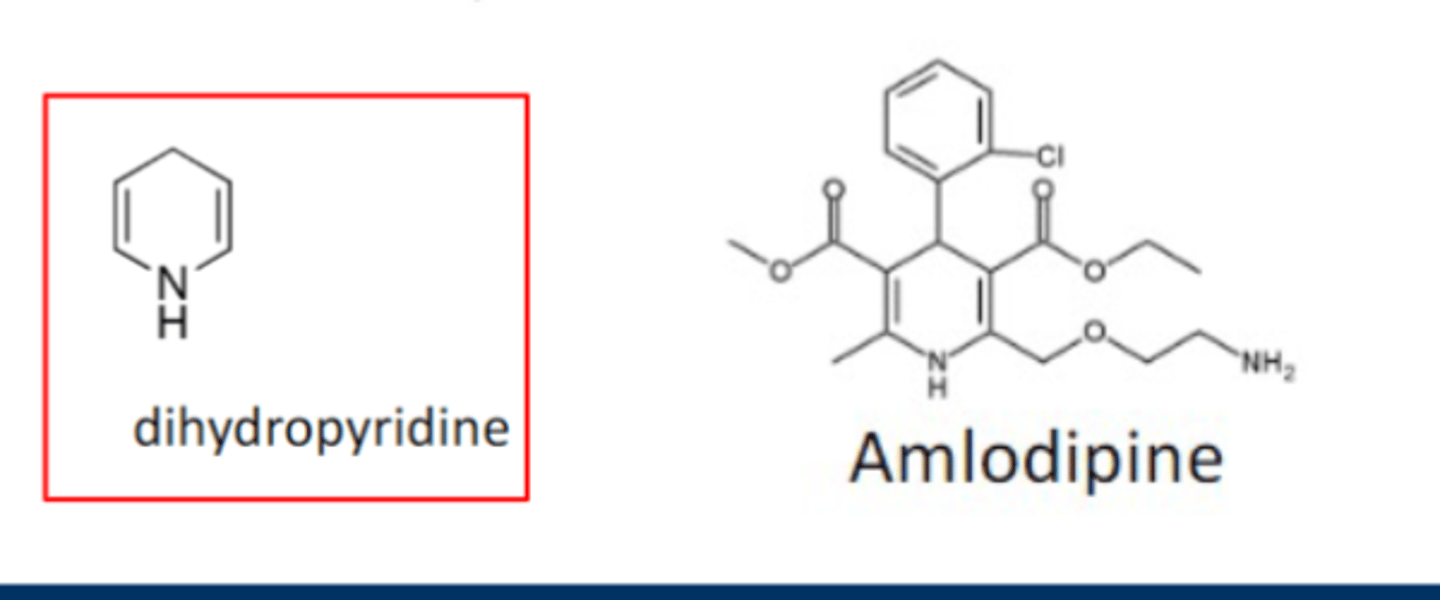
Dihydropyridines (DHPs) Drugs
CCBs like amlodipine, clevidipine, felodipine, isradipine, nicardipine, nifedipine, nisoldipine, nimodipine
2 Main Classes of CCBs
Non-DHPs and DHPs
Chronotropic (heart rate) is associated with ______ node and dromotropic (conduction velocity) is associated with ______ node.
SA; AV
Are DHPs used first line for supra-ventricular tachycardia or afib?
DHPs are also not used as first line for supraventricular tachycardia or atrial fibrillation due to lack of direct effects on L-type channels in cardiac tissue
DHPs
• Stronger vasodilators
• MOA: CCBs
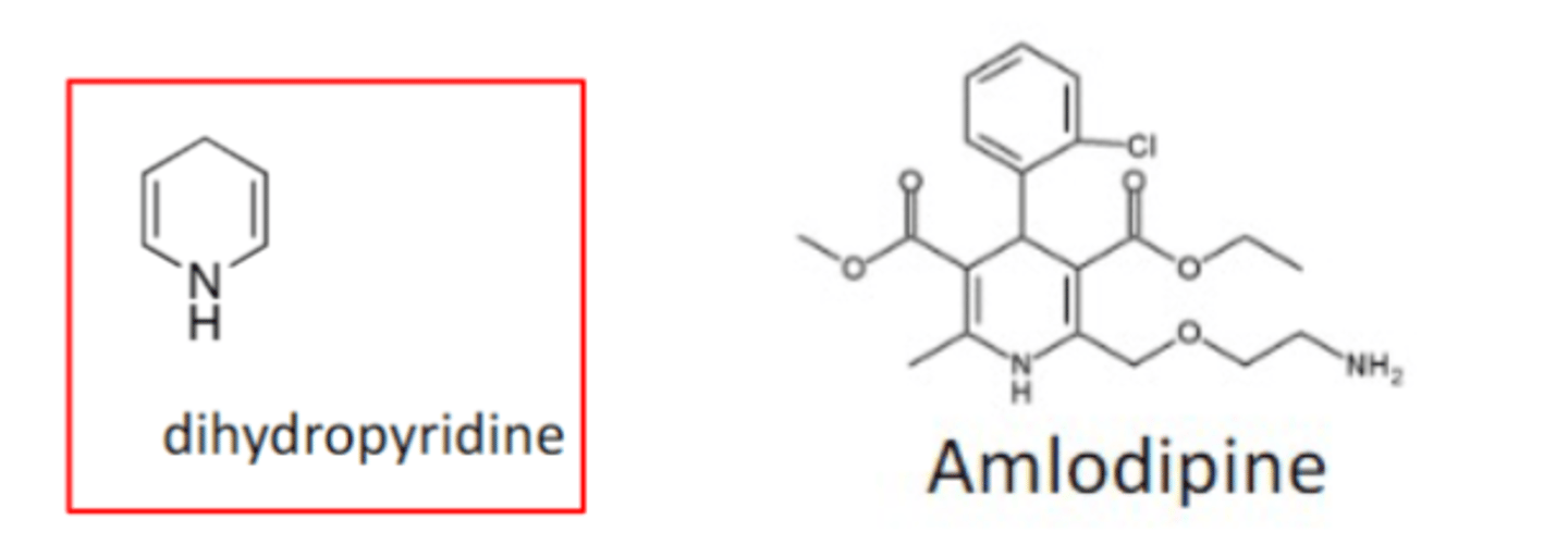
• Structure: dihydropyridine (6-membered ring with parallel double bonds with a N heterocycle)
- Esters at 3 and 5, asymmetry shows enhanced selecticity
- Phenyl ring at C4 is substituted at ortho and/or meta (kinda like the opposite of B1 selectivity for BBs)
• Additional Indications: Angina not first line (especially amlodipine, nifedipine, nicardipine)
• Precautions: Severe aortic stenosis, cardiogenic shock
• Metabolism: In the liver via CYP3A4
- Renal adjustment is not required tho, but beware of grapefruit juice
• Can enhance GERD due to CCBs lowering esophageal sphincter contraction
• AEs: Peripheral edema, N/V, diarrhea, anorexia, flushing, headache, dizziness
• Drug Interactions: BBs, anesthetics, amiodarone, digoxin, prednisone, CYP3A4 substrates, CYP3A4 inhibitors and inducers
• NO LABS REQUIRED
Non-DHPS
• Have more effect on CO (negative chronotropic, inotropic, and dromotropic) since it more selective for T-type
• MOA: CCBs
• Structure:
- Benzothiazepines (ditiazem)
- Phenylalklamines (verapamil)
• Precautions: Advanced heart block, severe aortic stenosis, cardiogenic shock, Beta1-Adrenergic receptor antagonists
• Contraindications: Beta-blockers, heart block, sick sinus syndrome; not recommended in pts with HF with reduced EF
• Metabolism: In the liver via CYP3A4, but are also inhibitors
• Can enhance GERD due to CCBs lowering esophageal sphincter contraction
• AEs: First-degree AV block, bradycardia, exacerbation of chronic HF or cardiac pulmonary edema, constipation (greatest with verapamil), N/V, diarrhea, anorexia, dizziness, flushing, headache, peripheral edema
• Drug Interactions: BBs, anesthetics, amiodarone, digoxin, prednisone, CYP3A4 substrates, CYP3A4 inhibitors and inducers

What CCB causes constipation?
Verapamil (non-DHPs do since there is a reduction in GI motility caused by inhibition of GI smooth muscle contraction)
Is it a good idea to combine non-DHPs with BBs?
NO! They both cause bradycardia and can lead to hypotension
CYP3A4 Inhibitors
G-PACMAN + SSRIs
Grapefruit
Protease Inhibitors (antivirals -virs)
Azole Antifungals
Cyclosporine, Cobicistat
Macrolides (Erythromycin/Clarithromycin except Azithromycin)
Amiodarone (and Dronedarone)
Non-DHP CCBs (Diltiazem, Verapamil)
CYP3A4 Inducers
Rifampin, St. John's Wort
Adrenergic Receptors
• α1 receptors (constrict)
- Skin, GI, urogenital, renal, liver
• α2 receptors found on presynaptic terminals of sympathetic postganglionic neurons
• β1 receptors (dilate)
• Found in the heart (nodal cells, myocytes in atria and ventricles) to increase contractibility and HR and decrease conduction time
• Found in the kidney to increase renin release
• β2 receptors (dilate)
• Skeletal muscle vessels and Respiratory Bronchi
Effects of SNS on Heart Rate
• Nodal Cells: Increase Na+ permeability in Phase 4 by accelerating activation of If channels and Ca+ permeability in Phase 1 to facilitate APs
• Cardiomyocytes: Increase Ca++ influx to cause more forcefully contractions and increase SV + phosphorylation/inactivation of phospholamban causes its inhibition, allowing SERCA to pump Ca++ into the SR and allow faster clearance for muscle relaxation and Ca++ cycling (efficient and forceful contractions)
Why is it important that the brain and heart have little SNS innervation?
Flow to these smaller essential organs are primarily regulated by metabolic need.
We don't want too much flow that would be too quick to deliver oxygen, or that may over dilate vessels in soft brain tissue. We don't want to little flow to reduce removal of CO2 and not deliver O2 quick enough
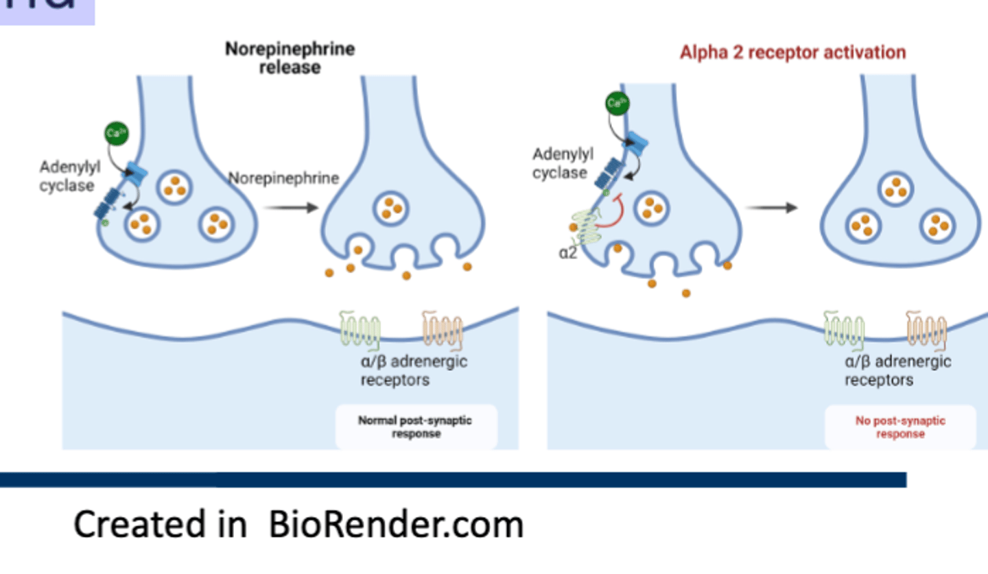
α2s Receptors
• Commonly expressed on presynaptic terminals of sympathetic postganglionic neurons.
• Activation inhibits adenylyl cyclase and leads to inhibition of NE release (feedback inhibition)
• Reduce SNS outflow from medulla
Intrinsic Sympathomimetic Activity (ISA)
The ability to work as a partial agonist for β1 even while blocking the effects of endogenous NE.
Ex. Pindolol, Acebutolol, Labetalol, Carteolol

Membrane Stabilizing Activity (MSA)
Refers to an ability to inhibit or reduce action potentials in cells expressing adrenergic receptors
Ex. Propranolol, Acebutolol, Metoprolol (at high doses), Carvediol
What beta-blocker is good for neurogenic HTP?
Propranolol, since it is highly lipid soluble. It is also membrane stabilizing
What BB can be used for intraocular pressure?
Betaxolol (Med Chem mentioned Carteolol, Madolol and Timolol)
What are the first-line agents for HTP?
Thiazide diuretics, CCBs, ACEi/ARBs
Beta-blockers
• 3 different generations; -lols
• MOA: BBs
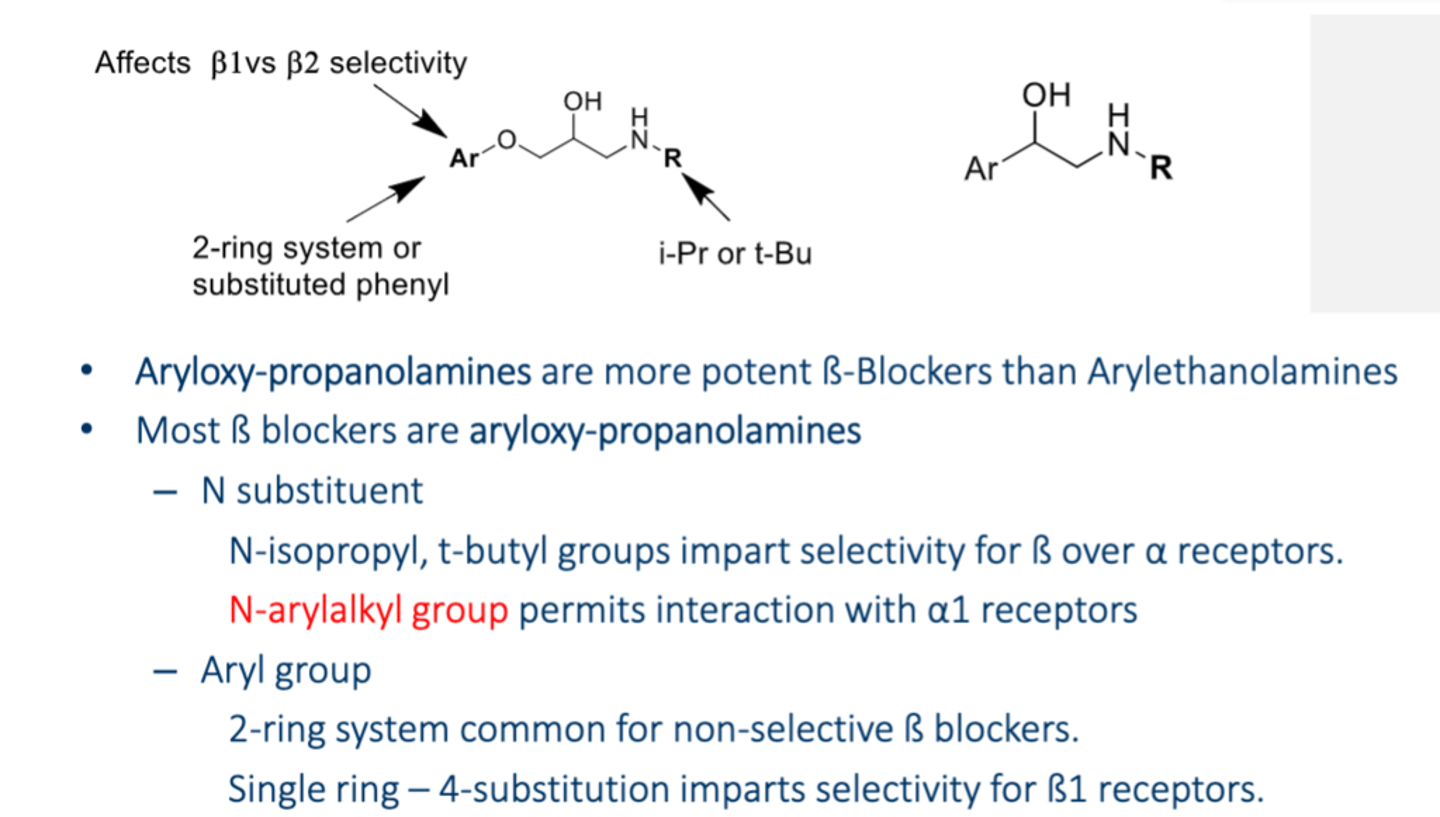
• Structure:
- Bulkier compared to agonist
- Aryloxy-propanolamines (S) - more potent
- Arylethanolamines (R)
- Get B1 selectivity is you use a para-substituted phenyl ring since the B1 N310 is not H-bonding with B1 F325, so the N310 is targeted
- Arylaklyl group permits interaction with α1 receptors
• Indications:
- Prevention of MACE in CCD in patients with HF (<50%) use metoprolol, bisoprolol or carvedilol or a recent history of MI
- HTP (NOT 1st LINE)
- Glaucoma (betaxolol)
- Angina/acute MI
- Some cardiac arrhythmias
- Congestive HF
- Migraine
- Hyperthyroidism
• Precautions: Advanced heart block, severe aortic stenosis, cardiogenic shock, Beta1-Adrenergic receptor antagonists
• Contraindications: Non-DHPs, SA or AV node dysfunction, decompensated HF, severe bronchospastic disease, STOPPING ABRUPTLY (need to taper out)
• Metabolism: CYP2D6
- Lipophillic propranolol is cleared by the liver while the rest is cleared by the kidney
• AEs:
- Bronchoconstriction (if not selective, it may block beta 2 of lungs)
- Hypo/hyperglycemia
- Impaired peripheral circulation
- Depression (due to decreased NE)
- Elevated triglycerides (just like loop diuretics)
• Drug Interactions: Non-DHPs (CCBs), α2-agonist (Clonidine)
What HTP agents cause hypertriglycerediemia?
Beta blockers and Loops Diuretics and Thiazide diuretics
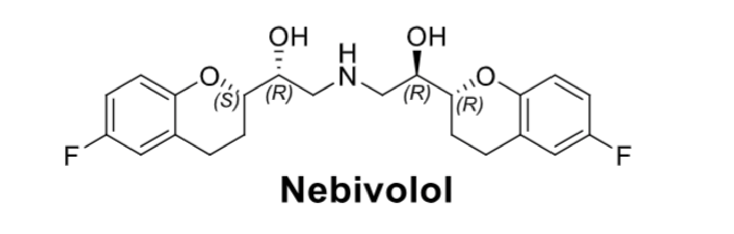
Nebivolol
– ß blocking effect almost exclusively SRRR isomer - looks like a mustache and is almost symmetrical
– NO potentiating vasodilatory effect by stimulating endothelial NO synthesis → ↓peripheral resistance and blood pressure
- Highly ß1 selective at low dose, but loses its cardioselectivity at higher doses.
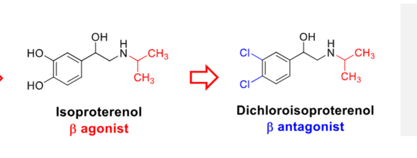
How were Beta antagonists developed from Beta agonists?
• Replace catechol hydroxyls in isoproterenol with chlorines → Dichloroisoproterenol as ß-antagonist.
• Later inserted an oxygen (ether) and converted chlorine into a cyclic structure
α1-Antagonists
• Prazosin, doxazosin, terazosin, alfuzosin (-zosins)
• MOA: α1-Antagonists
• Structure:
- Substituted quinazolines with acylated piperazine ring
- Reduction of the furan in prazosin into terazosin significantly increases the duration of action
• Indications:
- Resistant or neurogenic hypertension
• Precautions: Orthostatic hypotension
• Contraindications:
• Metabolism:
• AEs:
• Drug Interactions:

α2-Agonists
• Guanfacine, clonidine, methyldopa (prodrug) - GMC
- Methyldopa is a prodrug
• MOA: α2-Antagonists
• Structure:
- Phenyl with two Cl attached ortho
• Indications:
- Resistant or neurogenic hypertension
- Methyldopa used for hypertension during pregnancy
• Precautions: Orthostatic hypotension
• Contraindications: BBs
• Metabolism:
• AEs:
• Drug Interactions: BBs

What drug is used for HTP during pregnancy?
Methyldopa
How does our behavior affect Renin Release?
• High Salt Diet inhibits renin release
• Chronic High Stress increases renin release
What class of drugs are mostly prodrugs?
ACE
Which class of HTP drugs have the least side effects?
ARBs
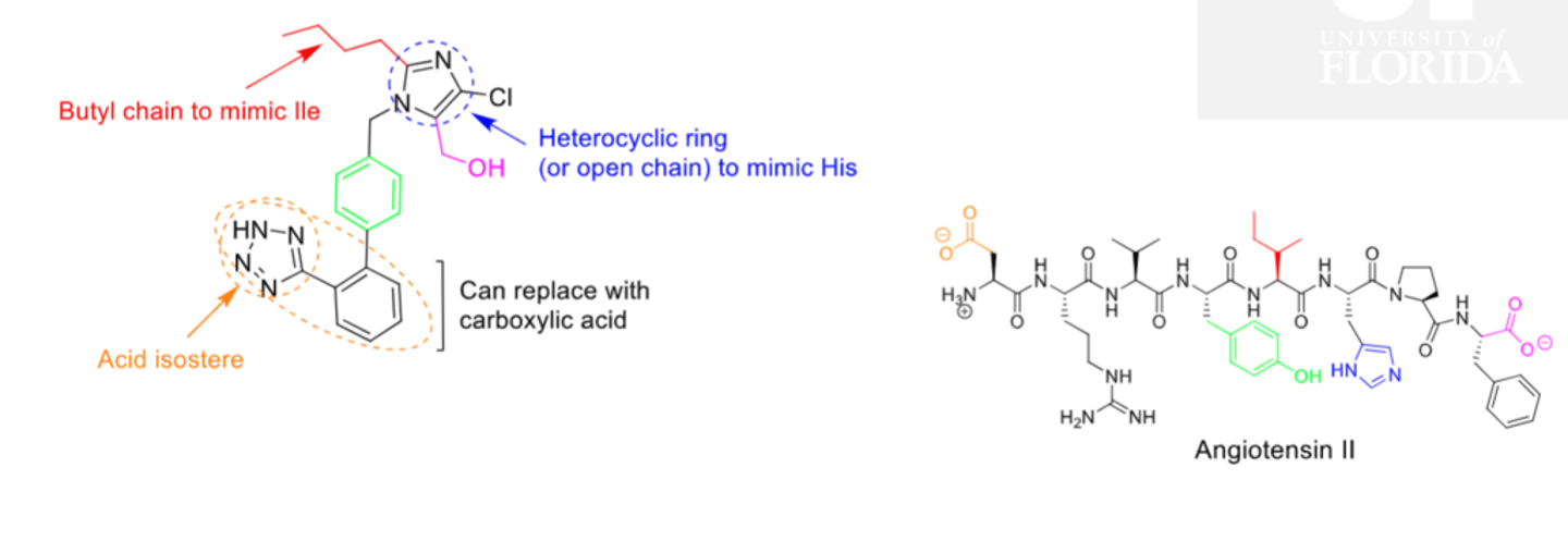
ARBs
• -sartans; have the lowest incidence of side effects compared with other antihypertensive; three or them are prodrugs (candesartan, olmesartan and azilsartan)
• MOA: ARBs; causes Ang II to be redirected to a "cardioprotective" pathway that includes AT2R and the ACE2 enzymes that convert Ang II to Ang-(1-7)
• Structure:
- Imidazole-5-acetic acid analogs
- Imidazole + Biphenyl group that is Acid or acid isostere (tetrazole, oxadiazole) – orthoposition optimal substitution
• Indications:
- HTP
• Precautions: If a patient has angioedema with an ACE, an ARB can be used after 6 weeks ago
• Contraindications: Pregnancy, history of angioedema with an ARV, bilateral artery stenosis
• Metabolism:
• AEs: Acute renal failure, orthostatic hypotension, hyperkalemia
• Drug Interactions: Do not use in combination with ACE inhibitors or direct renin inhibitor
• Monitoring: Check SCr and K within 2 weeks of starting and/or dose titration (30% increase in SCr is acceptable)
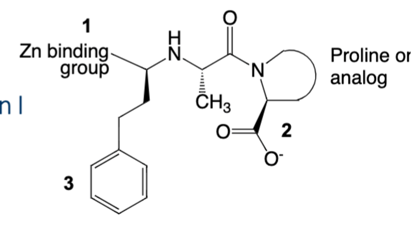
ACEi
• -prils; lots of them are prodrugs with the exception of lisinopril and captopril
• MOA: ACE inhibitors; Rampril is triphasic
• Structure:
1. Has a Zn binding moiety that is a Dicarboxylic acid or phosphonic acid or thiol
2. Carboxylic acid that mimics substrate C-terminus - proline analog that is usually a bulky bicyclic ring
3. Hydrophobic group to mimic Phe residue
• Indications:
- HTP
- Prevent of MACE in CCD patients with diabetes, HTN, renal disease, MI or LV dysfunction (LVEF < 40%)
• Precautions: ACE Escape via chymase, cathepsin, trypsin
• Contraindications: Pregnancy, Angioedema, bilateral artery stenosis
• Metabolism:
• AEs: Dry cough (more common in African Americans) and angioedema, acute renal failure, orthostatic hypotension, hyperkalemia, increase in SCr
• Drug Interactions: Do not use in combination with ARBs or direct renin inhibitor
• Monitoring: Check SCr and K within 2 weeks of starting and/or dose titration (30% increase in SCr is acceptable)
What ACE inhibitors are NOT prodrugs?
Captopril and Lisinopril are not prodrugs (do not have to be metabolized by the liver.). They are choices for those who have liver failure
Fosinopril
Only phosphinate-containing ACE inhibitor that has a phosphinic acid as the zinc binding site
ACE Escape
Via Chymase, Cathepsin, Trypsin
Renin inhibitors
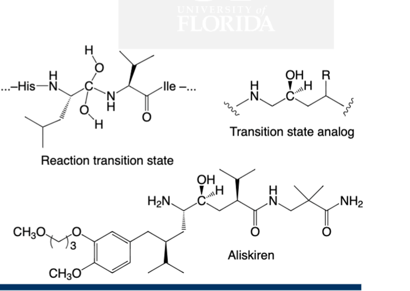
• Aliskiren; NO ACE escape
• MOA: Inhibit Renin; No ACE escape; mimics tetrahedral transition state
- Is an aspartyl protease, so two aspartic acid residues facilitate attack of water on a labile amide bond between Leu and Val
• Structure:
- Tetrahedral carbon
• Indications:
- HTP
• Precautions: Do not use with ACE inhibitor or ARB; do not use with cyclosporine or itraconazole
• Contraindications: Pregnancy
• Metabolism:
• AEs: Less risk of dry cough, hyper kalemia, angioedema
• Drug Interactions: Do not use with ACE inhibitor or ARB; do not use with cyclosporine or itraconazole
• Monitor K; high fat meals decrease absorption
Why does Na+ flow into the tubular cells?
• Since there is low intracellular [Na+] and there is a negative intracellular charge
• There are transporters in the apical membrane and also in the basolateral membrane (Na+, K+ ATPase)
![<p>• Since there is low intracellular [Na+] and there is a negative intracellular charge</p><p>• There are transporters in the apical membrane and also in the basolateral membrane (Na+, K+ ATPase)</p>](https://assets.knowt.com/user-attachments/0717724a-fc10-43b9-bde8-37eb91cf1808.png)
CA Inhibitors
Acetazolamide
Dorzolamide
Brinzolamide
Modest effect aka doesn't work well (short acting)
Increases K+ excretion
Mannitol
An osmotic diuretic, but it is not used for treatment of hypertension. It is FDA approved to reduce intracranial pressure associated with cerebral edema. It can be used in acute kidney injury to promote rapid diuresis before irreversible damage occurs

Loop Diuretics
• Furosemide, bumetanide, torsemide; -mides
• More potent than other diuretics
• MOA: Targets NKCC2 (25%)
• Structure:
- Structurally diverse
• Indications:
- Preferred for HF (and when CrCL <30)
• Precautions:
• Contraindications: Hypokalemia, hypersensitivity
• Metabolism:
• AEs: Hypokalemia, hypertriglyceridemia/hypercholesterolemia, ototoxicity, metabolic alkalosis
• Drug Interactions:
• Monitor K, SCr within 7 days of starting and with each dose adjustment

What HTP agent can cause ototoxicity?
Loop diuretics
Thiazide Diuretic
• HCTZ
• MOA: Targets NCC (4-7%)
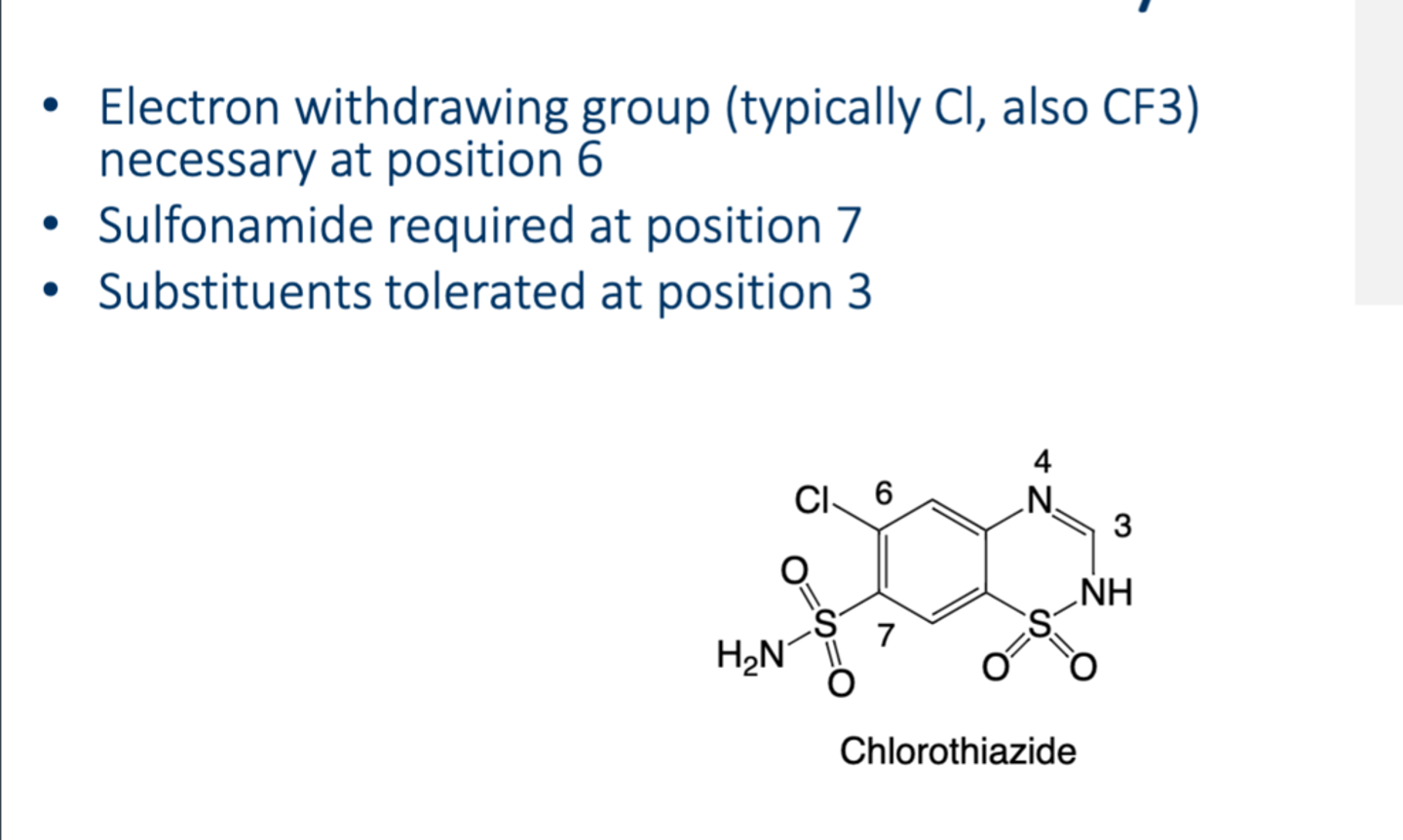
• Structure:
- Bicyclic benzothiadiazine (has two sulfonamides)
• Indications:
- HTP (first line)
• Precautions:
- Need to be treated in caution in pts with gout
• Contraindications:
- CrCl < 30 mL (except for metolazone)
- Sulfa allergies!
• Metabolism:
• AEs : Hyponatremia (due to increased B2o movement in DCT compiled with loss of Na+), glucose intolerance, hypertriglyceridemia/hypercholesterolemia, Metabolic alkalosis, hyperuricemia
• Drug Interactions:
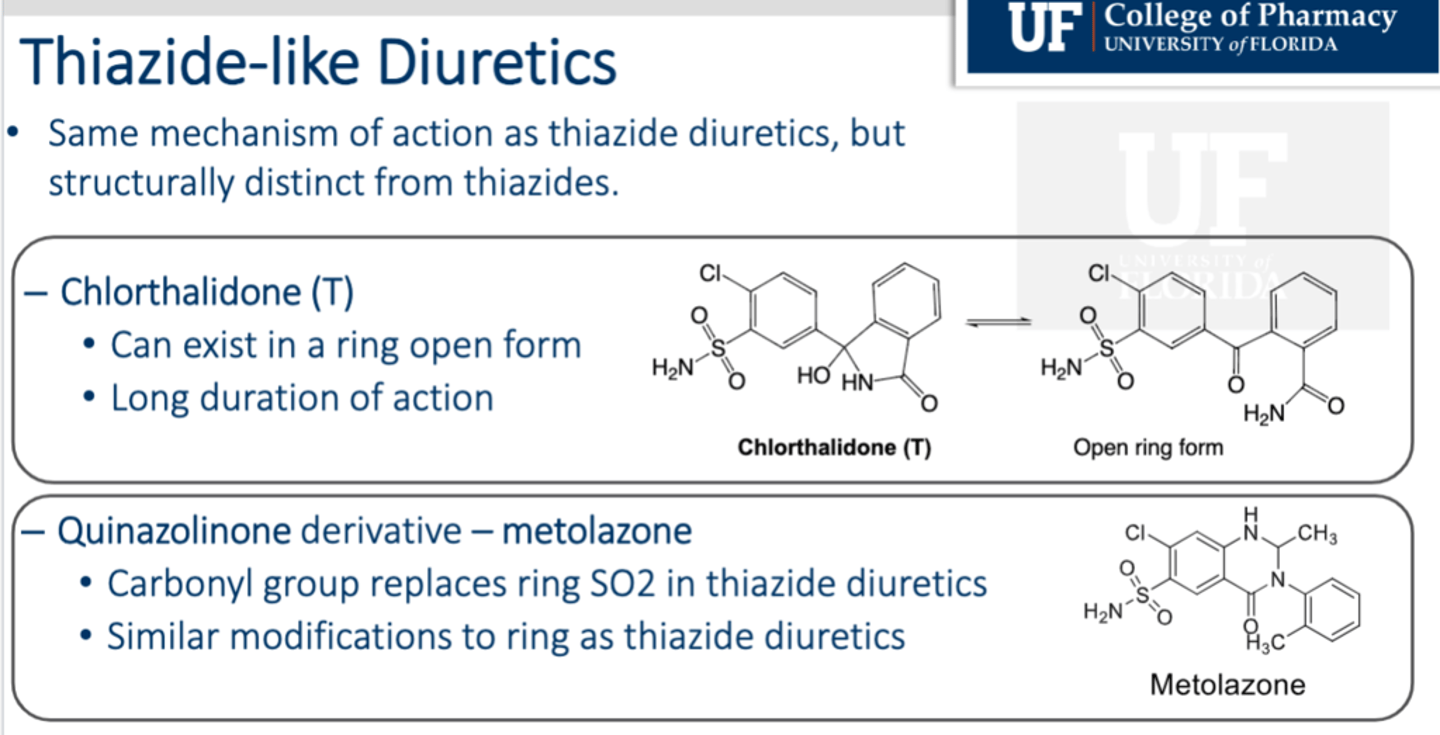
Thiazide-Like Diuretics
- Include chlorthalidone, metalazone and indapamide (indapamide cannot be used with CCBs)
- Have only 1 sulfonamide with some of the other characteristics
What drugs cause hyperkalemia?
ARBs, ACEi, K+ Sparring, Aldosterone antagonists
This can trigger metabolic acidosis
What drugs cause hypokalemia?
Loop diuretics, Thiazide diuretics, CA inhibitors
This can trigger metabolic alkalosis
What HTP drugs can cause hyperglycemia?
Beta blockers and Thiazide diuretics
K+ Sparring Diuretic
• By blocking Aldosterone actions at the MR, ENaC is targeted for internalization and degradation leading to less transport into the cell
• Can be combined with a thiazide
• Amiloride, triamterene
• MOA: Target ENaC and stabilize NaK ATPase
• Structure:
-
• Indications:
- HTP
• Precautions: Avoid in pts with CrCl <45
• Contraindications: Avoid in pts with CrCl <45
• Metabolism:
• AEs :Hyperkalemia, and Hyperchloremic Metabolic acidosis
• Drug Interactions: