9/15 - Diagnostic Anesthesia and Imaging in Horses
 Call Kai
 Learn
Learn Practice Test
 Spaced Repetition
 Match
Match Flashcards
 Knowt Play
1/16

There's no tags or description
Looks like no tags are added yet.
Name | Mastery | Learn | Test | Matching | Spaced | Call with Kai | Chat |
|---|
No analytics yet
Send a link to your students to track their progress
17 Terms
Mepivicaine, Bupivicaine
Which two local anesthetics are most commonly used in equine diagnostics? Lidocaine is also used but has more tissue reactivity, and they differ in onset/effect duration. Lidocaine and M are fast, and B is intermediate onset but longer duration (3-6 hours).
No
If there is a suspected incomplete fracture, cellulitis, or an uncooperative horse horse, should you use diagnostic anesthesia?
Flare
What term refers to severe lameness that occurs after a block, usually 30-60 minutes later? Synovial sepsis can occur 12-24 hours after the block.
Subchondral
Pain originating from what bone location can prevent adequate response to local anesthesia? Poor technique, inadequate volume, and severe pain can also cause this. Pain originating more proximal and neurological/mechanical pain will also cause this.
Palmar Digital
What nerve block is most distal and is done first, just proximal to the collateral cartilage and abaxial to the edge of the DDFT? It needs 23/25G 5/8'‘ needle and 1.5-2mL.

Abaxial Sesamoid
Which nerve block is second and is palmar to the neurovascular bundle at the abaxial surface of the PSB? It requires 23/25G 5/8” needle and 2mL.

Low 4/6
What nerve block is third, and in the forelimb is done at the L4 with 23G needle and 2-3mL per site? In the hindlimb it is done at the L6 with a 23G needle and 2-3mL per site.

Proximal
The nerve block that blocks which part of the metacarpus/metatarsus (proximal or distal) is referred to as the high 4/6 point block, and blocks the lateral palmar and deep branch of the lateral palmar nerves? It is trying to rule out proximal suspensory desmitis.

Scatter
What term refers to deflection of x-rays which worsens safety/image quality? It is worse in the upper limb and axial skeleton due to increased tissue thickness, but is reduced with good collimation and grid.
40
A change of what percentage of bone density may be required before it is apparent visually on radiograph?
Frequency
What component of ultrasonography determines depth and resolution? This is mainly used to image soft tissues. Linear transducers are most common and most useful.
0-3
The fibre alignment score (FAS) in ultrasonography covers what range, from 76-100% at the lowest number to 0-25% at the highest number?
NS
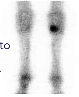
What imaging technique (abbreviated) uses gamma “nuclear” rays to image radioisotopes (Tc) and bone markers injected into the animal? The image shows locations where there has been radiopharmaceutical uptake in bone (hotspots). This allows detection of bone activity changes before radiographs but is not always as specific as it is sensitive. It is indicated when diagnostic analgesia cannot be safely performed, if a fracture is suspected, if the animal is stressed, if there is multiple limb lameness, if there is an axial skeleton problem, and if there is obscure/intermittent lameness or poor performance.
CT
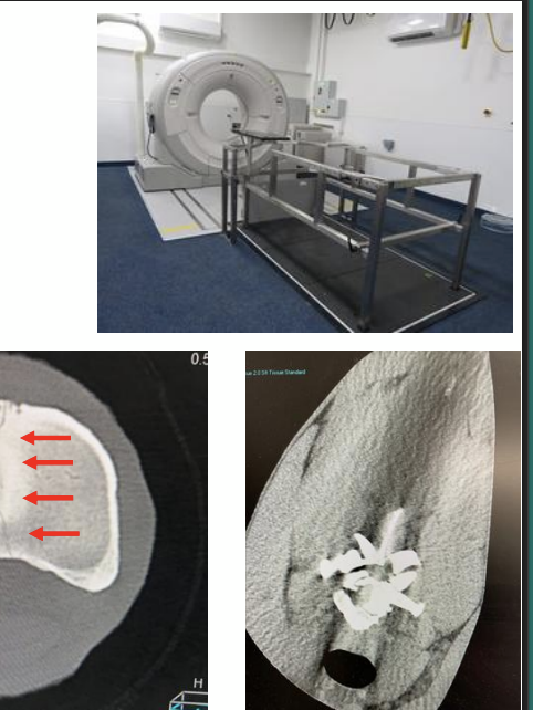
What imaging technique (abbreviated) produces a 3D image using x-rays taken around a single axis of rotation? It allows detailed visualization of interior structures like bone, but is less effective for soft tissue. Standing or GA are required. It is indicated for sinus/teeth/neuro cases, neck ataxia/trauma, limb fractures/subchondral problems/OA/OCLL, and for OA/SI disease in small horses and ponies. It is expensive and requires expert training, and involves radiation.
MRI
What imaging technique (abbreviated) uses signals from protons detected in a magnetic field to create an image? It requires GA for high field and standing sedation for low field. It provides good imaging of soft and hard tissues, and is most useful on the lower limb, such as for foot penetrations. It requires a long time and expert training, and is expensive.

Thermography
What imaging technique is a non-invasive representation of surface temperatures, which reveals superficial inflammation? This is useful in finding laminitis, abscesses, corns, buckled shins (cannon bone problems), tendonitis, desmitis, SI abnormalities, and muscle injury.

Arthroscopy
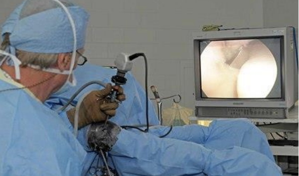
What imaging technique is used for evaluation of cartilage, bone, and soft tissues of synovial cavities, causing minimal trauma/morbidity, good visibility, and good cosmesis? It allows simultaneous diagnosis and treatment, but requires GA, high cost, and expert training.