Theme A - Cells A2.2 Cell structure
 Call Kai
 Learn
Learn Practice Test
 Spaced Repetition
 Match
Match Flashcards
 Knowt Play
1/66

There's no tags or description
Looks like no tags are added yet.
Name | Mastery | Learn | Test | Matching | Spaced | Call with Kai | Chat |
|---|
No analytics yet
Send a link to your students to track their progress
67 Terms
What is a cell?
the basic structural unit of all living organisms
Outline cell theory (3)
1. All living organisms are composed of cells
2. Cells are the basic structural unit of all living organisms
3. All cells arise from pre-existing cells
The cell theory was developed by _____________ __________ (several specific observations of cells making up the living tissue of organisms were used to form a general conclusion that all life is cellular) but is applied in a __________ ____________ (the general conclusion that all life is cellular is used to predict that all living organisms are composed of cells).
inductive reasoning
deductive manner
Summarise how to make and stain temporary mounts of cells and tissues. (6)
Temporary mounts are commonly referred to as wet mounts
● Place the cells on the slide, in a layer not more than one cell thick.
● Add a drop of water or stain.
● Stains help structures that are pale or transparent to show up more clearly.
● Carefully lower a cover slip on to the drop.
● Try to avoid trapping air bubbles.
● Remove excess fluid or stain by putting the slide inside a folded piece of paper towel and
pressing lightly on the cover slip.
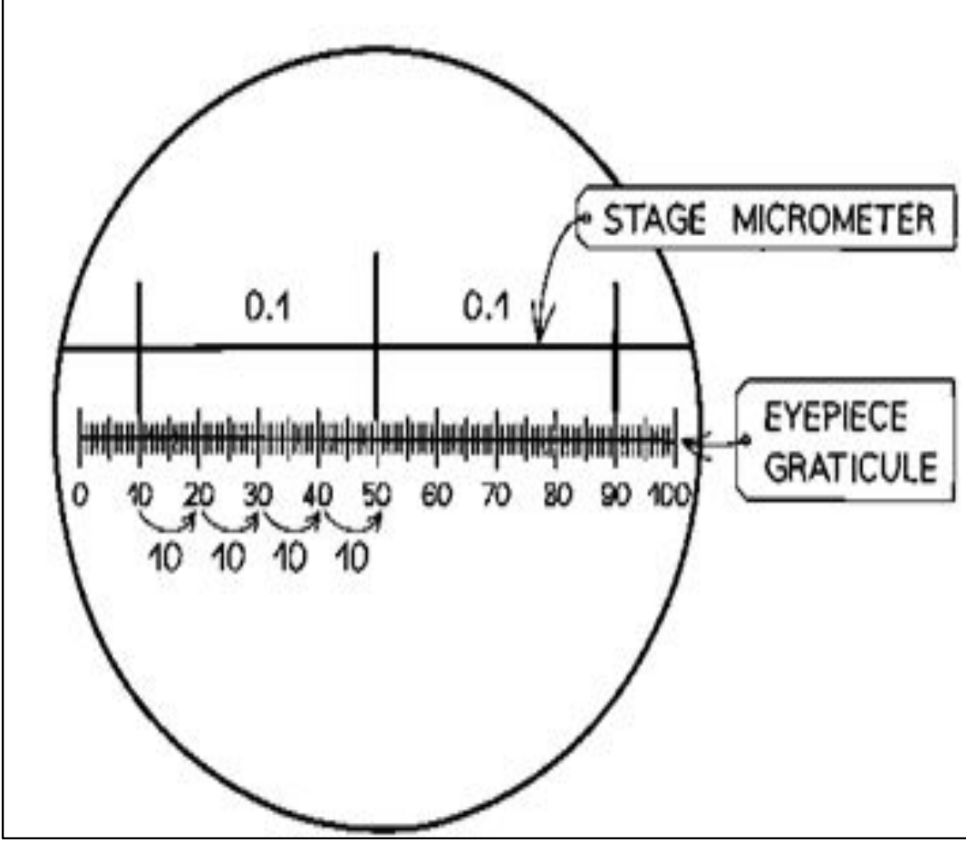
Describe how to use an eyepiece graticule and stage micrometre to measure
sizes of a specimen. (2)
●The stage micrometre shows the actual size of the image using divisions
that are each 100 μm (0.1 mm) apart.
● Each 100 μm division of the stage micrometre is equivalent to 20 eyepiece
graticule divisions, which means that one graticule division is equal to 5 μm.
Make sure that all your measurements are in the same SI units before you carry out calculations.
Outline the applications of electron microscopy. (4)
● Rather than passing light through a specimen, electron microscopes pass a beam of electrons through a specimen.
● Electrons will be absorbed by the denser parts of the sample, and scattered or able to pass through less dense areas, after which they are picked up by an electron detector and used to form an image.
● Because electrons have a much shorter wavelength than light, electron microscopes have a much higher resolution than light microscopes.
● The resolution of a light microscope is 200 nm compared with 0.1 nm for an electron microscope.
Describe the application of techniques that are commonly used in microscopy.
(technique name, microscopy type, resolution, method, advantages) (4)

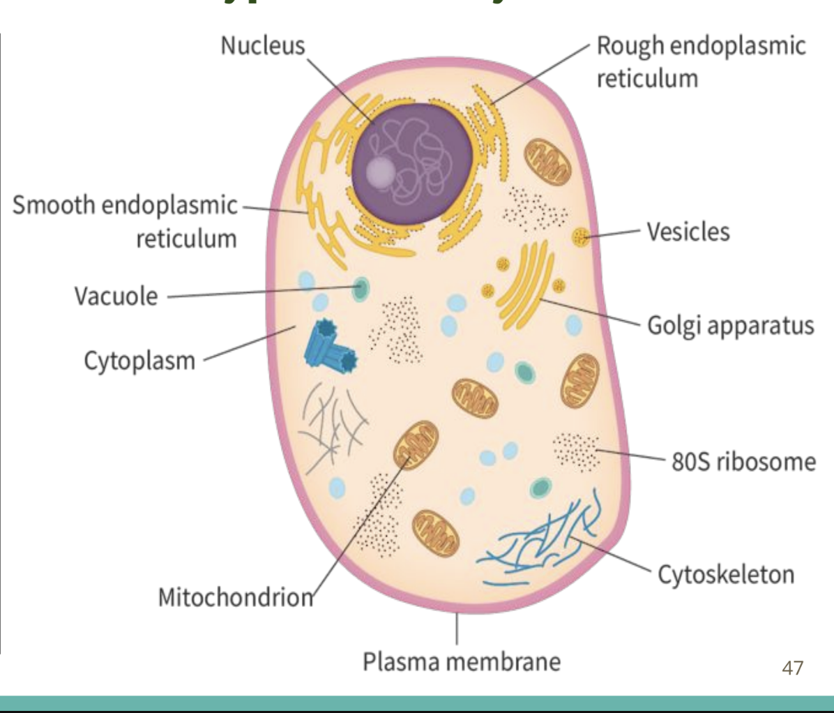
Describe the structure and components of a typical
cell. And the reasons for these structures (3)
1. DNA: the genetic information needed by living organisms to carry out all functions of life. The location of DNA differs between organisms.
2. Cytoplasm: composed mainly of water, includes all organelles except the nucleus within the cytosol (a mixture of substances dissolved in water).
3. Plasma membrane: composed of a phospholipid bilayer, it encloses the cell and controls entry of substances in/out of the cell.
Describe the structure of a typical prokaryotic cell and the function of prokaryotic cell structures and components.
exceedingly small
Cell wall: a thick and strong layer composed of carbohydrates and proteins that encloses the plasma membrane to provide protection and maintain the cell’s shape.
Plasma membrane: the arrangement of the plasma membrane with the cell wall varies between prokaryotes, like in Gram-positive and Gram-negative bacteria.
Gram-positive bacteria: a single lipid bilayer of the plasma membrane is covered by an outer cell wall. When stained, they appear purple under light microscopy. Bacillus and Staphylococcus are examples of Gram-positive bacteria.
No Outer Membrane: Do not draw a second lipid membrane outside the cell wall (this is only for Gram-negative bacteria).
No Membrane-Bound Organelles: Do not draw a nucleus, mitochondria, or chloroplasts, as these are eukaryotic structures.
Cytoplasm: composed of the cytosol but lacks compartmentalization (no membrane-bound organelle of eukaryotes) due to the absence of any membrane-bound organelles in prokaryotes.
Naked DNA: Nucleoid: a non-compartmentalized region within the cytoplasm that contains continuous circular DNA (NOT associated with histones).
no true nucleus
70S ribosomes: responsible for protein synthesis and are smaller than eukaryotic ribosomes.

Describe the structure of a typical eukaryotic cell and the function of
eukaryotic cell structures and components. (11)
Plasma membrane: selectively permeable, encloses a compartmentalized cytoplasm, create distinct parts of cells; compartmentalization, separate the contents of the cell from the external medium, controls the entry and exit of molecules
Cytoplasm: compartmentalized cytosol with membrane-bound organelles.
80S ribosomes: responsible for protein synthesis and are larger than prokaryotic ribosomes. They can either be bound to the ER or free within the cytoplasm.
Nucleus: largest organelle, encloses DNA for storage by the nuclear envelope (a porous double-membrane). Eukaryotic DNA is found as multiple linear strands (chromosomes), and is bound to histone proteins that form nucleosomes, which are involved in gene expression.
Membrane-bound cytoplasmic organelles
Mitochondria: oval-shaped double-membranous organelles with their own circular DNA and ribosomes. Outer membrane is smooth boundary & inner membrane is folded to form cristae, containing an aqueous solution of metabolites and enzymes (matrix) They are primarily responsible for ATP synthesis in aerobic respiration.
Endoplasmic reticulum (ER): extensive network of branching tubules and flattened sacs that extend from the nucleus to the plasma membrane.
Smooth ER: does not contain bound ribosomes and is involved in lipid biosynthesis. & site of storage of calcium ions
Rough ER: closer to the nucleus and contains bound ribosomes for protein biosynthesis that are packaged in the vesicles and then discharged from the cell by exocytosis (e.g.) digestive enzymes discharged this way)
Golgi apparatus: collection of fused cisternae (flattened sacs that are less tubule-like than the ER) that form two major networks; the Cis Golgi network (first cisternal structure facing the nucleus) and the Trans Golgi network (final cisternal structure facing the cell plasma membrane). Golgi cisternae are not contiguous and their widths are not uniform. The Golgi apparatus is involved in packaging, modification, and sorting of proteins and lipids within the cell.
Vesicles: spherical sacs composed of a single lipid bilayer that function in storage and transport of substances within the cytoplasm. They are transient, small, highly mobile, and fuse with other membrane-bound organelles. (some vesicles may form lysosomes, some develop into non-permanent vacuoles)
Vacuoles: spherical sacs composed of a single lipid bilayer that function in storage of water and other molecules in (mostly) plants. They are permanent, large, relatively stationary, and do not fuse with other membrane-bound organelles.
Lysosomes: round organelles composed of single lipid bilayers that contain hydrolytic enzymes for digesting biomolecules in low pH conditions
Ribosomes: no membrane, consisting of protein and nucleic acid RNA, free in cytoplasm/bound to RER + also occur within mitochondria and in chloroplasts (here, ribosomes are smaller, 70S), site of protein synthesis
Cytoskeleton: network of actin filaments, intermediate filaments, and microtubules, that aid in cell movement, reinforcement of cell shape, cell division, and organelle movement/anchoring. (NOT AN ORGANELLE BUT PROTEIN FIBER)
lengthening and shortening of microtubules helps with movement of cell components (chromosomes) & facilitated by motor proteins to their destination
Microtubules also support and maintains the shape of cell
Name the eight processes that all living things carry out. (8)
Movement: the ability to move
Reproduction: the ability to produce offspring sexually or asexually
Homeostasis: the ability to maintain constant internal conditions
Metabolism: the ability to carry out chemical reactions
Growth: the ability to develop in size or complexity.
Sensitivity (response to stimuli): ability to perceive stimuli and respond appropriately to them
Excretion: the ability to release waste or toxic materials into the environment
Nutrition: the ability to obtain energy needed to sustain metabolism
MR HM GREN
Draw a diagram of a paramecium (unicellular) (9) and explain how a paramecium carries out all the functions of life in a single cell
Metabolism: most metabolic reactions happen in the cytoplasm and specialized organelles, controlled by internal enzymes.
Reproduction: The nucleus can divide to support cell division by mitosis; reproduction is often asexual
Homeostasis: the contractile vacuole fills up with water and expel through the plasma membrane to manage the water content
Movement: moves by using thousands of tiny, hair-like structures called cilia that cover the outside of its body
Growth: after consuming and assimilating biomass from food, the paramecium will get larger until it divides
Response: the wave action of the cilia moves the paramecium in response to changes in the environment, e.g. towards food
Excretion: the plasma membrane controls the entry and exit of substances, including expulsion of metabolic waste
Nutrition: food vacuoles contain organisms the paramecium has consumed

Explain how does Chlamydomonas (unicellular) carry out all MR HM GREN
Metabolism: Most metabolic reactions happen in the cytoplasm and specialized organelles, controlled by internal enzymes.
Reproduction: The nucleus can divide to support cell division by mitosis; reproduction is often asexual, forming identical zoospores inside the cell wall.
Homeostasis: The contractile vacuole fills up with water and expels it through the plasma membrane to manage the water content.
Movement: Moves by using two whip-like, hair-like structures called flagella located at the front of its body to swim through water.
Growth: After creating and assimilating biomass from photosynthesis, the chlamydomonas will get larger until it divides.
Response: The beating action of the flagella moves the chlamydomonas in response to changes in the environment, e.g., using an eyespot to move towards light.
Excretion: The plasma membrane controls the entry and exit of substances, including the expulsion of metabolic waste products like carbon dioxide via diffusion.
Nutrition: Chloroplasts contain chlorophyll to produce organic molecules (like glucose) through photosynthesis using light, carbon dioxide, and water.
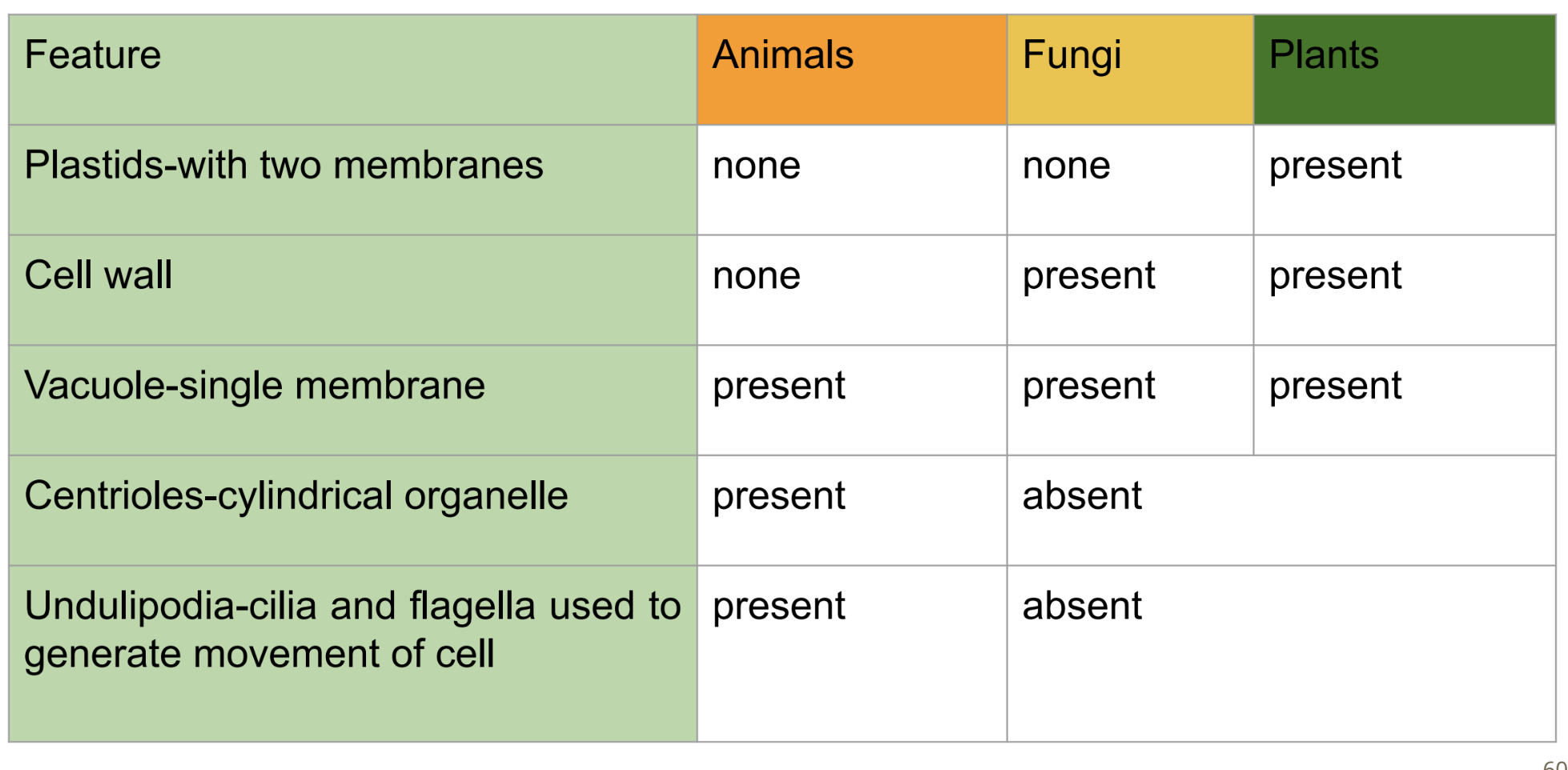
Outline how there may be differences in the cell structure of plant, animal

and fungal cells.
What does it mean by atypical cell?
Cells that do not have a feature/organelle for the standard structure and function expected for that type of organism
Give examples of eukaryotic cells with atypical cell structure. (4)
Aseptate fungal hyphae: lack septa (internal cell walls), creating a continuous multinucleate cytoplasm.
Skeletal muscle: muscle fibers are composed of columns of cells that have fused together to produce multinucleate structures.
Erythrocyte (RBC): mature RBCs lack a nucleus, so they cannot produce more proteins or repair themselves.
Phloem sieve tube elements: contain pores within their walls and minimal organelles(no nucleus), depending on companion cells for staying alive as they are specialized for transport in plants.

Annotate and identify prokaryote, plant or animal cell
Animal cell


Annotate and identify prokaryote, plant or animal cell (11)
Plant cell


Annotate and identify prokaryote, plant or animal cell
Prokaryote
Draw animal cell (13)

Draw plant cell (12)

Draw prokaryotic cell (9)
If asked to draw prokaryotic cell, please draw bacterial cell
If asked to draw unicellular, you can draw paramecium cell

Distinguish between resolution and magnification.
Magnification is how many times larger an image is compared to the real-life object, while Resolution is the ability to distinguish between two separate points that are close together as distinct objects
Electron microscopy
uses electrons to make a magnified image
show particles & wave properties: need to travel through a vacuum → the biological material must be dead
the limit of resolution = 5nm
much greater resolvingpower than light microscopes,
used to resolve the fine structure of cell, ultrastructure
Types of electron microscopy
transmission electron microscopy (TEM): electron beam pass through an extremely thin section of fixed, biological material
membranes and other structures are stained with heavy metal ions so they stand out as dark areas in the image
scanning electron microscopy (SEM): narrow electron beam scanned back and forth across the surface of the specimen & converted into a three-dimensional image
List the features of cells that can be observed by electron microscopy that are not visible by light microscopy.
ribosomes, membrane-bound organelles (such as mitochondria, rough and smooth endoplasmic reticulum, and the Golgi apparatus), and the detailed internal architecture (like mitochondrial cristae or chloroplast thylakoids)
State two problems that arise in electron microscopy because of the nature of an electron in relation to the living cell.
Vacuum required: Electrons are scattered by air molecules → electron microscope requires vacuum.
Specimen: Must be dead; water/volatile substances evaporate → cells collapse.
Radiation damage: High-energy electrons → ionisation, bond breaking, heat → specimen damaged/destroyed.
Result: Cannot observe living cells or dynamic processes.
What can be observed with a light microscope?
The nucleus is the largest substructure (organelle) of a cell and may be observed with a light microscope
Cryogenic electron microscopy
flash-freezing solutions of proteins or other biomolecules and then exposing them to electrons to produce very high-resolution images of individual molecules.
used to reveal how proteins work, how they malfunction in disease and how to target them with drugs.
Advantage: cryogenic electron microscopes use frozen samples, less-intense electron beams can be used and the evaporation of water (destroying molecules) is no longer a problem.
Freeze fracture
preparation of specimens for electron microscope examination by freezing, fracturing along natural structural lines and preparing a replica.
Fluorescence microscopy
fluorescent dyes absorb light at one wavelength and emit it at another longer wavelength
reveal their cellular location
first set filters the light before it reaches the specimen
The first set filters the light before it reaches the specimen, passing only those wavelengths that excite the specifically chosen fluorescent dye. The second filter blocks out this light and passes only those wavelengths emitted when the dye fluoresces.
be coupled to antibody molecules, which then act as highly specific and versatile staining reagents that bind selectively to targeted biomolecules
Immunofluorescence
antibodies chemically labelled with fluorescent dyes to visualize molecules under a light microscope.
allowed doctors to make a diagnosis on whether cancer is present based on small samples of tissue
What is differentiation
process by which cells become specialized, when some genes and not others are expressed in a cell’s genome.
(Newly formed cells grow and enlarge. A growing cell can normally divide into two cells. Cell division is very often restricted to unspecialized cells before they become modified for a particular task)
give one example of prokaryotic cell and explain how it carries out all characteristics of organisms
Escherichia coli is shown. E. coli is a common bacterium that occurs in huge numbers in the lower intestine of humans and other endothermic (once known as ‘warm-blooded’) vertebrates, such as mammals. It is a major component of the faeces of these animals.
Movement: Flagellum rotates → E. coli moves toward favourable conditions.
Respiration: Aerobic respiration → glucose + O₂ → ATP. Can also respire anaerobically.
Homeostasis: Regulates internal conditions, e.g. water and ion concentrations.
Metabolism: Enzymes catalyse reactions; nutrients converted into ATP and cell materials.
Growth: Takes up nutrients → synthesises proteins, DNA and cell components → increases in size.
Reproduction: Binary fission → DNA replicates → cell divides into two genetically identical cells.
Excretion: Removes metabolic waste, e.g. CO₂ and other waste products.
Nutrition: Absorbs nutrients/organic molecules from its environment for energy and growth

distinguish between the chromosomes of prokaryotic and
eukaryotic cells.

Centrioles
Centriole: 9 sets of 3 microtubules; hollow cylinder.
Location: Animal cells; 2 centrioles at right angles → centrosome.
Before cell division: Centrioles replicate.
Function: Organise/grow spindle fibres → chromosome movement during nuclear division.
limitation of cell theory
striated muscle cells contain many nuclei whereas most eukaryotic cells have one nucleus
red blood cells have no nucleus whereas most eukaryotic cells have one nucleus
multicellular fungi, such as Mucor, have multicellular hyphal cells
sieve tube elements in phloem tissue do not contain a nucleus, and rely on companion cells to provide metabolic support
viruses have some characteristics of living organisms but are not cells.
If all cells come from pre-existing cells, where did the first one come from?