2.80-2.92 Coordination and response
 Call Kai
 Learn
Learn Practice Test
 Spaced Repetition
 Match
Match Flashcards
 Knowt Play
1/86

There's no tags or description
Looks like no tags are added yet.
Name | Mastery | Learn | Test | Matching | Spaced | Call with Kai | Chat |
|---|
No analytics yet
Send a link to your students to track their progress
87 Terms
What is the nervous system pathway?
Receptor
Coordinator
Effector
What is a stimulus?
A change in the environment
What does the receptor do?
The receptor detects the stimulus
How is the signal transferred from the receptor to the coordinator?
Via sensory neurons
What does the coordinator do?
The coordinator processes information
What does the effector do?
The effector carries out the response
What are the three parts of the nervous system?
The brain, the spinal cord and the peripheral nerves
What is the system that includes the brain and the spinal cord called?
Central nervous system
What are the parts of the neurone (LH to RH)?
nerve endings
axon
myelin sheath
dendrites
cell body
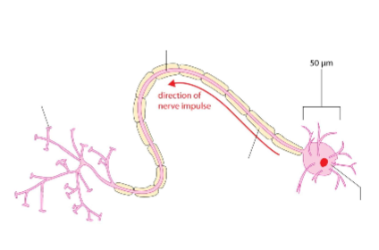
What is the role of dendrites?
They carry the nerve impulses towards the cell body
What is the role of the myelin sheath?
It insulates the axon so the nerve impulse travels faster
What is the role of the nerve endings?
They carry the nerve impulses to the other neurones
What is the role of the cell body?
It contains all the usual organelles of a cell, eg mitochondria, nucleus
What is the role of the axon?
It carries nerve impulses away from the cell body
What are the two types of neurone?
Motor neurone and sensory neurone
What is a sensory neurone?
Carries nerve impulses from receptors to the CNS
What is a motor neurone?
Carries nerve impulses from the CNS to effectors
What are the similarities between the motor neurone and the sensory neurone?
both have cell body
dendrites, axon, myelin sheath, nerve endings
What are the differences between the motor neurone and the sensory neurone?
cell body position differs
motor neurone axon is longer
motor neurone dendrites are shorter
motor neurone dendrites are branched
sensory neurone dendrites have a myelin sheath
What is a reflex arc?
A pathway which a nerve impulse follows for a reflex action through the spinal cord
What are the parts of the reflex arc?
sensory neurone
receptor
synapse
relay neurone
motor neurone
effector
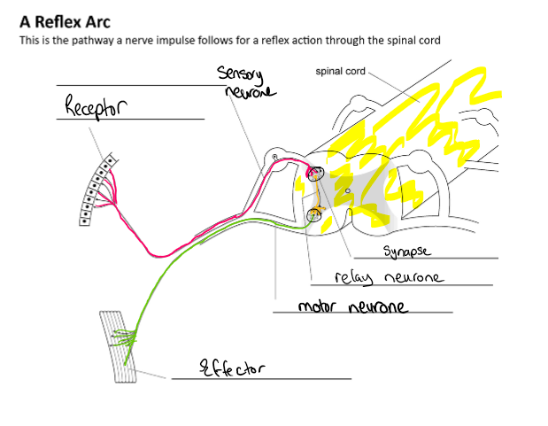
Describe the process of a reflex action.
Fill in the gap:
The _ (a pin prick)
Pain _ are stimulated
Nerve impulse travels along the _ neurone
Nerve impulse enters the spinal cord
Nerve impulse passes across a _ into a relay neurone
Nerve impulse passes into a _ neurone and travels along this out of the spinal cord
Nerve impulse reaches the effector (the finger muscle) and the muscle _, pulling the finger away
The stimulus (a pin prick)
Pain receptors are stimulated
Nerve impulse travels along the sensory neurone
Nerve impulse enters the spinal cord
Nerve impulse passes across a synapse into a relay neurone
Nerve impulse passes into a motor neurone and travels along this out of the spinal cord
Nerve impulse reaches the effector (the finger muscle) and the muscle contracts, pulling the finger away
What are 3 key features of a reflex action?
bypasses the brain
fast
unconscious, innate
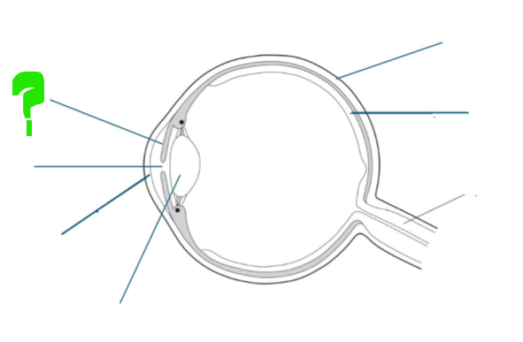
What is this part of the eye?
Iris
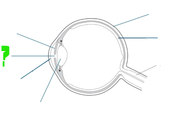
What is this part of the eye?
Pupil
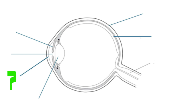
What is this part of the eye?
Cornea
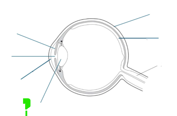
What is this part of the eye?
Lens
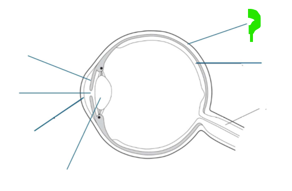
What is this part of the eye?
Sclera
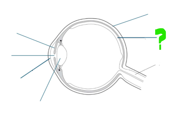
What is this part of the eye?
Retina
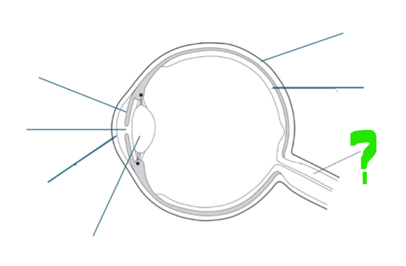
What is this part of the eye?
Optic nerve
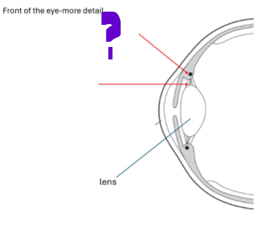
What is this part of the front of the eye?
The ciliary muscle
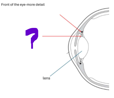
What is this part of the front of the eye?
Suspensory ligaments
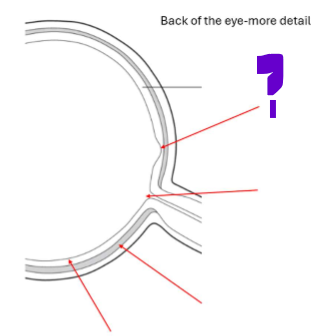
What is this part of the back of the eye?
Fovea
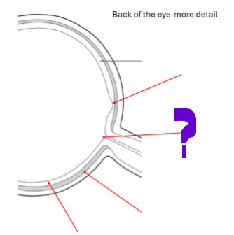
What is this part of the back of the eye?
Blind spot
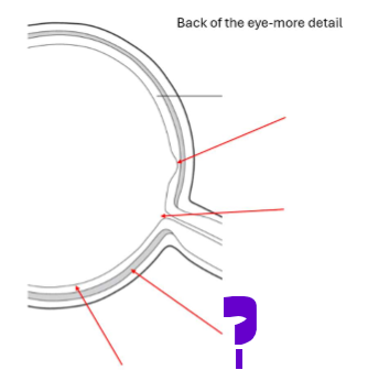
What is this part of the back of the eye?
Choroid
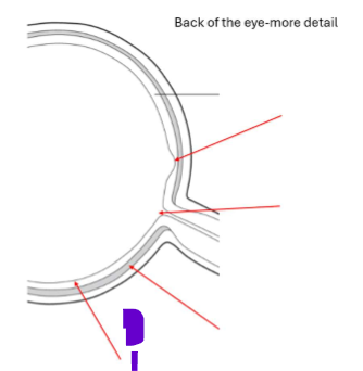
What is this part of the back of the eye?
Retina
Where is there the highest concentration of receptors?
In the fovea
Describe the choroid:
It is a black layer that sits under the retina
It absorbs excess light
Describe the cornea:
Transparent coating on the front of the eye
What is the function of the cornea?
The cornea protects the eye and refracts light entering the eye
Describe the pupil:
Central hole in the iris
What is the function of the pupil?
Allows light to enter the eye
Describe the iris:
Coloured ring of muscle tissue
What is the function of the iris?
Alters pupil size by contracting or relaxing
Describe the lens:
Transparent biconvex lens
What is the function of the lens?
Focuses light clearly onto the retina
Describe the ciliary muscle:
Ring of muscle tissue
What is the function of the ciliary muscle?
Alters the shape of the lens
Describe the suspensory ligaments:
Ligament tissue
What is the function of the suspensory ligaments?
Connects the ciliary muscle to the lens
Describe the optic nerve:
Nervous tissue
What is the function of the optic nerve?
Carries nerve impulses to the brain
In DIM LIGHT, what do the radial and circular muscles do?
The radial muscles contract and the circular muscles relax
In DIM LIGHT, what happens to the pupil size?
It increases, and the light entering the eye increases (dilation)
In BRIGHT LIGHT, what do the radial and circular muscles do?
Radial muscles relax and circular muscles contract
In BRIGHT LIGHT, what happens to the pupil size?
It decreases, and the amount of light entering the eye decreases (constriction)
What does antagonistic mean?
Equal and opposite muscle movements which work together for a movement - whilst one relaxes, one contracts
How is light focused?
Light is refracted by the cornea and the lens and then focused on the fovea
What is the relative angle of light rays reaching the eye when focusing on a near object?
Wide angle
What is the degree of refraction needed when focusing on a near object?
High degree
What is the shape of the lens when focusing on a near object?
Short and fat - more curved to increase refraction
Describe the ciliary muscle when focusing on a near object?
Contracts towards the lens inwards
Describe the suspensory ligaments when focusing on a near object?
Slacken, no longer pulling tight on the lens
What is the relative angle of light rays reaching the eye when focusing on a far object?
Almost parallel
What is the degree of refraction needed when focusing on a far object?
Low degree (as the cornea has already refracted most of the light)
What is the shape of the lens when focusing on a far object?
Longer and thinner
Describe the ciliary muscle when focusing on a far object?
Relaxes away from the lens, pulling suspensory ligaments taut
Describe the suspensory ligaments when focusing on a far object?
Taut, pulling tight on the lens
What is a synapse?
A junction between 2 neurones across which nerve impulses pass via neurotransmitter molecules
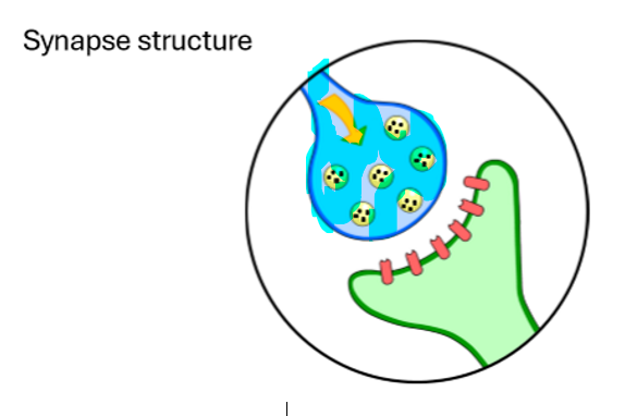
What is this highlighted part of the synapse?
Presynaptic neurone
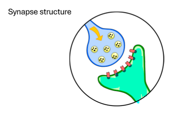
What is this highlighted part of the synapse?
Postsynaptic neurone
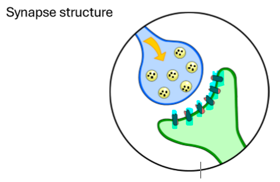
What is this highlighted part of the synapse?
Complimentary receptors
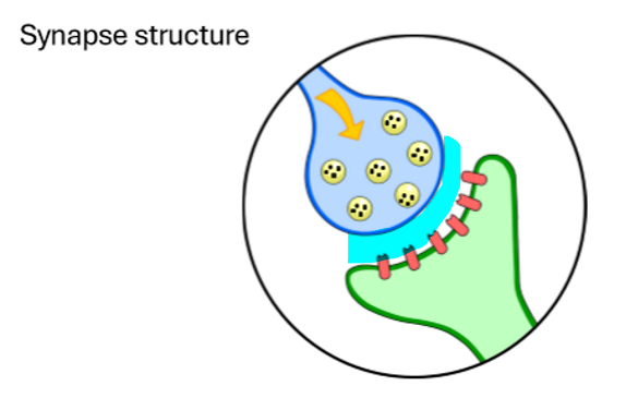
What is this highlighted part of the synapse?
Synaptic cleft
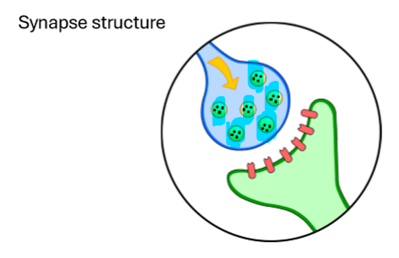
What is this highlighted part of the synapse?
Vesicles
What are inside of the vesicles?
Neurotransmitter molecules
How are neurotransmitters used in a synapse function?
Fill in the gap:
The nerve impulse arrives at the first neurone called the _ neurone
Vesicles (sacs) containing _ move towards the membrane of the neurone
The vesicles _ with the _ of the _ and release their contents into the synapse
The neurotransmitter molecules _ across the synapse
The neurotransmitter molecules bind to _ on the membrane of the postsynaptic neurone
The receptor molecules stimulate the generation of a _ _ impulse in the postsynaptic neurone
The nerve impulse arrives at the first neurone called the presynaptic neurone
Vesicles (sacs) containing neurotransmitters move towards the membrane of the neurone
The vesicles fuse with the membrane of the neurone and release their contents into the synapse
The neurotransmitter molecules diffuse across the synapse
The neurotransmitter molecules bind to receptors on the membrane of the postsynaptic neurone
The receptor molecules stimulate the generation of a new nerve impulse in the postsynaptic neurone
What is positive phototropism?
When the shoot grows towards light
What is negative phototropism?
When the root grows away from light
What is positive geotropism?
When the root grows towards the direction of gravity
What is negative geotropism?
When the shoot grows away from the direction of gravity
What is positive hydrotropism?
When the root grows towards water
What does the hormone auxin do?
Auxin stimulates new cells to grow longer or elongate
What happens to auxin when light shines all around the tip?
Auxin is distributed evenly throughout and the cells in the shoot grow at the same rate
What happens to auxin when the light shines on the shoot from one side?
Auxin produced in the shoot tip concentrates on the shaded side so:
shaded side cells elongate and grow faster than the cells on the sunny side
Shoot grows towards the direction of light, max psis
What are gibberellins important for?
They are plant hormones that are important in initiating seed germination, inducing flowering and fruit growth
What is ethene?
A gas released by plants which controls cells division and ripening of fruits
What is the gel