Critical Care Exam 1
 Call Kai
 Learn
Learn Practice Test
 Spaced Repetition
 Match
Match Flashcards
 Knowt Play
1/208

Earn XP
Description and Tags
Module 1, 2 and 3
Name | Mastery | Learn | Test | Matching | Spaced | Call with Kai | Chat |
|---|
No analytics yet
Send a link to your students to track their progress
209 Terms
Define acute rsp failure
sudden decrease in arterial oxygenation with/without CO2 retention (hypoxemia w/ or w/out hypercapnia)
PaO2 falls quickly
PaCO2 may be norm, low, or high
What can be described as a special, severe form of acute rsp failure with major oxygenation problems?
ARDS
What is considered type 1 respiratory failure?
hypoxemia respiratory failure or “lung failure”
what is the primary problem in type 1 respiratory failure?
arterial oxygenation (low PaO2)
problems with gas exchange in the lung
Oxygenation problems in lung (type 1 rsp failure) caused by poor matching of gas and blood examples:
pulmonary shunt
low V/Q ratios
diffusion problems (early pulmonary fibrosis)
hypoventilation (which increase PACO2 and reduces PAO2/PaO2)
Clinical causes of type 1 rsp failure
atelectasis
pneumonia
pulmonary edema
Rsp failure signs/symptoms for mild hypoxia (rsp, cardiac, neuro)
rsp: increase rr, mild SOB, early rsp distress
cardiac: increase HR, mild hypertension, peripheral vasoconstriction
neuro: overconfidence, restlessness, anxiety, euphoria, lightheadedness, nausea, dizziness, fatigue
Rsp failure signs/symptoms for mod hypoxia (rsp, cardiac, neuro)
rsp: tachypnea, increased MV/hyperventilation, accessory muscle use, intercostal retractions
cardiac: tachycardia, arrhythmias, hypertension
neuro: agitation, impaired judgement, confusion, decreased night vision, disorientation, lightlessness, headache, tingling, loss of coordination
Rsp failure signs/symptoms for severe hypoxia (rsp, cardiac, neuro)
rsp: severe dyspnea, slowed/irregular breathing, cyanosis, rsp arrest
cardiac: hypertension → hypotension, tachycardia → bradycardia, cardiac arrest
neuro: confusion, somnolence, severe headache, unconsciousness, vision disturbances, slowed reaction time, coma
Rsp failure signs/symptoms for hypercapnia (rsp, cardiac, neuro)
rsp: dyspnea, increased WOB, diaphoresis, accessory muscle use, intercostal retractions, decreased Vt, rapid shallow breathing
cardiac: increase or decrease HR, BP changes, arrhythmias
neuro: restlessness, anxiety, headache, dizziness, altered mental status, confusion, somnolence, unresponsiveness, coma
normal PaO2
80-100
mild hypoxemia PaO2
60-79
mod hypoxemia PaO2
50-59
mod-severe hypoxemia
40-49
very severe hypoxemia
<40
How do you assess for rsp failure?
assessment of oxygenation (SpO2), ventilation (rr, pattern, Vt, WOB), acid-base (ABG), cardiac/circulatory status (BP, HR, cap refill, cyanosis, arrhythmias), neurologic status (lvl off consciousness, orientation, mental status), nutritional status (BMI, visual test)
what is considered type 2 rsp failure?
hypercapnic rsp failure, pump failure, or ventilatory failure
what is the primary problem of type 2 rsp failure?
ventilation (bulk movement of gas in and out of lungs)
decreased ventilation → increased PaO2 (hypoventilation)
what’s the best index of alveolar ventilation?
PaCO2
what is an example of a chronic disease w/ acute problem
COPD exacerbation
ventilatory failure may be acute, subacute, or chronic
Difference between acute ventilatory failure vs chronic ventilatory failure (ABG-wise)
Acute ventilatory failure = sudden increase in arterial PaCO2 with a corresponding decrease in pH
Chronic ventilatory failure = chronically elevated PaCO2, with a normal or near-normal pH owing to metabolic compensation
Acute ventilatory failure mechanism
sudden hypoventilation will result in an increased PaCO2 and decrease in pH (acute rsp acid)
Causes of acute ventilatory failure
decreased ventilatory drive (anesthesia, sedative/narcotic overdose, head trauma, stroke)
airway obstruction, neuromuscular disease (Guillain-Barré, myasthenia gravis, spinal cord injury)
increased WOB/respiratory muscle fatigue (severe pneumonia, ARDS, CHF, pulmonary edema, shock, trauma, smoke/chemical inhalation, aspiration, near drowning)
if a pt’s PaCO2 is >45 and pH is <7.25 what is recommended
mechanical ventilation
mechanism of chronic ventilatory failure
long-standing hypoventilation leads to kidneys retaining HCO3 and pH normal or near normal
common causes of chronic ventilatory failure
COPD (not all COPD pts)
late-stage cystic fibrosis
severe interstitial lung disease
obesity hypoventilation syndrome
what is an example of an acute ventilatory failure superimposed on chronic ventilatory failure and what equipment could you use to help this pt?
COPD pt w/ an acute exacerbation
use NIV or HFNC
can be hard to wean these pts off vent
signs/symptoms of rsp failure
increased HR
increased RR
accessory muscle use
intercostal retractions
nasal flaring
diaphoresis
oxygen desaturation
excitement
restlessness
anxiety
headache
altered mental status
confusion
somnolence
coma
rapid shallow breathing
severe rsp failure may be signaled by
slowed/irregular breathing
reduced chest expansion
cardiac arrhythmias
hypotension
rsp acidosis ABG
low pH, high PaCO2, normal or slightly high HCO3
causes of rsp acidosis
hypoventilation from CNS depression (sedatives, opioids)
neurologic disease
decreased metabolic rate, pain, chronic CO2 retention
rsp alk ABG
high pH, low PaCO2, HCO3 normal or slightly low
causes of rsp alk
hyperventilation from hypoxemia
metabolic acidosis
pain
anxiety
decreased BP
inappropriate vent
metabolic acidosis ABG
low pH, low HCO3, norm or low PaCO2 (if compensated from hyperventilation)
causes of metabolic acid
lactic acidosis (sepsis)
keotacidosis
renal failure
GI losses of bicarb (diarrhea)
ingestion of acids
metabolic alk ABG
high pH, high HCO3, norm or high PaCO2 (compensating with hypoventilation)
causes of metabolic alk
vomiting
NG suction
renal H+ loss
hypokalemia
hypovolemia
excess bicarb administration
changes made on NPPV based off ABG
Essentially, matching oxygenation and ventilation problems to FiO2, EPAP, IPAP, and flow:
IPAP primarily affects ventilation (Vt, PaCO2), so ABG shows hypercapnia (high PaCO2); then increase IPAP to increase Vt and lower PaCO2
EPAP functions like PEEP; it improves oxygenation by recruiting alveoli and improving V/Q, so if ABG shows hypoxemia (low PaO2), increase EPAP
avoid EPAP < 5 cmH2O
Adjust FiO2 to maintain target SpO2/SaO2
Flow (L/min) is auto-generated on this device
changes made on HFNC based off ABG
Titrate FiO2 based on PaO2/SpO2; hypoxemia → increase FiO2
Higher flows (30-60L/min) help wash out dead space, provide small positive pressure, and improve oxygenation
If pt is tachypneic w/ high WOB → increase flow to reduce WOB and improve oxygenation
indications for HFNC
primary hypoxemic rsp failure (pneumonia, early ARDS, pulmonary edema) w/ minimal hypercapnia
pts who need high FiO2 but cannot tolerate tight masks or NIV
pts needing improved secretion clearance and humidification
setting changes for HFNC
start w/ moderate flow (30-40 L/min) and FiO2 to achieve target SpO2 (>90%)
if hypoxemia persists → increase FiO2 and/or flow
If WOB is high → high flow to provide more support
always reassess ABG and clinical status; failure to improve should consider NIV or intubation
benefits of HFNC
precise FiO2 up to 100%
High flows (30-60 L/min) with heated humidification → improved comfort, mucociliary clearance, less mucus plugging
Provides low-level positive airway pressure, improving oxygenation and reducing atelectasis
allows eating, drinking, talking, and better mobility compared to tight NIV masks
can slightly aid CO2 removal via dead space washout
BiPAP vs CPAP
BiPAP
2 pressures:
IPAP (inspiratory) → ventilation (Vt, PaCO2)
EPAP (expiratory → oxygenation (PEEP effect)
used for hypercapnic rsp failure, mixed hypoxemic/hypercapnic failure
helps both oxygenation and ventilation
CPAP
1 constant pressure throughout respiratory cycle
primarily improves oxygenation by splinting airways and recruiting alveoli
commonly used for OSA and hypoxemic rsp failure when ventilation is adequate
when to use single limb circuits
NIV-specific devices
used with dedicated NPPV machines
1 limb with a leak port (in-circuit or mask) to allow CO2 washout
requires a mask with a leak port or exhalation valve to prevnt suffocation
Best for noninvasive ventilation outside ICU, chronic NIV, sleep labs

when to use dual limb circuit
critical care ventilators
inspiratory and expiratory limbs with exhalation valves
less risk of CO2 rebreathing
used in ICU, can deliver NIV via full-face mask without built-in leak port
more sophisticated monitoring, but more expensive and often restricted to ICU

what type of mask would a pt in acute respiratory distress or failure need
full-face or total face mask (covers nose and mouth) for optimal gas exchange
what type of mask would a pt with chronic or nocturnal (OSA) need
nasal masks or nasal pillows may be appropriate
can depend on pt comfort
what must all masks have
a proper seal and, for single-limb NIV devices, a leak port/safety valve
contraindications for NPPV
inability to protect airway (low consciousness, high aspiration risk)
active vomiting or nausea
massive secretions or inability to clear secretions (aspiration risk)
hemodynamic instability or cardiac arrest
facial trauma or inability to fit mask
uncooperative or agitated patients who cannot tolerate mask
recall when esophageal sphincter muscles matter
when peak inspiratory pressures approach or exceed 20-25 cmH2O, the esophageal sphincter can open
this can cause gastric insufflation, vomiting, and aspiration risk
goal Vt in initial BiPAP settings
target Vt: ~6mL/kg PBW
adjust IPAP-EPAP difference to achieve this Vt while keeping peak pressures < 30 cmH2O
general risks of NIV
gastric insufflation → vomiting → aspiration
vent-induced lung injury (excessive Vt or pressures)
skin breakdown from masks
CO2 rebreathing if leak/flow inadequate
delayed intubation if NIV is failing but not recognized
pt discomfort, anxiety, claustrophobia
what you need to asses when placing pt on NIV
gas exchange: ABGs
WOB: rr, accessory muscle use, intercostal retractions, chest expansion
hemodynamics: hr, bp, arrhythmias
mental status: ability to cooperate, protect airway, remove mask if needed
mask fit and skin integrity: check for leaks and pressure sores
response to settings: adjust IPAP, EPAP, FiO2, based on ABG and clinical response
readiness to discontinue NIV: once initial indication resolves, consider trials off NIV/CPAP
where does gas exchange occur
alevoli
elements that make up the conducting airways
upper airways (nose, pharynx, larynx)
larynx/trachea
lower airways (bronchi, bronchioles
conducting airway problems that may affect oxygenation and/or ventilation
airway obstruction
increased secretions
airway mucosal edema
bronchospasm
problems that can occur in the upper airways
upper respiratory tract infection
upper airway edema
tumor, abscess
foreign body inhalation
laryngeal abnormalities (vocal cord paralysis, laryngeal tumor, subglottic stenosis edema)
laryngeal edema may be caused by
infection (croup, epiglottitis)
artificial airways (cuff overinflated)
trauma
inhalation of noxious gases/flames
angioedema (swelling under the skin caused by an allergic reaction)
OSA is caused by
decrease in upper airway muscle tone during sleep resulting in soft tissue obstruction and periods of apnea
lower airway problems
airway inflammation
mucosal edema
excess secretions
mucus plugging
bronchospasm
emphysema
chronic bronchitis
asthma
cystic fibrosis
bronchiectasis
bronchospasm, mucosal edema and asthma increase
airway resistance
physical principles of gas exchange
diffusion follows concentration/partial pressure gradients (gas moves where there is less concentration/pressure)
partial pressure is proportional to concentration of gas in a mixture
each gas contributes to total pressure in proportion to its fraction
CO2 is ~20 times more soluble than O2, so it diffuses more easily across barriers
air is humidified in the airways, giving a water vapor pressure of 47 mmHg, which affects inspired PO2
diffusion of gas barriers
fluid and surfactant layer lining the alveolus
alveolar epithelium
epithelial basement membrane
interstitial space
capillary basement membrane
capillary endothelium into the blood and then into RBC’s
Fick’s law equation
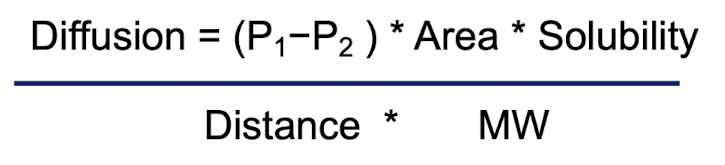
Determinants of diffusion
pressure gradient
area
distance
solubility and MV are fixed
inspired oxygen formula

total pressure is the sum of partial pressures of what 4 gases
O2, N2, CO2, H2O
a decrease in inspired oxygen concentration (FiO2) or a decrease in barometric pressure (PB) will reduce… and may cause…
reduce inspired oxygen tension (PIO2) and may cause ambient hypoxia
a special case of ambient hypoxia d/t reduced barometric pressure at altitude
altitude hypoxia
norm PIO2 is 150 mmHg at sea level results in what PaO2
normal PaO2 in the range of 80-100 mmHg
Inspired oxygen tension (PIO2) must be
sufficient
PIO2 is deteremined by
barometric pressure and FiO2
the effectiveness of gas exchange and oxygen transfer across the lung can be assessed by
measurement of arterial oxygen tension (PaO2)
pulse oxi (SpO2)
direct measurement of arterial oxygen sat (SaO2)
what can tell if there is adequate tissue for gas exchange
intrapulmonary shunt fraction (Qs/Qt)
calculated using clinical shunt equation
blood oxygen content equation

the blood oxygen content equation helps you break down the question of
“where is the problem?”
normal CaO2
~20mL O2/100mL blood
factors that effect Hb or SaO2 may have significant effect on
CaO2 and O2 delivery to tissues
impairments to diffusion
V/Q mismatch
reduction in the available surface area for diffusion
increase in diffusion distance
diffusion limitation causes
interstitial lung disease (ILD): inflammation, edema fibrosis → increase distance, stiff lungs
pulmonary vascular disease
emphysema: destruction of alveolar walls → decrease surface area
alveolar or interstitial edema: fluid in interstitium/alveoli → increase distance
hypoxemia from diffusion defects can appear during exercise which can be reversed with
administration of low-moderate O2
causes of V/Q mismatching
hypoventilation
ventilation-perfusion mismatch (low V/Q and right-to-left shunt)
diffusion limitations
V/Q <1.0 but >0 means
under ventilation with respect to perfusion
responds well to low-mod O2
V/Q = 0 means
absent ventilation with respect to perfusion
shunt → atelectasis
V/Q > 1.0 means
over ventilation with respect to perfusion
ventilation > perfusion
often related to low blood flow
V/Q = n/o or undefined
ventilation without perfusion
dead space units
no ventilation and no perfusion
silent unit
causes of low V/Q
asthma
bronchiectasis
bronchospasm
bronchiolar mucosal edema
COPD
cystic fibrosis
decreased Vt
focal pneumonia
partial airway obstruction
regional increases in fibrotic tissue
retained secretions
causes of pulmonary capillary shunt (V/Q = 0)
ARDS
atelectasis
complete airway obstruction
consolidative pneumonia
pneumothorax (large)
pulmonary edema (with complete alveolar filling)
pts with significant pulmonary capillary shunt may experience
refractory hypoxemia
tx for refractory hypoxemia
requires PEEP/CPAP and mechanical ventilation to recruit alveoli
high FiO2 will barely increase PaO2 in this case
causes of alveolar dead space
pulmonary embolus (w/ complete occlusion of pulmonary vessel)
obliteration of the pulmonary capillaries (emphysema)
what lung goes down to increase PaO2 in unilateral lung disease
“good” lung goes down
what is inhaled nitric oxide (iNO)
pulmonary vasodilator
colorless, odorless, highly diffusible gas
increases cGMP → vascular smooth muscle relation → pulmonary vasodilation
improves blood flow to ventilated alveoli, enhancing V/Q matching
FDA approved use of iNO
improve oxygenation and reduce need for ECMO in term and near-term neonates w/ hypoxic rsp failure and pulmonary hypertension
adult/off-label uses of iNO
ARDS (no mortality benefit)
pulmonary artery hypertension, right ventricular failure
persistent pulmonary hypertension of newborn
primary pulmonary hypertension
post-cardiac surgery
cardiac transplant
acute PE
COPD
congenital diaphragmatic hernia
sickle cell disease
testing pulmonary vascular responsiveness
what’s the most common type of bronchoscopy
flexible bronchoscopy
flexible vs rigid bronchoscopy
working channel for tools in flexible and separate channel for ventilation in rigid
what is the flexible bronchoscopy used for
diagnostic (inspection, biopsies, washings)
therapeutic (removing mucus plugs, secretions, small foreign bodies)
what is the rigid bronchoscopy used for
large or difficult foreign body removal
better control of airway and control of excessive bleeding