Swine infectious diseases: Polysystemic diseases
1/103
There's no tags or description
Looks like no tags are added yet.
Name | Mastery | Learn | Test | Matching | Spaced |
|---|
No study sessions yet.
104 Terms
What type of virus is porcine circovirus?
DNA non-enveloped, smallest virus to infect mammals
How many syndromes is porcine circovirus associated with?
10
PCV2 has been identified as causing or being associated
with disease pathogenesis of many porcine syndromes
including Porcine multisystemic wasting disease (PMWS), porcine dermatitis and nephropathy syndrome (PDNS), PCV-associated disease (PCVAD),
porcine respiratory disease complex (PRDC), acute
pulmonary edema (APE), PCV2-associated neuropathy
(PAN), reproductive failure, granulomatous enteritis,
necrotizing lymphadenitis, and exudative epidermitis.
What two serotypes of circovirus often coinfect?
PCV2a and 2b often result in coinfection
What pathogenesis is typically seen with PCV2a and 2b coinfection?
2b with 2b infection being
associated with pulmonary edema, granulomatous
enteritis, as well as lymphoid necrosis and depletion
Is PCV2a prevalent, and how is it shed?
yes, found in 50% of feral pigs. PCV has pro
longed shedding in respiratory and oral secretions and
is highly resistant within the environment.
PCV is transmitted via feces, urine, and direct transmission as well as transplacentally, through the colostrum, and via seminal flui
Are young immune to PCV?
Animals are typically 4 weeks old before clinical
signs appear, suggesting that maternal antibodies are
protective
Which serotype is more severe PCVa or PCVb?
PCVb and is becoming more prevalent and vaccines are not effective
When is PCV typically pathogenic? What typically needs to be present to have clinical manifestation?
Lesions are often only seen when animals are
coinfected with other agents such as porcine parvovirus
(PPV), porcine reproductive and respiratory syndrome
virus (PRRSV), or Mycoplasma hyopneumoniae,
which prime or activate the immune system.
What cells in the body does PCV infect, what area of the body does it replicate, and what specific fetal cells does PCS infect?
Macrophages and endothelial cells
Bone marrow and thymus
Fetal cardiocytes and hepatocytes
Which breed of pig is more predisposed to severe PCV?
Landrace are more susceptible to lesions and disease
Three criteria for PCV diagnosis?
Diagnosis of a PCV related syndrome requires presence of three criteria: (1) compatible clinical signs, (2) characteristic microscopic lesions, and (3) PCV2 within lesions
Is there a vaccine for PCV and if so what strain?
Several inactivated subunit vaccines based on PCV2a are commercially available and are effective in controlling and preventing disease, but continued evolution of the virus may evade current vaccines
What are key pathogenic traits of Post Weaning Polysystemic Wasting Syndrome?
anemia, lymphopenia
Thymic depletion and atrophy
Clinical signs of PMWS?
clinical disease includes progressive
weight loss, lethargy, jaundice, respiratory disease, diarrhea,
lymphadenitis, and anemia. PMWS is an acquired immunodeficiencY
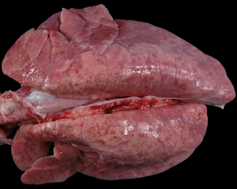
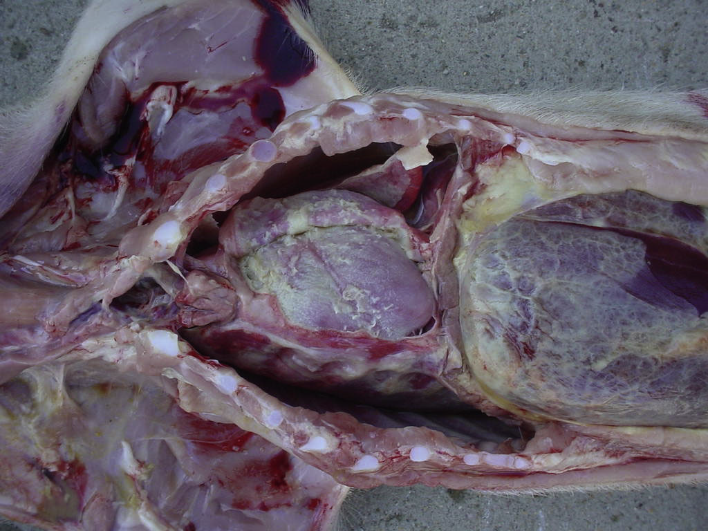
What viral syndrome is likely the etiology for this photo?
Postweaning multisystemic wasting syndrome
Key pathogenic findings on necropsy for PMWS?
Macroscopic lesions consist of generalized
lymphadenopathy, hepatitis with icterus, edema,
nephritis, and pneumonia (Krakowka et al., 2001). Lungs
fail to collapse and are mottled white to tan (Gillespie
et al., 2009). Enlarged lymph nodes have lymphoid
depletion with histiocytic replacement (Gillespie et al.,
2009; Rosell et al., 2000).
Other common lesions include
interstitial pneumonia (Figs 16.10, 16.11) and interstitial
nephritis, granulomatous inflammation in the liver,
spleen, tonsil, thymus, and Peyer’s patches (Chae, 2004).
Intracytoplasmic botryoid inclusion bodies (Fig. 16.12)
are common in epithelial cells of the bronchi, renal
tubules, and bronchial glands as well as within macrophages
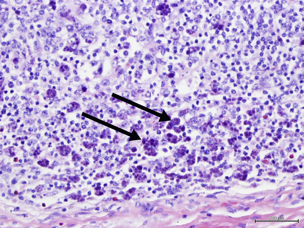
What viral syndrome is the cause of these botryoid inclusion bodies indicated by the black arrows?
PMWS
What is the pathogenesis of Porcine Dermatitis and Nephropathy Syndrome?
The current hypothesis is that immune complex deposition is involved in pathogenesis. This disease is not always associated with PCV2, but may also be caused by coinfection of PRRSV and TTV
Clinical features of PDNS?
fever, lethargy, and raised purple lesions on the skin,
especially the rear legs
Histologic presentation of PDNS?
Gross and histological lesions are present in the skin and kidney.
Systemic vasculitis is a hallmark lesion of PDNS. Histologically,
the vasculitis is similar to type III hypersensitivity.
Clinical signs of Porcine Respiratory Disease Complex?
Animals with PRDC have a decreased growth rate, decreased feed efficiency lethargy, anorexia, fever, cough, and dyspnea
Necropsy findings for PRDC?
The hallmark lesion is granulomatous
bronchointerstitial pneumonia with peribronchial and
peribronchiolar fibrosis
Pathogenesis of Reproductive Failure Circovirus?
Infection of piglets is transplacental
from PCV2-infected sows (Mateusen et al., 2007). Target
cells depend on age. Myocardiocytes are the target cell
of fetuses, whereas lymphoid tissues are targeted in
neonates
Clinical presentation of RFCV (Reproductive failure circovirus), what ages are affected? Does gestation time of infection matter?
If the sow is infected at 57 days of gestation, there
is increased viral replication with edema, hepatomegaly,
and congestion of the fetus, whereas if infected around
90 days of gestation, there are increased reproductive
abnormalities (Gillespie et al., 2009). PVC2 infection of
pregnant sows can cause fetal death leading to mummification
or late-term abortion, stillbirths, mummification
(often in the same litter), and preweaning mortality.
In piglets, the heart is the most common organ affected and superficial lymph nodes may be enlarged
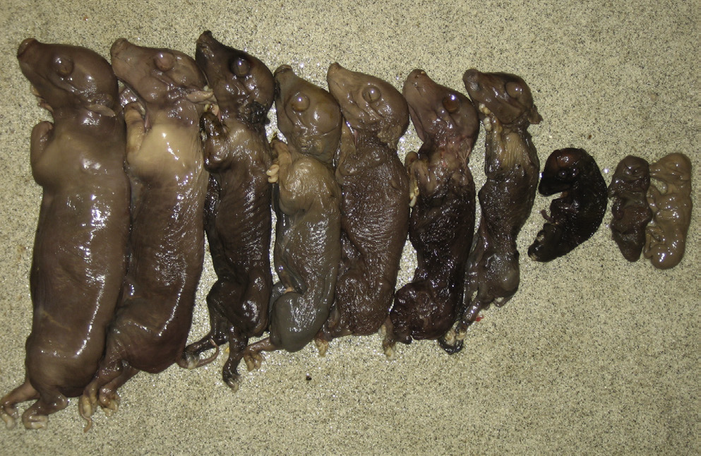
Which pathogen causes abortion at different gestational ages?
Reproductive failure circovirus

What potential virus could cause the associated lesions?
PDNS
What is the clinical manifestation of PCV2-associated granulomatous enteritis and what age pig does it predominate?
Clinically, PCV2-associated granulomatous enteritis affects 2- to 4-month old pigs and resembles chronic ileitis with diarrhea,
unthriftiness, decreased growth, and increased mortality
What infectious agent is PCV2-associated granulomatous enteritis indistinguishable from?
Lesions are of a necrotizing ileitis and
colitis indistinguishable grossly from proliferative ileitis
caused by L. intracellularis
What is the clinical presentation of PCV associated Acute Pulmonary Edema Syndrome?
Animals display a rapid onset of respiratory
distress followed nearly immediately by death
with no indications of previous disease
Common necropsy finding of PCV associated Acute Pulmonary Edema Syndrome?
Clear fluid accumulates within the thorax with wet, heavy lungs and expansion of interlobular septae with edema
Common necropsy finding of PCV2-Associated Neuropathy?
acute hemorrhages and edema of cerebellar meninges and parenchyma
Which salmonella is the most antimicrobial resistant found in pigs?
S. enterica ser. Choleraesuis var. kunzendorf
Most common Salmonella found in pigs of north america
?
S. enterica ser. Typhimurium is the
most frequently isolated serotype in North America and
typically causes enterocolitis.
Which salmonella in pigs is
is associated with localized epizootics characterized by
chronic wasting, caseous lymphadenitis, diarrhea, and
pneumonia?
S. enterica ser. Typhisuis
Transmission of salmonella in pigs?
The source of
S. enterica ser. Choleraesuis for swine is typically other
swine and environments contaminated by swine (or
other animals).
Transmission is
both vertical and horizontal
Stress:Some form of stress, including shipping, food deprivation, concurrent diseases, research protocols, and mixing pigs from different sources, usually precedes clinical disease. Stress also increases shedding by inapparent carriers.
What compounds in Salmonella are important for pathogenesis?
Lipid A and LPS damage and microvascular thrombosis and endothelial necrosis which often lead to mucosal ischemia.
What are the other names for Glassers disease?
Glasser’s Disease (Haemophilus, Porcine
Polyserositis, Infectious Polyarthritis, Fibrinous
Polyserositis, and Arthritis)
What is the etiologic agent for Glassers disease in pigs?
Haemophilus parasuis, gram - pleomorphic coccibacilii
Where on the pig is H. parasuis commonly isolated from?
cultured from the nasal cavities of swine
Who does H. parasuis typically coinfect with?
This organism is an opportunistic pathogen with PRRSV, PCV2, and B. bronchiseptica.
Acute and chronic manifestation of H. parasuis?
In susceptible herds, the clinical
signs occur within a week after exposure and consist of
some or all of the following: pyrexia 40–41.7°C, anorexia,
coughing, depression, swollen joints with lameness, neurological
signs, dyspnea, and sudden death. Long-term sequelae include
abortion and chronic arthritis.
Can H. parasuis be treated?
Yes with high dose penicilin
Pathology of H. parasuis?
Gross lesions may include cyanosis of the
ears and tail, polyarthritis of one or more joints, fibrinous
pleuritis, pericarditis, peritonitis, and leptomeningitis
Causitive agent of Swine erysipelas?
caused by Erysipelothrix rhusiopathiae, gram positive bacillus
What are the two most common serotypes of Swine erysipelas?
The majority of isolates from
swine are serotypes 1 and 2.
Which species is the primary reservoir for erysipelas?
The domestic pig is the primary reservoir of E. rhusiopathiae,
Where in the pig does erysipelas live and spread from?
These pigs harbor the bacteria in lymphoid tissues (tonsils, Peyer’s patches) and shed it in nasal secretions, saliva, and feces.
Contact with which species…hint hint bird…. increases the risk of pigs acquiring erysipelas?
Additionally, contact with infected sheep,
turkeys, chickens, ducks, and emus is a potential source
of infection for swine
What age group of pigs are most likely to present with clinical erysipelas?
Swine older than 3 months and younger than 3 years of age are most likely to develop clinical disease. Passive
How is erysipelas transmitted?
The organisms gain entry to the body
via the palatine tonsils or gut-associated lymphoid tissue, and through skin wounds
Typical clinical presentation of erysipelas?
Animals with acute SE
may have no clinical signs or have a combination of
classical rhomboid or diamond-shaped urticarial (pink
to purple) skin lesions on the snout, ears, abdomen,
and thighs; fever of 40–42°C; anorexia; depression;
stiff, stilted gait; sitting posture; abortion; and sudden
death
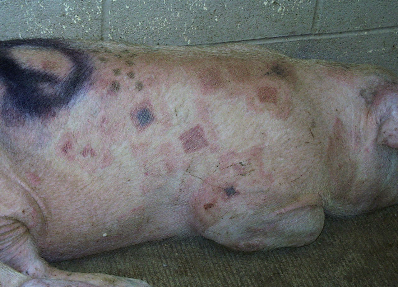
What is the likely pathogen?
erysipelas
What is the likely pathogen? Presented with limping
H. parasuis?
Why would a pig be dog sitting with erysipelas?
Because of the arthtitis it causes
Treatment of choice for Erysipelas?
Penicillin
Is there a vaccine for erysipelas and are they effective?
Yes and not for eliminating chronic SE
What are the microscopic lesions of erysipelas look like on a pigs skin?
Microscopic
lesions in the acute phase are the result of damage done
to endothelial cells in capillaries and venules
Is erysipelas long lived or short lived in the environment?
long lived
Is erysipelas zoonotic?
Yes
Streptococcosis (Streptococcal Meningitis) is caused by what bacteria?
Streptococcus suis lancfield group D, capsular type 2 being the most common.
S. equisimilis, much less common
Transmission of Strep suis?
Transmission between herds is via flies and carrier animals. Newborns are infected during parturition and suckling
What age group is strep sui typically seen in pigs?
the signs are usually first evident in
recently weaned young between 5 and 12 weeks of
age.
Where do subclinical carriers retain Strep suis in the body?
Subclinical carriers harbor the organism in their tonsilar crypts, nasal cavity, and reproductive and gastrointestinal tracts
Where does pathogenesis start for Strep suis in the pig?
Pathogenesis of S. suis is believed
to begin via colonization of the palatine and pharyngeal
tonsils.
What is the most well-characterized virulence factor for Strep suis?
Hemolysin (suilysin)
is one of the best-characterized virulence factors and
is toxic to epithelial, endothelial, and phagocytic cells
Most common clinical sign of strep suis?
Manifestations of meningitis are the most characteristic signs of S. suis type 2 infections in swine, and swine aged 5–16 weeks are most commonly affected. Pyrexia to 42.5°C is usually the initial sign, followed by anorexia, depression, ataxia, paddling, opisthotonus, convulsions, and death. Additional signs of S. suis infection include pneumonia, rhinitis, polyarthritis, and less commonly, stillbirths, abscesses, vaginitis, and myocarditis
Diagnosis of strep suis?
Isolation of the organism from an area other than the nasal or oral cavity (where organism is part of normal flora) is required,
Best control and prevention strategy for strep suis?
depopulation with subsequent repopulation
with clean animals is a feasible method of control
and prevention. S. suis is susceptible to common disinfectants.
Is streptococcus suis zoonotic?
S. suis type 2 is zoonotic to humans
Causitive agent of Aujeszky’s disease (pseudorabies)
Suid herpes virus 1
What is pseudorabies?
DNA enveloped virus, The disease is caused by suid her1 in the genus Varicellovirus, family Alphaherpesvirinae, Herpesviridae. Herpesviridae are known for the ability to
establish latent infections, particularly in the sensory
ganglia of the nervous system.
Is pseudorabies reportable?
Yes
What year was the US declared disease free of pseudorabies?
in 2005 was declared to be free of the disease
What other animals can pseudorabies affect?
pigs, cattle, sheep, goats, dogs, cats, rodents,
macaques, and marmosets.
How does presentation differ between pigs and other mammals with suid herpes virus 1 (pseudorabies)?
Pigs may host subclinical and latent infections
whereas infection of all other animals results in death. Animals other than pigs, which are considered dead-end hosts, typically die within 3 days of being infected.
Transmission of pseudorabies?
Transmission can occur via direct contact,
fomites, insemination, inhalation of aerosolized particles,
or transplacental transmission.
Pathogenesis of suid herpes virus 1 (pseudorabies) and what nerve does it prefer to cause latent infection in?
the virus enters via the mucosal epithelium in the nasopharynx and tonsils and replicates in the epithelium. resulting in latent
infection of the trigeminal ganglia
What are the clinical signs of pseudorabies (suid herpes virus 1):
The virus predominantly impacts the respiratory and nervous systems.
Affected pigs will tremble, hypersalivate, stumble, and exhibit nystagmus and opisthotonus, often with epileptiform-like seizures.
Because of posterior paresis, the animals may
be observed sitting like a dog
Once CNS signs start, death usually follows within 24–36 h, and mortality approaches 100% in neonates and young pig
How does the dog sitting clinical sign differ between erysipelas and pseudorabies infection?
Erysipelas due to pain, Pseudorabies due to neurologic disease
How does pseudorabies vary by age in disease progression?
Respiratory signs characterized by sneezing,
nasal discharge, and cough become the hallmark
of pigs that are infected at greater than 9 weeks of
age.
CNS signs in younger pigs / neonates, 100% mortality. Mortality may decrease to 50% by 4 weeks of age.
Diagnosis of pseudo rabies?
Virus isolation allows for a definitive diagnosis, with the brain, tonsils, and lung being the organs of choice
Does the vaccine work and how does it work?
Modified live, killed, and gene-deleted vaccines with foreign-gene insertions are available to aid in the control of PRV. The vaccines protect pigs against clinical signs and mortality but do nothing toward eradicating the disease; the vaccine does not eliminate the virus in infected animals, nor does it prevent animals from becoming infected with the virus
Gross lesions of pseudorabies?
a fibrinonecrotic rhinitis; necrotic foci in the tonsils, liver, spleen, lungs, intestines, and adrenals;
Common histopath finding in neural tissue of pseudorabies positive pigs?
Eosinophilic intranuclear inclusions
What is the etiology of encephalomyocarditis virus?
Encephalomyocarditis virus (EMCV) is found in the genus Cardiovirus, family Picornaviridae. RNA non-enveloped
What species is the reservoir of EMC?
Rodents are thought to be a reservoir
Does EMC commonly cause clinical disease?
No not typically
What is the clinical presentation of EMC and in what pig age group?
1.An acute myocarditis and reproductive failure in sows
In young pigs the only clinical sign may be sudden death.
Which area of the heart is most affected by EMC?
Myocardial lesions are most prominent in the right
ventricle and are grayish-white in color.
Where is the highest risk of zoonosis for EMC?
The risk of human infection
may increase if porcine-to-human xenografts are
performed.
What is porcine teschovirus?
Teschen disease is caused by porcine
teschovirus, a member of the family Picornaviridae,
genus Teschovirus. RNA Non-enveloped.
IS Porcine teschovirus host specific?
Yes
Where does Porcine Teschovirus replicate?
The virus replicates in the tonsil
and intestinal tract.
Most common clinical manifestation of Porcine Teschovirus is?
Teschen disease is caused by a highly virulent PTV-1
strain: All ages are affected and the main presentation
is polioencephalomyelitis
What is talfan disease and how does it differ from Porcine Teschovirus?
Talfan disease is caused by a less virulent PTV-1 strain.
Signs are milder than in Teschen disease and typically
consist of benign enzootic paresis.
Is Porcine Teschovirus reportable?
Yes